

The Journal of Practical Medicine ›› 2025, Vol. 41 ›› Issue (19): 3026-3033.doi: 10.3969/j.issn.1006-5725.2025.19.010
• Clinical Research • Previous Articles
Xuman LU1,2,Zhengyi SHI3,Yuanrui LEI2,Haibin HUANG3,Renmiao DENG3,Xudong DONG3,Yuliang HUANG3,Fanbiao KONG3,Xiaotong. WANG2( )
)
Received:2025-06-18
Online:2025-10-10
Published:2025-10-10
Contact:
Xiaotong. WANG
E-mail:008.wxt@163.com
CLC Number:
Xuman LU,Zhengyi SHI,Yuanrui LEI,Haibin HUANG,Renmiao DENG,Xudong DONG,Yuliang HUANG,Fanbiao KONG,Xiaotong. WANG. The expression of PCBP1 in gastric cancer and its relationship with ferroptosis factor STUB1[J]. The Journal of Practical Medicine, 2025, 41(19): 3026-3033.
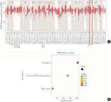
Fig.1
PCBP1 bioinformatics analysis results"
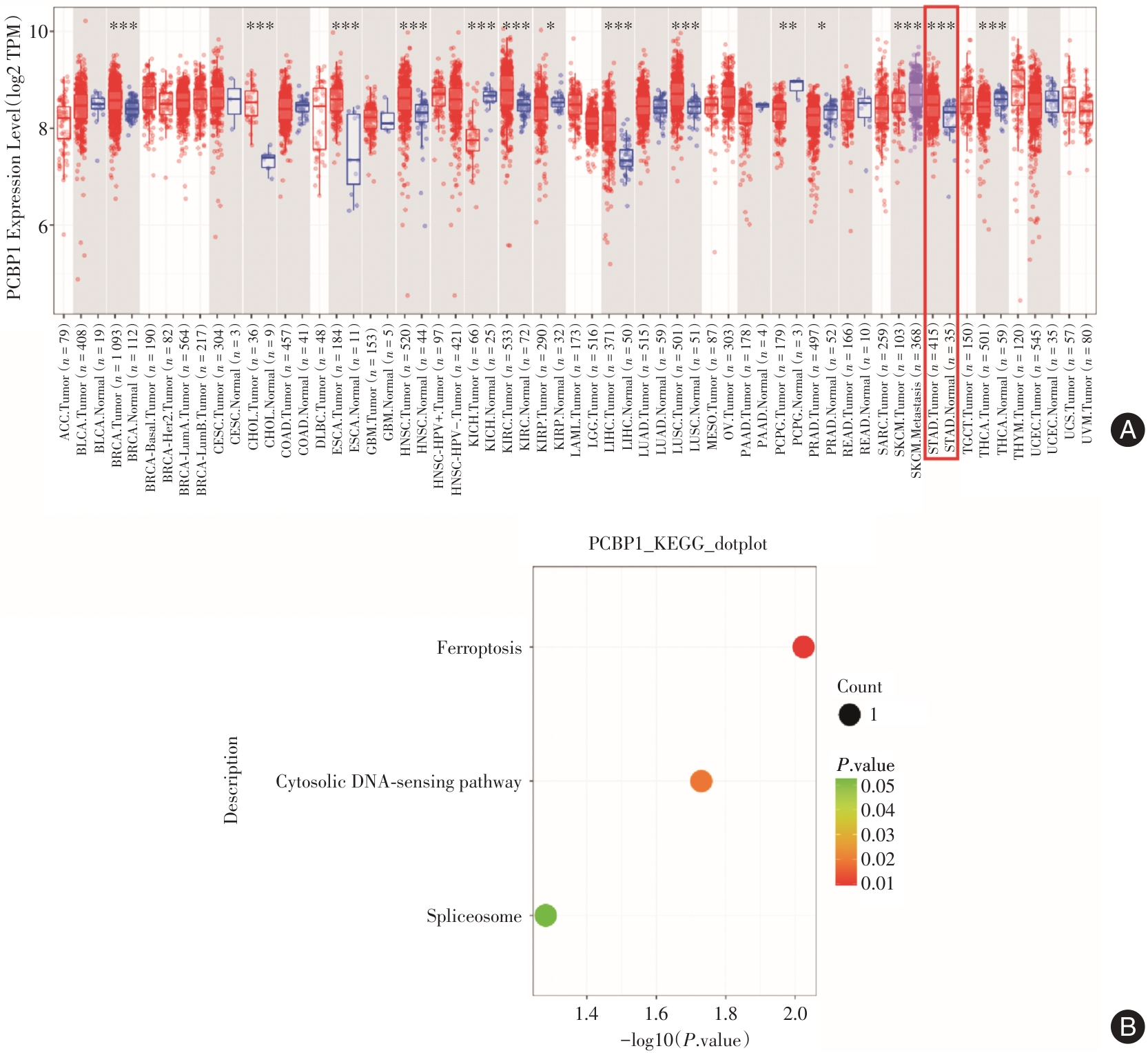
Fig.2
Immunohistochemical results of PCBP1 and STUB1"
Tab.1
The relationship between the expression of PCBP1 ( cancer ) and STUB1 ( cancer ) and clinicopathological features of gastric cancer"
| 临床病理特征 | 例数 | PCBP1(癌) | χ2值 | P值 | STUB1(癌) | χ2值 | P值 | ||
|---|---|---|---|---|---|---|---|---|---|
| 阳性/例 | 阳性率/% | 阳性/例 | 阳性率/% | ||||||
| 年龄 | 0.762 | 0.311 | 0.047 | 0.556 | |||||
| ≤ 60岁 | 16 | 10 | 62.5 | 6 | 37.5 | ||||
| > 60岁 | 17 | 13 | 76.5 | 7 | 41.2 | ||||
| 性别 | 2.158 | 0.148 | 1.528 | 0.204 | |||||
| 男 | 24 | 15 | 62.5 | 11 | 45.8 | ||||
| 女 | 9 | 8 | 88.9 | 2 | 22.2 | ||||
| 分化程度 | 13.295 | < 0.001 | 10.452 | 0.020 | |||||
| 低分化 | 19 | 18 | 94.7 | 3 | 15.8 | ||||
| 中分化 | 14 | 5 | 35.7 | 10 | 71.4 | ||||
| 浸润深度 | 0.120 | 0.512 | 4.891 | 0.031 | |||||
| T1—T2 | 15 | 10 | 66.7 | 9 | 60.0 | ||||
| T3—T4 | 18 | 13 | 72.2 | 4 | 22.2 | ||||
| 淋巴结转移 | 10.705 | 0.002 | 9.909 | 0.030 | |||||
| 有 | 23 | 20 | 87.0 | 5 | 21.7 | ||||
| 无 | 10 | 3 | 30.0 | 8 | 80.0 | ||||
| 临床分期 | 1.963 | 0.154 | 0.863 | 0.284 | |||||
| Ⅰ—Ⅱ期 | 17 | 10 | 58.8 | 8 | 47.1 | ||||
| Ⅲ—Ⅳ期 | 16 | 13 | 81.3 | 5 | 31.3 | ||||
| Lauren分型 | 9.081 | 0.011 | 15.917 | < 0.001 | |||||
| 肠型 | 10 | 4 | 40.0 | 9 | 90.0 | ||||
| 混合型 | 7 | 4 | 57.1 | 2 | 28.6 | ||||
| 弥漫型 | 16 | 15 | 93.8 | 2 | 12.5 | ||||
| 脉管侵犯 | 1.148 | 0.256 | 1.015 | 0.267 | |||||
| 有 | 11 | 9 | 81.8 | 3 | 27.3 | ||||
| 无 | 22 | 14 | 63.6 | 10 | 45.5 | ||||
| 神经浸润 | 4.591 | 0.053 | 0.346 | 0.442 | |||||
| 有 | 27 | 21 | 77.8 | 10 | 37.0 | ||||
| 无 | 6 | 2 | 33.3 | 3 | 50.0 | ||||
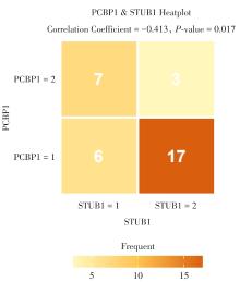
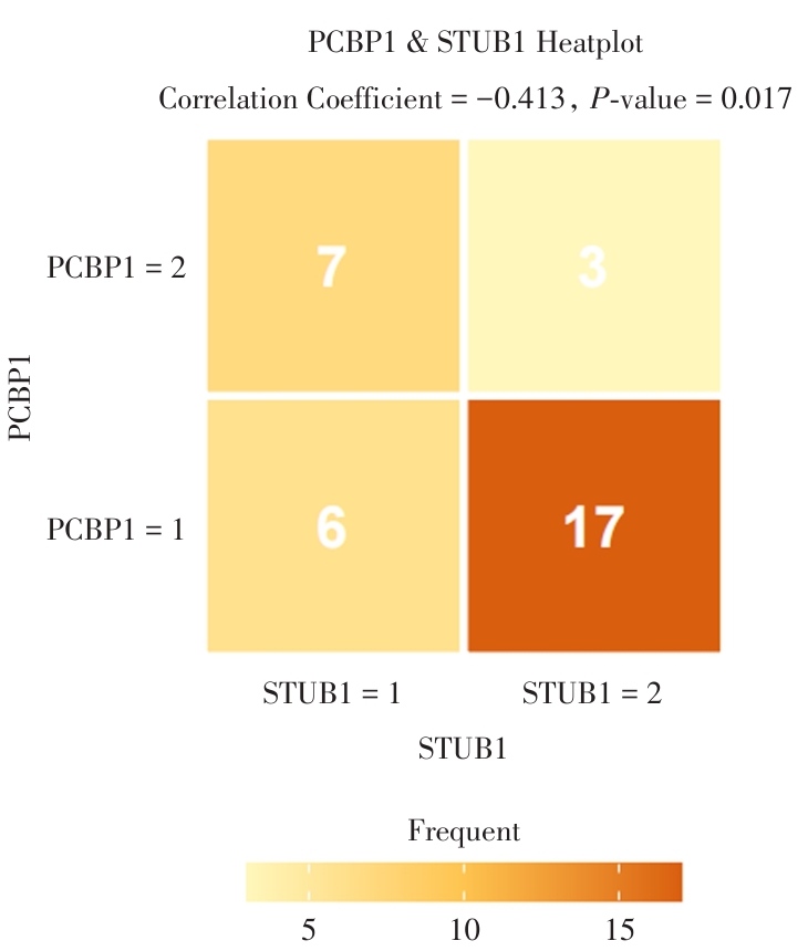
Fig.3
Correlation analysis of PCBP1 and STUB1 in gastric cancer tissues"
Tab.2
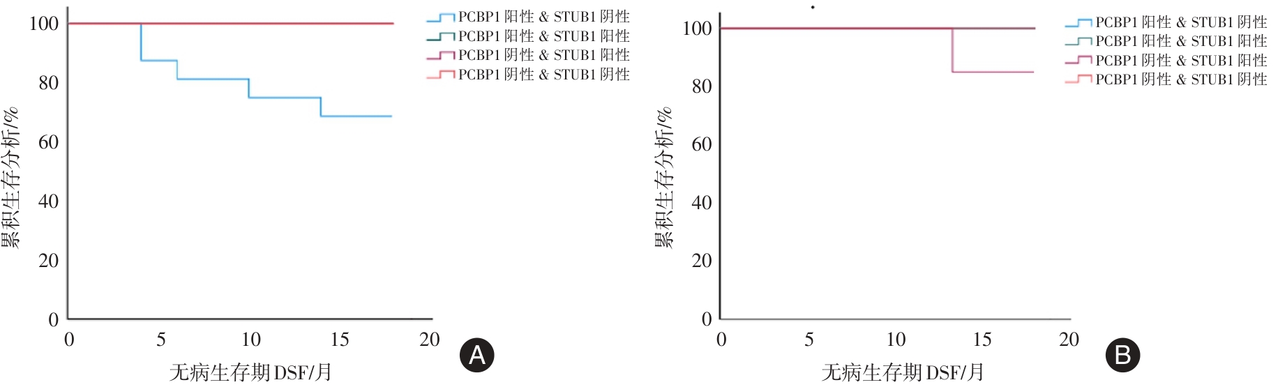
Follow-up of PCBP1 and STUB1 co-expression"
| 组合分组 | 例数 | 失访数 | 复发数 | 死亡数 |
|---|---|---|---|---|
| PCBP1阳性& STUB1阴性 | 17 | 1 | 5 | 0 |
| PCBP1阳性& STUB1阳性 | 6 | 0 | 0 | 0 |
| PCBP1阴性& STUB1阳性 | 7 | 0 | 0 | 1 |
| PCBP1阴性& STUB1阴性 | 3 | 0 | 0 | 0 |
| χ2值 | 7.863 | 3.158 | ||
| P值 | 0.049 | 0.368 |
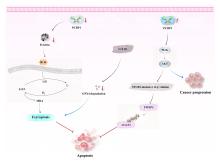
Fig.4
Regulatory mechanism of PCBP1 and STUB1 in gastric cancer"
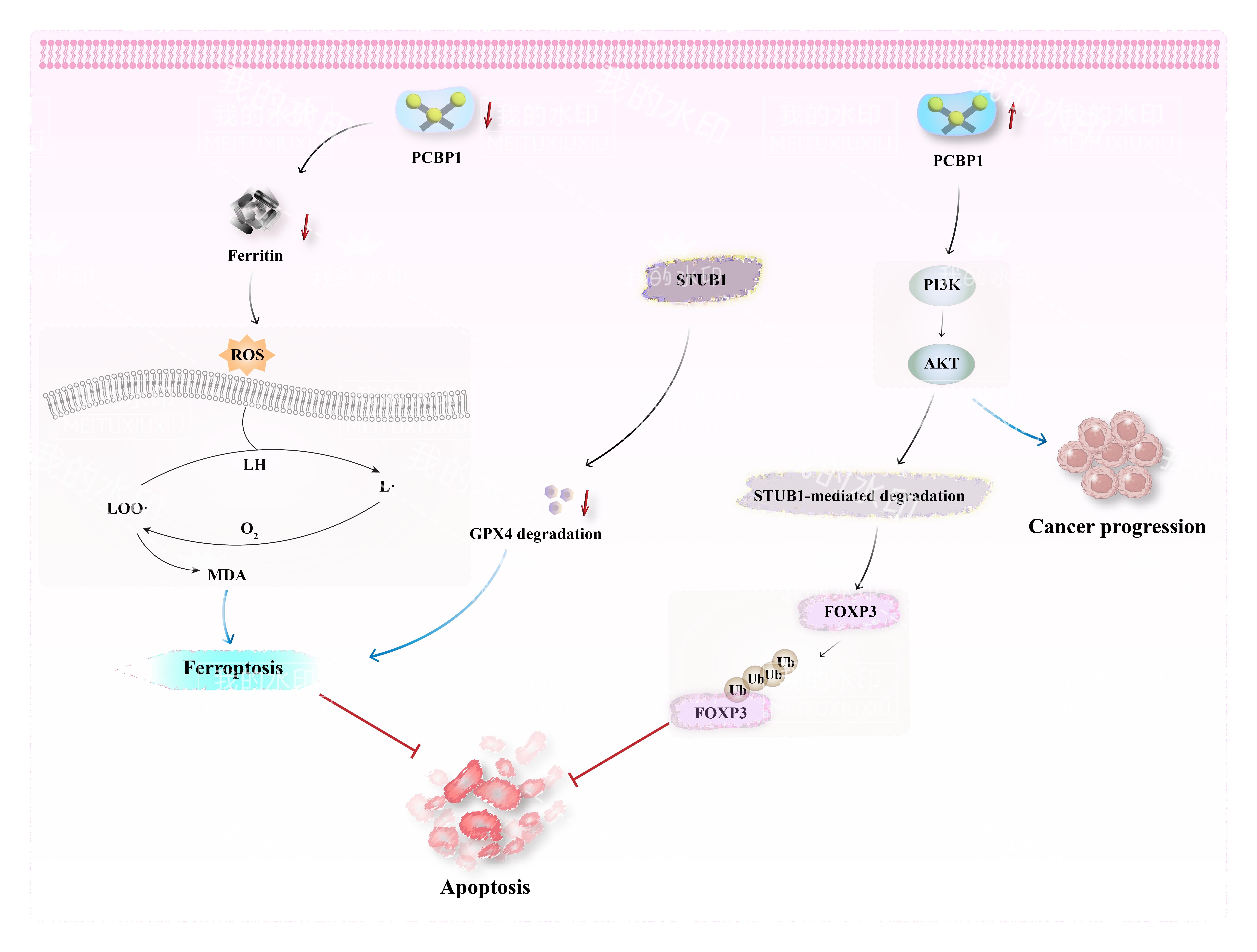
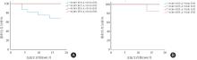
Fig.5
Survival curves of PCBP1 positive & STUB1 negative, PCBP1 positive & STUB1 positive, PCBP1 negative & STUB1 positive, PCBP1 negative & STUB1 negative patients"
| [1] |
BRAY F, LAVERSANNE M, SUNG H, et al. Global Cancer Statistics 2022: GLOBOCAN Estimates of Incidence and Mortality Worldwide for 36 Cancers in 185 Countries[J]. CA Cancer J Clin,2024,74(3):229-263. doi:10.3322/caac.21834
doi: 10.3322/caac.21834 |
| [2] | 丁平安,杨沛刚,田园,等. 胃癌伴左锁骨上淋巴结转移患者的临床病理特征及预后 [J]. 实用医学杂志, 2021, 37 (13): 1695-1700. |
| [3] |
NAYAK A, KUMAR S, SINGH S P, et al. Oncogenic potential of ATAD2 in stomach cancer and insights into theprotein-protein interactions at its AAA + ATPase domain and bromodomain[J].J Biomol Struct Dyn,2022,40(12):5606-5622. doi:10.1080/07391102.2021.1871959
doi: 10.1080/07391102.2021.1871959 |
| [4] | 贾冰冰, 叶梦, 霍丽蓉. PCBP1相关生物功能的研究进展[J]. 医学研究杂志, 2018, 47 (9): 176-179+135. |
| [5] |
SHI H, BENCZE K Z, STEMMLER T L,et al. A cytosolic iron chaperone that delivers iron to ferritin[J]. Science, 2008,320(5880):1207-1210. doi:10.1126/science.1157643
doi: 10.1126/science.1157643 |
| [6] |
WANG X Q, FAN A Q, HONG L. LncRNA MIR210HG promotes the proliferation of colon cancer cells by inhibiting ferroptosis through binding to PCBP1[J]. Sci Rep,2025,15(1):871. doi:10.1038/s41598-025-85321-7
doi: 10.1038/s41598-025-85321-7 |
| [7] |
WEI F, MENG Y, ZHU D X, et al. Mechanism and role of ferroptosis in the development of gastric cancer[J]. Clin Exp Med,2025,25(1):182. doi:10.1007/s10238-025-01722-y
doi: 10.1007/s10238-025-01722-y |
| [8] |
ZHANG L, LUO Y L, XIANG Y, et al. Ferroptosis inhibitors: Past, present and future[J]. Front Pharmacol,2024,15:1407335. doi:10.3389/fphar.2024.1407335
doi: 10.3389/fphar.2024.1407335 |
| [9] |
CUI S, SIMMONS G J, VALE G,et al. FAF1 blocks ferroptosis by inhibiting peroxidation of polyunsaturated fatty acids[J]. Proc Natl Acad Sci USA,2022,9:e2107189119. doi:10.1073/pnas.2107189119
doi: 10.1073/pnas.2107189119 |
| [10] |
SHAO M, QI K, WANG L,et al. E3 ubiquitin ligase CHIP interacts with transferrin receptor 1 for degradation and promotes cell proliferation through inhibiting ferroptosis in hepatocellular carcinoma[J]. Cell Signal,2024,118:111148. doi:10.1016/j.cellsig.2024.111148
doi: 10.1016/j.cellsig.2024.111148 |
| [11] | 张银亮,骆泽谭,赵睿,等. 血根碱通过调控STUB1/GPX4诱导直肠癌细胞发生铁死亡[J]. 南方医科大学学报,2024,44(8):1537-1544. |
| [12] | 王衍颜,张军,毛沛,等. 肺鳞状细胞癌组织PCBP1、GPSM2表达与EMT、临床病理特征和预后的关系[J]. 现代生物医学进展,2024,24(19):3654-3656+3666. |
| [13] |
郑荣寿,张思维,孙可欣,等. 2016年中国恶性肿瘤流行情况分析[J].中华肿瘤杂志,2023,45(3):212-220. doi:10.3760/cma.j.cn112152-20220922-00647
doi: 10.3760/cma.j.cn112152-20220922-00647 |
| [14] | 覃黎明,周子寒,曹骥,等. 2011—2018年广西胃癌流行特征及疾病负担分析[J]. 中国癌症防治杂志,2024,16(02):172-179. |
| [15] | 范文凭,林琳,刘江美,等.基于GBD数据分析1990—2019年中国恶性肿瘤疾病负担趋势[J].中国肿瘤,2024,33(1):20-26. |
| [16] |
CHOI H S, HWANG C K, SONG K Y,et al. Poly(C)-binding proteins as transcriptional regulators of gene expression[J]. Biochem Biophys Res Commun,2009,380(3):431-436. doi:10.1016/j.bbrc.2009.01.136
doi: 10.1016/j.bbrc.2009.01.136 |
| [17] | 杨杰,李卫,刘贺,等. 应用酵母双杂交筛选人胃癌耐药细胞SIVA-1基因相互作用蛋白[J]. 现代肿瘤医学,2023,31(5):806-811. |
| [18] |
KONG F B, SHI Z Y, HUANG Y L,et al. SIVA-1 interaction with PCBP1 serves as a predictive biomarker for cisplatin sensitivity in gastric cancer and its inhibitory effect on tumor growth in vivo[J]. J Cancer,2024,15(13):4301-4312. doi:10.7150/jca.92963
doi: 10.7150/jca.92963 |
| [19] |
LIU R Q, WU Y T, CHENG Y,et al.TBBPA induced hepatocyte ferroptosis by PCBP1-mediated ferritinophagy[J]. J Hazard Mater,2025,494:138515. doi:10.1016/j.jhazmat.2025.138515
doi: 10.1016/j.jhazmat.2025.138515 |
| [20] |
JAIN C, SHAH Y M. PCBP1 is essential for proper iron absorption[J]. Blood, 2023,142(19):1585-1587. doi:10.1182/blood.2023022267
doi: 10.1182/blood.2023022267 |
| [21] |
PENG K, CHEN X, LIN A,et al. PolyC-RNA-binding protein 1 (PCBP1) enhances tropomyosin 3 (TPM3) mRNA stability to promote the progression of esophageal squamous cell carcinoma[J]. Bioengineered,2022,13(4):8581-8592. doi:10.1080/21655979.2022.2053801
doi: 10.1080/21655979.2022.2053801 |
| [22] |
MEI W, WEI M, TANG C,et al. BCAT2 binding to PCBP1 regulates the PI3K/AKT signaling pathway to inhibit autophagy-related apoptosis and ferroptosis in prostate cancer[J]. Cell Death Dis,2025,16(1):337. doi:10.1038/s41419-025-07559-3
doi: 10.1038/s41419-025-07559-3 |
| [23] |
JIANG Y, ZHOU H Z, ZHANG J X,et al. LINC00926, Regulated by TCF12, Modulates the Ubiquitination of GPX4 to Regulate Ferroptosis by Interacting with STUB1 in HUVECs[J].Mol Biotechnol,2025,doi: 10.1007/s12033-025-01441-5 .
doi: 10.1007/s12033-025-01441-5 |
| [24] |
ZHANG X, ZHANG Y, LI R,et al. STUB1-mediated ubiquitination and degradation of NSUN2 promotes hepatocyte ferroptosis by decreasing m5C methylation of Gpx4 mRNA[J]. Cell Rep,2024,43(11):114885. doi:10.1016/j.celrep.2024.114885
doi: 10.1016/j.celrep.2024.114885 |
| [25] |
TIELIWAERDI A, AINI A, AMUTI M,et al. STUB1 promotes the degradation of HSPB1 and induces ferroptosis in lung cancer cells[J]. Environ Toxicol,2024,39(8):4156-4170. doi:10.1002/tox.24296
doi: 10.1002/tox.24296 |
| [26] |
SUN X, ZHANG Q, LIN X,et al. Imatinib induces ferroptosis in gastrointestinal stromal tumors by promoting STUB1-mediated GPX4 ubiquitination[J]. Cell Death Dis,2023,14(12):839. doi:10.1038/s41419-023-06300-2
doi: 10.1038/s41419-023-06300-2 |
| [27] |
ZHANG L, LI Q, XU J,et al. Cimetidine promotes STUB1-mediated degradation of tumoral FOXP3 by activating PI3K-Akt pathway in gastric cancer[J]. Ann Transl Med,2020,8(20):1304. doi:10.21037/atm-20-6070
doi: 10.21037/atm-20-6070 |
| [28] |
DAI H, CHEN H, XU J,et al.The ubiquitin ligase CHIP modulates cellular behaviors of gastric cancer cells by regulating TRAF2[J].Cancer Cell Int,2019,19:132. doi:10.1186/s12935-019-0832-z
doi: 10.1186/s12935-019-0832-z |
| [1] | Tong DUAN,Qi WU,Hui. LIU. Correlation between iron death⁃related gene expression level and myocardial dysfunction and prognosis in sepsis [J]. The Journal of Practical Medicine, 2025, 41(6): 846-851. |
| [2] | Zonglin LI,Chunlin FENG,Xin LIU,Xingming SHU,Min. SONG. CCCTC⁃binding factors promote the formation of oxaliplatin related gastric cancer drug-tolerant cells by resisting apoptosis [J]. The Journal of Practical Medicine, 2025, 41(4): 490-499. |
| [3] | Danping WANG,Yufeng CAI,Dehua HU,Liang. ZHANG. The role of LncRNA RMST in gastric cancer: Expression levels, diagnostic value, and prognostic implicationssion [J]. The Journal of Practical Medicine, 2025, 41(3): 409-413. |
| [4] | Weizun LI,Chen XING,Hengqing AN. STAT activation inhibitory protein 2 is involved in reprogramming of lipid metabolism by regulating the prostate cancer development [J]. The Journal of Practical Medicine, 2025, 41(18): 2844-2852. |
| [5] | Jian ZHAO,Min DAI,Ran SUN,Yajun XU. Analysis of the relationship between Hp infection and the expression of p27, CyclinD1, MMP⁃9 proteins and the related microRNAs and clinicpathological features in gastric cancer [J]. The Journal of Practical Medicine, 2025, 41(18): 2890-2897. |
| [6] | Zhen ZHANG,Ming CHENG,Zhaoshu JIANG,Jie YANG,Zhenliang LUO,Feng. CAO. Effects of Orexin-A/OX1R/OX2R on iron death and lipid peroxidation regulation in chronic unpredictable mild stress depressed rats [J]. The Journal of Practical Medicine, 2025, 41(16): 2507-2514. |
| [7] | Xifeng XU,Xia WANG,Jianliang WU,Jinlong. CHENG. Effect of immunohistochemical detection of omentin-1, SPP1 and MMR protein expression status on clinicopathological features and prognosis analysis of endometrial cancer [J]. The Journal of Practical Medicine, 2025, 41(16): 2521-2527. |
| [8] | Qian ZHANG,Yuntai CAO,Zhijie WANG,Boqi. ZHOU. Advances in deep learning for endoscopic image⁃based diagnosis of early gastric cancer [J]. The Journal of Practical Medicine, 2025, 41(14): 2160-2166. |
| [9] | Xiaomei CHEN,Anqi WANG,Jizhen YANG,Miao YU. Prognosis and immune correlation analysis of m1A/m5C/m6A/m7G regulated genes in gastric cancer [J]. The Journal of Practical Medicine, 2024, 40(9): 1230-1237. |
| [10] | Jianliang YAN,Zeyu XIE,Rongrong JING,Ming. CUI. Research on establishing gastric cancer lymph node metastasis prediction model based on machine learning and routine laboratory indicators [J]. The Journal of Practical Medicine, 2024, 40(6): 844-849. |
| [11] | Shu CHEN,Jinglei ZHANG,Kang RONG,Nan ZHANG,Weiyi SUN. Research progress of exosomes in distant metastasis and drug resistance of gastric cancer [J]. The Journal of Practical Medicine, 2024, 40(6): 870-876. |
| [12] | Li XU,Shanshan HU,Haiming. ZHAO. LncRNA GNAS⁃AS1 participates in the proliferation and migration of gastric cancer cells by regulating the miR⁃449a/Notch1 axis [J]. The Journal of Practical Medicine, 2024, 40(4): 483-489. |
| [13] | Jian ZHAO,Songjie LIU,Guanchao ZHANG,Yuhou SHEN,Fengchen LI,Bing XU. Expression of CENPF and miR⁃1⁃3p in the serum of patients with advanced gastric cancer and their correlation with prognosis [J]. The Journal of Practical Medicine, 2024, 40(3): 365-370. |
| [14] | Jun XU,Xiaoli WANG,Jingyi NI,Didi. ZHANG. Clinical efficacy and safety of disitamab vedotin in the treatment of advanced gastric cancer [J]. The Journal of Practical Medicine, 2024, 40(20): 2913-2917. |
| [15] | Chunyan NIU,Xiaoping WANG,Xiangyang ZHAO,Jiankang HUANG,Yue CHEN,Yongqiang SHI,Yongqiang SONG,Hui WANG,Xinguo WU,Yongdan BU,Jijin LI,Tao TAO,Jinhua WU,Changlin XUE,Fuyu ZHANG,Jinming YANG,Chunrong HAN,Juan YUAN,Yinling WU,Hongbing XIONG,Peng XIAO. A multicenter population investigation on precancerous lesions of gastric cancer in Lishui District, Nanjing [J]. The Journal of Practical Medicine, 2024, 40(20): 2929-2934. |
| Viewed | ||||||
|
Full text |
|
|||||
|
Abstract |
|
|||||