

The Journal of Practical Medicine ›› 2023, Vol. 39 ›› Issue (22): 2891-2897.doi: 10.3969/j.issn.1006-5725.2023.22.006
• Feature Reports:Breast tumors • Previous Articles Next Articles
Qicheng JIN,Jianghong LV,Lilong XU,Hongfen WEI,Tongfang TANG,Chuanju ZHANG,Shiyan. LI( )
)
Received:2023-07-17
Online:2023-11-25
Published:2023-12-11
Contact:
Shiyan. LI
E-mail:lishiyan@zju.edu.cn
CLC Number:
Qicheng JIN,Jianghong LV,Lilong XU,Hongfen WEI,Tongfang TANG,Chuanju ZHANG,Shiyan. LI. Impacts of different ultrasound imaging parameters on the differential diagnosis of benign and malignant breast nodules with S⁃Detect technology[J]. The Journal of Practical Medicine, 2023, 39(22): 2891-2897.
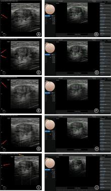
Fig.1
Two-dimensional ultrasound images of the same breast nodule under different parameter settings and their correspondingS-Detect analysis images."
Tab.1
Situation of the cases included in the study"
| 病理结果 | 合计 | |||
|---|---|---|---|---|
| 有 | 无 | |||
| 图像采集情况 | 采集完整 | 71 | 21 | 92 |
| SFLI未采集 | 39 | 12 | 51 | |
| 合计 | 110 | 33 | 143 | |
Tab.2
Comparison of the lesion shape with BPI under different imaging conditions"
| BPI | DFI( n = 143) | SFLI( n = 92) | DFLI( n = 143) | STDI( n = 143) | ||||||||
|---|---|---|---|---|---|---|---|---|---|---|---|---|
| O | R | I | O | R | I | O | R | I | O | R | I | |
| O | 63 | 0 | 10 | 40 | 2 | 1 | 62 | 5 | 6 | 62 | 4 | 7 |
| R | 3 | 3 | 1 | 2 | 3 | 1 | 2 | 4 | 1 | 4 | 3 | 0 |
| I | 3 | 1 | 59 | 5 | 2 | 36 | 11 | 1 | 51 | 21 | 4 | 38 |
| P值 | 0.080 | 0.392 | 0.431 | 0.012 | ||||||||
| K值 | 0.765 | 0.749 | 0.670 | 0.489 | ||||||||
Tab.3
Comparison of the lesion orientation with BPI under different imaging conditions"
| BPI | DFI( n = 143) | SFLI( n = 92) | DFLI( n = 143) | STDI( n = 143) | ||||
|---|---|---|---|---|---|---|---|---|
| P | NP | P | NP | P | NP | P | NP | |
| P | 119 | 8 | 74 | 5 | 118 | 9 | 125 | 2 |
| NP | 2 | 14 | 3 | 10 | 4 | 12 | 5 | 11 |
| P值 | 0.109 | 0.727 | 0.267 | 0.453 | ||||
| K值 | 0.698 | 0.663 | 0.598 | 0.732 | ||||
Tab.4
Comparison of the lesion posterior features with BPI under different imaging conditions"
| BPI | DFI( n = 143) | SFLI( n = 92) | DFLI( n = 143) | STDI( n = 143) | ||||||||||||
|---|---|---|---|---|---|---|---|---|---|---|---|---|---|---|---|---|
| NPF | E | S | CP | NPF | E | S | CP | NPF | E | S | CP | NPF | E | S | CP | |
| NPF | 42 | 8 | 2 | 0 | 29 | 4 | 3 | 1 | 41 | 8 | 3 | 0 | 37 | 10 | 4 | 1 |
| E | 9 | 53 | 0 | 2 | 5 | 27 | 1 | 0 | 14 | 49 | 1 | 0 | 16 | 46 | 1 | 1 |
| S | 1 | 0 | 12 | 2 | 3 | 0 | 9 | 2 | 1 | 0 | 12 | 2 | 2 | 0 | 13 | 0 |
| CP | 2 | 2 | 1 | 7 | 2 | 2 | 2 | 2 | 1 | 3 | 3 | 5 | 4 | 3 | 1 | 4 |
| P值 | 0.742 | 0.751 | 0.250 | 0.335 | ||||||||||||
| K值 | 0.687 | 0.595 | 0.612 | 0.536 | ||||||||||||
Tab.5
Comparison of the lesion echo pattern with BPI under different imaging conditions"
| BPI | DFI( n = 143) | SFLI( n = 92) | DFLI( n = 143) | STDI( n = 143) | ||||||||||||||||
|---|---|---|---|---|---|---|---|---|---|---|---|---|---|---|---|---|---|---|---|---|
| AE | CCS | HE | IE | H | AE | CCE | HE | IE | H | AE | CCS | HE | IE | H | AE | CCS | HE | IE | H | |
| AE | 4 | 0 | 1 | 0 | 0 | 4 | 0 | 0 | 1 | 0 | 4 | 0 | 0 | 1 | 0 | 5 | 0 | 0 | 0 | 0 |
| CCS | 0 | 2 | 1 | 2 | 0 | 0 | 1 | 1 | 0 | 0 | 0 | 1 | 2 | 1 | 1 | 0 | 2 | 3 | 2 | 0 |
| HE | 0 | 4 | 94 | 3 | 3 | 2 | 1 | 57 | 4 | 1 | 2 | 2 | 84 | 12 | 4 | 10 | 2 | 80 | 11 | 1 |
| IE | 0 | 0 | 5 | 16 | 0 | 0 | 0 | 2 | 13 | 0 | 0 | 0 | 2 | 19 | 0 | 2 | 0 | 3 | 16 | 0 |
| H | 0 | 0 | 3 | 1 | 4 | 6 | 3 | 61 | 19 | 3 | 0 | 0 | 3 | 2 | 3 | 17 | 4 | 91 | 30 | 1 |
| P值 | 0.390 | 0.579 | 0.075 | 0.001 | ||||||||||||||||
| K值 | 0.637 | 0.669 | 0.549 | 0.442 | ||||||||||||||||
Tab.6
Comparison of the judgment of the nature (benign of malignant) of lesions under different imaging conditions with BPI"
| BPI | DFI( n = 143) | SFLI( n = 92) | DFLI( n = 143) | STDI( n = 143) | ||||
|---|---|---|---|---|---|---|---|---|
| 可能良性 | 可能恶性 | 可能良性 | 可能恶性 | 可能良性 | 可能恶性 | 可能良性 | 可能恶性 | |
| 可能良性 | 68 | 9 | 44 | 2 | 69 | 8 | 76 | 1 |
| 可能恶性 | 4 | 62 | 2 | 44 | 8 | 58 | 15 | 51 |
| P值 | 0.267 | > 0.999 | > 0.999 | < 0.001 | ||||
| K值 | 0.818 | 0.913 | 0.775 | 0.771 | ||||
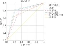
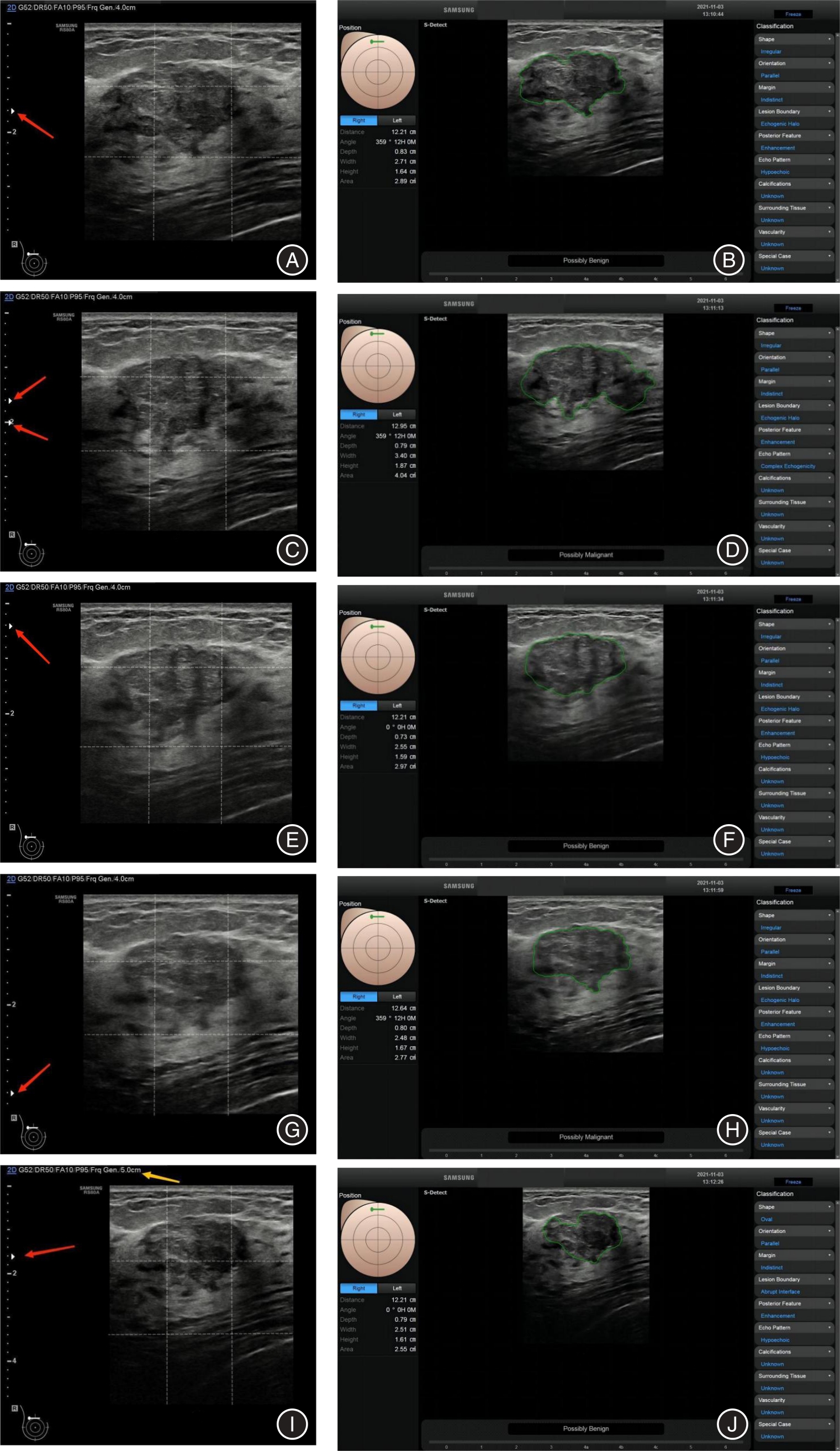
Fig.2
Comparison of diagnostic efficacy of the same breast nodule under BPI, DFI, DFLI, and STDI"
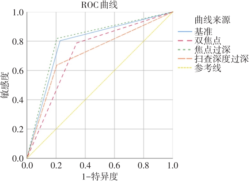
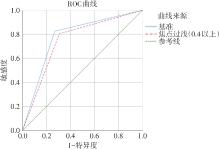
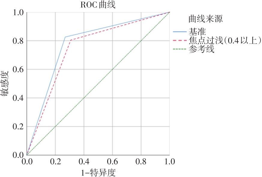
Fig.3
Comparison of diagnostic efficacy of the same breast nodule under BPI and SFLI"
| 1 | 沈松杰, 孙强, 黄欣, 等. 中国女性乳腺癌筛查指南(2022年版)[J]. 中国研究型医院, 2022, 9( 2): 6- 13. |
| 2 | 沈松杰, 孙强. 中国女性乳腺癌筛查现状及适宜模式探索[J]. 协和医学杂志, 2018, 9( 4): 298- 302. |
| 3 | BARCZYNSKI M, STOPA-BARCZYNSKA M, WOJTCZAK B, et al. Clinical validation of S-Detect(TM) mode in semi-automated ultrasound classification of thyroid lesions in surgical office[J]. Gland Surg, 2020, 9: S77- S85. |
| 4 | KIM H L, HA E J, HAN M R, et al. Real-World Performance of Computer-Aided Diagnosis System for Thyroid Nodules Using Ultrasonography[J]. Ultrasound Med Biol, 2019, 45: 2672- 2678. |
| 5 |
SZCZEPANEK-PARULSKA E, WOLINSKI K, DOBRUCH-SOBCZAK K, et al. S-Detect Software vs. EU-TIRADS Classification: A Dual-Center Validation of Diagnostic Performance in Differentiation of Thyroid Nodules[J]. J Clin Med, 2020, DOI: 10.3390/jcm9082495 .
doi: 10.3390/jcm9082495 |
| 6 | WEI Q, ZENG S E, WANG L P, et al. The value of S-Detect in improving the diagnostic performance of radiologists for the differential diagnosis of thyroid nodules[J]. Med Ultrason, 2020, 22: 415- 423. |
| 7 | XIA S J, YAO J J, ZHOU W, et al. A computer-aided diagnosing system in the evaluation of thyroid nodules-experience in a specialized thyroid center[J]. World J Surg Oncol, 2019, 17: 210. |
| 8 | 杨磊, 唐灿. 人工智能在乳腺癌超声诊断的应用价值[J]. 实用医学杂志, 2022, 38( 1): 106- 110. |
| 9 | 孙芳, 许永波, 崔广和, 等. 基于超声特征构建机器学习模型预测浸润性乳腺癌Luminal分型[J]. 实用医学杂志, 2022, 38( 18): 2279- 2283. |
| 10 | DI SEGNI M, DE SOCCIO V, CANTISANI V, et al. Automated classification of focal breast lesions according to S-detect: validation and role as a clinical and teaching tool[J]. J Ultrasound, 2018, 21: 105- 118. |
| 11 | 周敏, 朱峰, 王小燕, 等. 乳腺癌早期诊断中乳腺超声检查的漏诊、误诊病例特征及影响因素的回顾性分析[J]. 实用临床医药杂志, 2021, 25( 24): 35- 38. |
| 12 | ZHANG D, JIANG F, YIN R, et al. A Review of the Role of the S-Detect Computer-Aided Diagnostic Ultrasound System in the Evaluation of Benign and Malignant Breast and Thyroid Masses[J]. Med Sci Monit, 2021, 27: e931957. |
| 13 | 贺芳, 肖际东, 文欢, 等. S-detect技术辅助超声鉴别诊断最大径≤ 2 cm乳腺良恶性肿块型病灶[J]. 中国医学影像技术, 2018, 34( 8): 1207- 1210. |
| 14 | 戚瑞祥, 朱罗茜, 方建华, 等. S-Detect技术在乳腺非肿块型病变中的诊断价值[J]. 中国超声医学杂志, 2021, 37( 3): 251- 255. |
| 15 | 周永刚, 袁丽君, 邢长洋, 等. 超声S-Detect分类技术在乳腺包块良恶性诊断中的应用价值[J]. 中华超声影像学杂志, 2017, 26( 12): 1053- 1056. |
| 16 | 冯杰, 吴宏, 王心怡, 等. 人工智能辅助BI-RADS分类指导乳腺肿物活检的初步研究[J]. 中国超声医学杂志, 2020, 36( 4): 325- 328. |
| 17 | 闫虹, 李响, 程慧芳, 等. S-Detect技术应用于超声诊断乳腺包块的影响因素及与超声医师联合诊断的分析[J]. 中国临床医学影像杂志, 2020, 31( 1): 24- 29. |
| 18 | MUNOZ S R, BANGDIWALA S I. Interpretation of Kappa and B statistics measures of agreement[J]. J Appl Stat, 1997, 24( 1): 105- 111. |
| 19 | 徐子琴. 医用B型超声诊断仪超声源剂量和图像质量控制[J]. 中国计量, 2006,( 11): 71- 72. |
| 20 | 王艳丽, 金宝荣. 影响超声图像质量的变量因素及控制对策[J]. 数理医药学杂志, 2007,( 1): 85- 86. |
| 21 | 祝清华, 张会新, 严帅, 等. 相控阵超声扇形扫描影响因素分析[J]. 无损检测, 2021, 43( 11): 13- 17. |
| 22 | 张歌, 宋宏萍, 杨珊灵, 等. 自动乳腺超声诊断系统结合计算机辅助检测乳腺恶性肿瘤敏感度的影响因素分析[J]. 中华医学超声杂志(电子版), 2019, 16( 9): 665- 670. |
| 23 | XIA Q, CHENG Y M, HU J H, et al. Differential diagnosis of breast cancer assisted by S-Detect artificial intelligence system[J]. Math Biosci Eng, 2021, 18( 4): 3680- 3689. |
| 24 | LIANG Y P, ZHANG J, PING Z, et al. Evaluation of the Quadri-Planes Method in Computer-Aided Diagnosis of Breast Lesions by Ultrasonography:Prospective Single-Center Study[J]. JMIR Med Inform, 2020, 8: e18251. |
| 25 | ZHAO C Y, XIAO M S, JIANG Y X, et al. Feasibility of computer-assisted diagnosis for breast ultrasound: the results of the diagnostic performance of S-detect from a single center in China[J]. Cancer Manag Res, 2019, 11: 921- 930. |
| 26 | BARTOLOTTA T V, ORLANDO A A M, DI VITTORIO M L, et al. S-Detect characterization of focal solid breast lesions: a prospective analysis of inter-reader agreement for US BI-RADS descriptors[J]. J Ultrasound, 2021, 24: 143- 150. |
| 27 | BARATOLOTTA T V, ORLANDO A A M, SPATAFORA L, et al. S-Detect characterization of focal breast lesions according to the US BI RADS lexicon: a pictorial essay[J]. J Ultrasound, 2020, 23: 207- 215. |
| 28 | KIM K, SONG M K, KIM E K, et al. Clinical application of S-Detect to breast masses on ultrasonography: a study evaluating the diagnostic performance and agreement with a dedicated breast radiologist[J]. Ultrasonography, 2017, 36: 3- 9. |
| [1] | Rui ZHANG,Ying ZHOU,Wenji NI,Ya HUANG,Dandan LI,Tao JIN,Yong. ZHONG. Application value of artificial intelligence⁃basedretinal microvascular analysis in diagnosis of diabetes complications [J]. The Journal of Practical Medicine, 2024, 40(8): 1142-1147. |
| [2] | Liangjiang HUANG,Dewen MAO,Jinghui ZHENG,Minggang WANG,Chun YAO. Application of artificial intelligence in HE risk prediction modelling and research advances [J]. The Journal of Practical Medicine, 2024, 40(3): 289-294. |
| [3] | Gaofeng GUO,Xiaoguo RUAN,Yangyang WANG,Jiaqiang. ZHANG. Predictive value of bedside diaphragmatic ultrasound for pulmonary complications after thoracoscopic lobectomy [J]. The Journal of Practical Medicine, 2024, 40(2): 207-212. |
| [4] | Li′na LIU,Heming WU,Zhiyuan ZHENG,Shuxian HUANG,Lingna. SHE. Application of ultrasound evaluation of NT thickening and nasal bone dyscalcification combined with CMA in prenatal diagnosis of fetuses [J]. The Journal of Practical Medicine, 2024, 40(19): 2755-2759. |
| [5] | Jingfang WAN,Kehong. CHEN. Advances in research on optimal dialysate bicarbonate concentration for maintenance hemodialysis patients [J]. The Journal of Practical Medicine, 2024, 40(18): 2520-2524. |
| [6] | Qingdong YAO,Chengbing ZHANG,Jun FU,Peng WANG,Bin LONG,Haifeng. LIU. The correlation analysis of coronary artery plaque AI quantitative parameter with FFR-CT in coronary CT angiography [J]. The Journal of Practical Medicine, 2024, 40(17): 2489-2494. |
| [7] |
LI Cong, KONG Lingcong, HU Lianting, YUAN Haiyun, REN Yun, ZHAO Hanpeng, WANG Yan, CHEN Xuanhui, LIU Huazhang, KUANG Yu, LIANG Huiying, YU Honghua, YANG Xiaohong. .
A clinical study on intelligent prediction of perioperative outcomes in congenital heart disease based on optical coherence tomography angiography technology [J]. The Journal of Practical Medicine, 2022, 38(9): 1136-1140. |
| [8] |
WU Zhuangzhuang, ZHANG Xiaojuan, SHI Zehong, SHI Yao, YUAN Shaol⁃ing..
The diagnostic value of ultrasonography,mammography and their combination in high⁃grade and middle⁃ low⁃grade ductal carcinoma in situ [J]. The Journal of Practical Medicine, 2022, 38(5): 560-564. |
| [9] |
TANG Xianpeng, LI Xigui, XIAO Yaocheng, HUANG Shaobin, SHU Guoliang, ZHANG Yanfen.
A study on the diagnostic accuracy of transrectal ultrasonography versus magnetic resonance in locally advanced rectal cancer [J]. The Journal of Practical Medicine, 2022, 38(22): 2850-2854. |
| [10] |
LIAO Shuting, YU Xiangrong. .
Application of spectral CT and artificial intelligence in the diagnosis of thyroid cancer [J]. The Journal of Practical Medicine, 2022, 38(2): 129-1133. |
| [11] |
WANG Shuang, FENG Yanhong..
Evaluation of right ventricular systolic function by fully automated three ⁃ dimensional echocardiography right ventricular quantification software in patients with chronic pulmonary heart disease [J]. The Journal of Practical Medicine, 2022, 38(19): 2476-2480. |
| [12] |
YANG Linlin, LIU Gang, YI Rui, WANG Juan, XIE Zhenhua, LIU Chengqiong, CHEN Jie.
Research progress on risk prediction of acute renal injury
[J]. The Journal of Practical Medicine, 2022, 38(18): 2367-2372.
|
| [13] | LUO Gang, PAN Si⁃lin, QIAO Sibo, PANG Shanchen, CHEN Taotao, SUN Lingyu, DONG Yukun.. Deep learning technology for automatic recognition of fetal echocardiography images [J]. The Journal of Practical Medicine, 2022, 38(14): 1830-1833. |
| [14] |
YANG Lei, TANG Can..
CAD machine diagnosis system in ultrasonic diagnosis of breast cancer
[J]. The Journal of Practical Medicine, 2022, 38(1): 106-110.
|
| [15] | HE Luoyi, LIU Pinjing, LI Liping, TANG Zhanhong.. Clinical study of bedside ultrasound evaluation of blood volume status in sepsis patients with mechanical ventilation [J]. The Journal of Practical Medicine, 2021, 37(6): 722-725. |
| Viewed | ||||||
|
Full text |
|
|||||
|
Abstract |
|
|||||