实用医学杂志 ›› 2026, Vol. 42 ›› Issue (18): 3339-3347.doi: 10.3969/j.issn.1006-5725.2026.18.012

• 论著·机制与实践 • 上一篇    

MST1/2抑制剂XMU-MP-1阻断Hippo信号通路对高氧诱导新生大鼠支气管肺发育不良肺损伤的影响

唐亮1,2, 谢欣1, 范茜1, 张窈嘉3, 李建党2()   

  1. 1. 广州医科大学附属第三医院新生儿科 (广东 广州 510150 )
    2. 中国人民解放军总医院第七医学中心儿科研究所 (北京 100010 )
    3. 首都医科大学燕京医学院 (北京 101300 )
  • 收稿日期:2026-06-12 修回日期:2026-08-03 接受日期:2026-08-04 出版日期:2026-09-25 发布日期:2026-10-19
  • 通讯作者: 李建党
  • 基金资助:
    广东省医学科研基金项目(B2026116); 广州市卫生健康科技项目(20241A011090); 广州市卫生健康科技项目(20221A011089)

The impact of MST1/2 inhibitor XMU-MP-1 blocking the Hippo signaling pathway on lung injury induced by hyperoxia in neonatal rats with bronchopulmonary dysplasia

Liang Tang1,2, Xin Xie1, Qian Fan1, Yaojia Zhang3, Jiandang Li2()   

  1. 1. Department of Neonatology,the Third Affiliated Hospital of Guangzhou Medical University,Guangzhou 510150,Guangdong,China
    2. Institute of Pediatrics,Seventh Medical Center,Chinese PLA General Hospital,Beijing 100010,Beijing,China
    3. Yanjing Medical College,Capital Medical University,Beijing 101300,Beijing,China
  • Received:2026-06-12 Revised:2026-08-03 Accepted:2026-08-04 Online:2026-09-25 Published:2026-10-19
  • Contact: Jiandang Li
  • About author:

    Tang Liang performed the experiments, analyzed the data, and wrote the article. Xie Xin and Fan Xi participated in the animal model construction and Western blot analysis. Zhang Yaojia contributed to data analysis. Li Jiandang designed the study, supervised the research, and reviewed the article. All authors read and approved the final manuscript as submitted. 【Conflict of interest】:The authors declare no conflict of interest.

摘要:

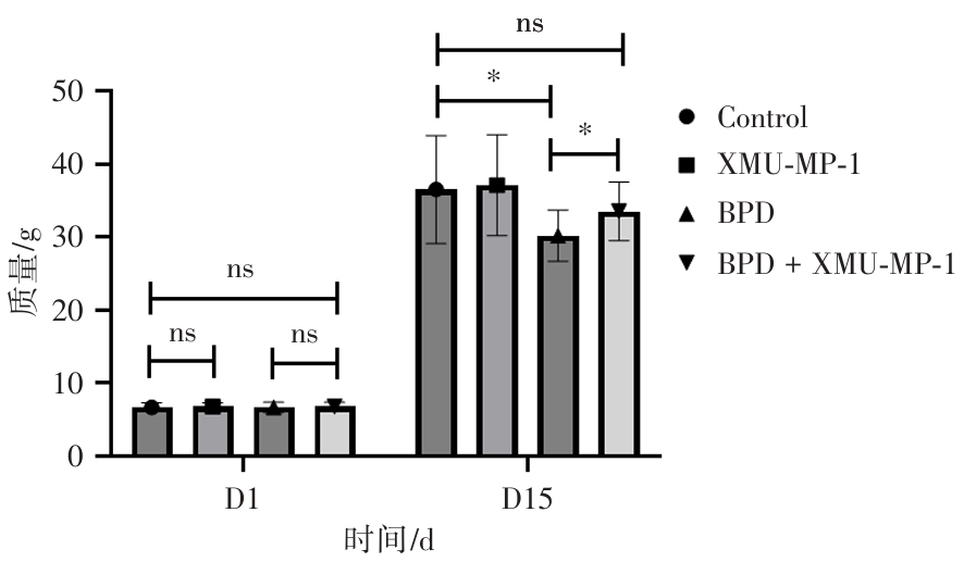
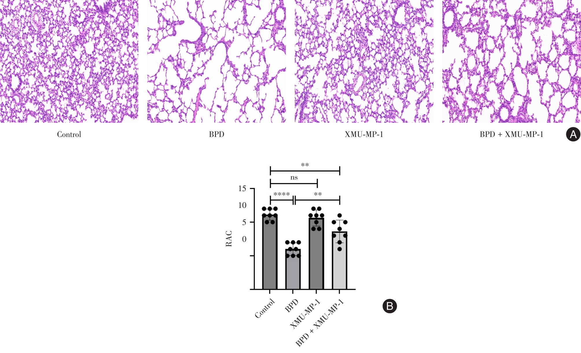
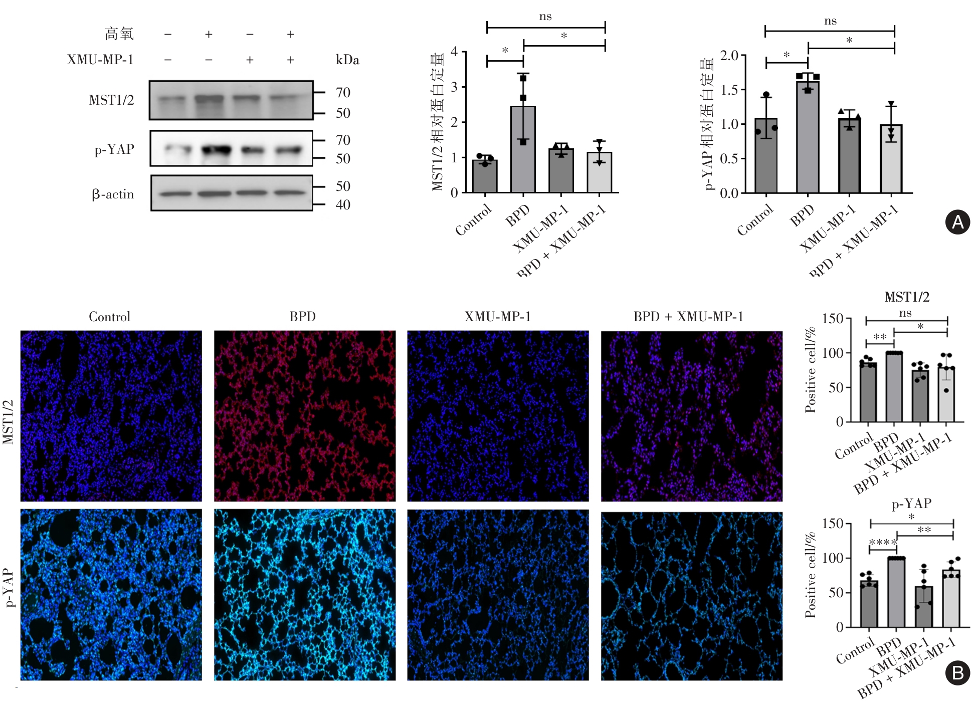
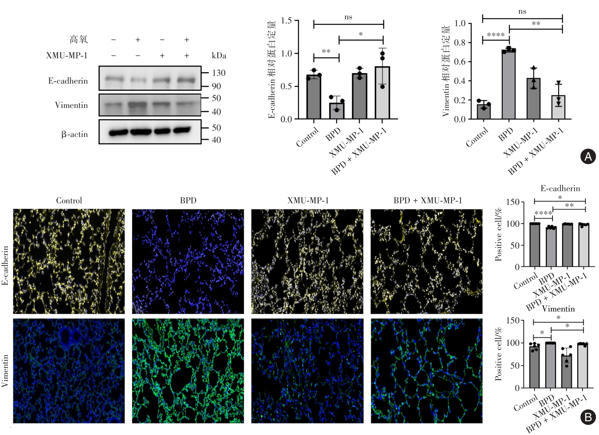
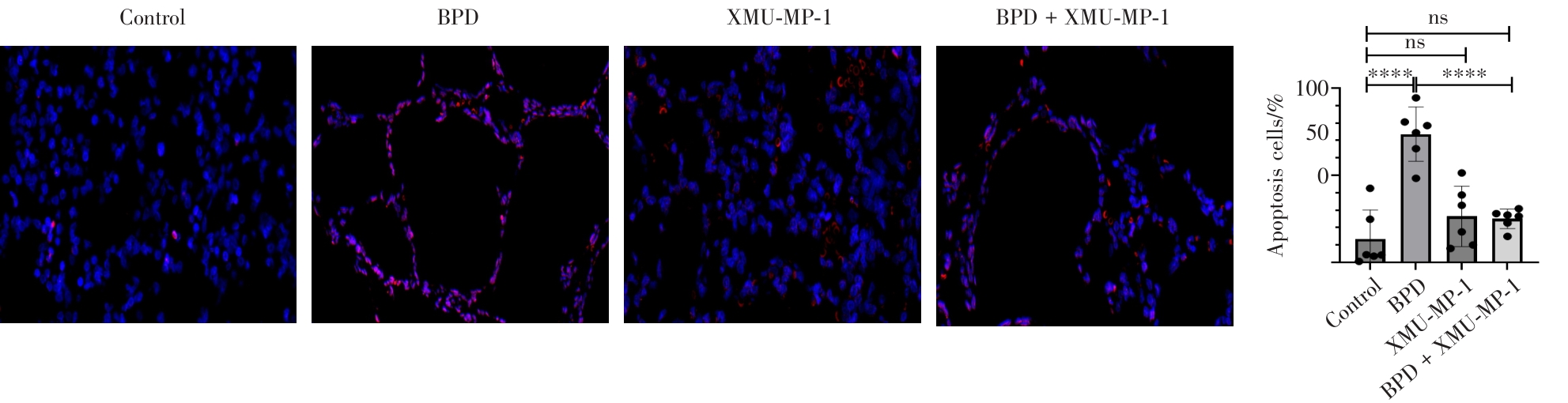
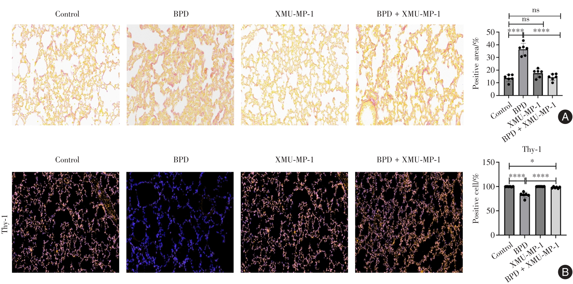
目的 探究MST1/2抑制剂XMU-MP-1阻断Hippo信号通路对高氧诱导新生大鼠支气管肺发育不良(BPD)肺损伤的影响。 方法 将新生大鼠随机分为对照组、XMU-MP-1组、BPD模型组和BPD + XMU-MP-1组,每组10只。BPD模型组及BPD + XMU-MP-1组置于85% O₂高氧箱中持续暴露14 d以建立BPD模型,对照组及XMU-MP-1组置于正常空气中。自造模第1天起,XMU-MP-1组和BPD + XMU-MP-1组每天腹腔注射MST1/2激酶抑制剂XMU-MP-1(1 mg/kg),对照组和BPD模型组注射等体积的生理盐水,连续7 d。造模期间监测大鼠体质量变化。造模结束后采用HE染色评估肺组织病理改变,并通过辐射状肺泡计数 (RAC)定量分析评估肺泡化程度;采用Western blot和免疫荧光染色检测肺组织Hippo信号通路蛋白(MST1/2、p-YAP)、上皮-间质转化相关标志物(Vimentin、E-cadherin)和成纤维细胞活化标志物Thy-1的表达水平;采用TUNEL染色检测肺组织细胞凋亡;采用天狼星红染色检测肺组织胶原沉积。 结果 与对照组相比,BPD模型组大鼠体质量显著降低(P < 0.05);肺组织呈现典型BPD病理改变,表现为RAC显著减少;肺组织细胞凋亡率、胶原沉积面积显著增加(均P < 0.05);MST1/2、p-YAP及Vimentin表达显著上调,E-cadherin及Thy-1表达显著下调(均P < 0.05)。与BPD模型组相比,BPD + XMU-MP-1组大鼠体质量显著恢复 (P < 0.05),上述肺组织病理损伤、蛋白表达异常、细胞凋亡及胶原沉积均被显著改善(均P < 0.05)。 结论 Hippo信号通路在高氧诱导的新生大鼠BPD模型中被异常激活,参与肺组织细胞凋亡、上皮-间质转化及胶原沉积;MST1/2抑制剂XMU-MP-1阻断该通路可减轻BPD损伤,提示Hippo通信是BPD防治的潜在靶点。

关键词: 支气管肺发育不良, Hippo信号通路, 细胞凋亡, 上皮-间质转化, 胶原沉积

Abstract:

Objective To investigate the effects of MST1/2 inhibitor XMU-MP-1 blocking the Hippo signaling pathway on hyperoxia-induced lung injury neonatal rats with bronchopulmonary dysplasia (BPD). Methods Neonatal rats were randomly divided into four groups: control group, XMU-MP-1 group, BPD model group, and BPD + XMU-MP-1 group, with 10 rats in each group. Rats in the BPD model group and BPD + XMU-MP-1 group were exposed to 85% O₂ in a hyperoxia chamber for 14 consecutive days to establish the BPD model, while those in the control group and XMU-MP-1 group were kept in normal air. From the first day of modeling, rats in the XMU-MP-1 group and BPD + XMU-MP-1 group received daily intraperitoneal injections of the MST1/2 kinase inhibitor XMU-MP-1 (1 mg/kg), whereas those in the control group and BPD model group received equal volumes of normal saline, for 7 consecutive days. Body weight was monitored during the modeling period. At the end of the modeling period, lung tissue sections were stained with hematoxylin and eosin (HE) for histopathological evaluation, and the extent of alveolarization was quantified by radial alveolar count (RAC). Western blot and immunofluorescence staining were used to detect the expression levels of Hippo signaling pathway proteins (MST1/2, p-YAP), epithelial-mesenchymal transition-related markers (E-cadherin, Vimentin), and the fibroblast activation marker Thy-1. TUNEL staining was employed to detect pulmonary cell apoptosis, and Sirius Red staining was used to assess pulmonary collagen deposition. Results Compared with the control group, the BPD model group exhibited significantly decreased body weight (P < 0.05); typical BPD pathological changes, characterized by significantly decreased RAC (P < 0.05); significantly increased pulmonary cell apoptosis rate and collagen deposition area (all P < 0.05); and significantly upregulated protein expression of MST1/2, p-YAP and Vimentin, as well as significantly downregulated expression of E-cadherin and Thy-1 (all P < 0.05). Compared with the BPD model group, the BPD + XMU-MP-1 group showed significantly restored body weight (P < 0.05), and the aforementioned pulmonary pathological damage, protein expression abnormalities, cell apoptosis, and collagen deposition were all significantly ameliorated (all P < 0.05). Conclusions The Hippo signaling pathway is abnormally activated in hyperoxia-induced BPD model of neonatal rats, participating in lung cell apoptosis, epithelial⁃mesenchymal transition and collagen deposition. Blocking this pathway with the MST1/2 inhibitor XMU-MP-1 alleviates BPD lung injury, suggesting that the Hippo pathway is a potential therapeutic target for BPD.

Key words: bronchopulmonary dysplasia, Hippo signaling pathway, apoptosis, epithelial-mesenchymal transition, collagen deposition

中图分类号: