

实用医学杂志 ›› 2026, Vol. 42 ›› Issue (14): 2488-2504.doi: 10.3969/j.issn.1006-5725.2026.14.002
• 心脑血管疾病专栏 • 上一篇
蒋晓路1,文红福1,沈媛1,张俊1,李黎妮1,许仁露1,张文1,王晓明2,季一飞3,李卫东4( )
)
收稿日期:2026-04-22
出版日期:2026-07-25
发布日期:2026-08-05
通讯作者:
李卫东
E-mail:lwd10000@126.com
基金资助:
Xiaolu JIANG1,Hongfu WEN1,Yuan SHEN1,Jun ZHANG1,Lini LI1,Renlu XU1,Wen ZHANG1,Xiaoming WANG2,Yifei JI3,Weidong LI4()
Received:2026-04-22
Online:2026-07-25
Published:2026-08-05
Contact:
Weidong LI
E-mail:lwd10000@126.com
摘要:
目的 探讨F-box/富含亮氨酸重复蛋白4(FBXL4)通过调控动力蛋白相关蛋白1(Drp1)介导的线粒体动力学抑制氧化应激和凋亡,保护心脏骤停(CA)后心肺复苏(CPR)致脑缺血再灌注损伤(CIRI)的分子机制。 方法 选取48只雄性SD大鼠随机分为4组(每组6只):Control组、Control + 过表达FBXL4(OE-FBXL4)组、CA/CPR组、CA/CPR + OE-FBXL4组,另设生存分析,每组6只。自主循环恢复(ROSC)后24 h处死取材,检测神经功能缺损评分(NDS)、7 d生存率;采用RT-qPCR和Western blot检测FBXL4、Drp1 mRNA及蛋白表达;Western blot检测线粒体动力学蛋白;免疫共沉淀验证FBXL4与Drp1相互作用;HE染色和尼氏(Nissl)染色观察脑组织病理;透射电镜观察线粒体超微结构;生化法检测丙二醛(MDA)、超氧化物歧化酶(SOD)、谷胱甘肽(GSH)、过氧化氢酶(CAT)评估氧化应激水平;Western blot检测凋亡相关蛋白B细胞淋巴瘤-2相关X蛋白(Bax)、B细胞淋巴瘤-2(Bcl-2)、半胱氨酸天冬氨酸蛋白酶-3/8(Caspase-3/8)表达。 结果 (1)与Control组相比,CA/CPR组大鼠NDS评分显著降低(P < 0.01),7 d生存率下降;脑组织FBXL4mRNA及蛋白表达显著下调(均P < 0.01),Drp1、Fis1、MFF表达显著上调(均P < 0.01),OPA1、MFN1、MFN2表达显著下调(均P < 0.01);HE和Nissl染色显示皮层及海马CA1、CA3区神经元排列紊乱、胞体萎缩、尼氏小体溶解,存活神经元数量减少;TEM显示线粒体肿胀、空泡化、嵴断裂,呈碎片化;MDA含量升高(P < 0.01),SOD、GSH、CAT活性降低(均P < 0.01);Bax/Bcl-2比值升高(P < 0.01),Caspase-3、Caspase-8表达增加(均P < 0.01)。(2)与CA/CPR组相比,CA/CPR + OE-FBXL4组NDS评分显著提高(P < 0.01),7 d生存率明显上升;FBXL4蛋白维持高水平表达;Drp1、Fis1、MFF蛋白水平下降(均P < 0.01),OPA1、MFN1、MFN2蛋白水平上升(均P < 0.01);HE和Nissl染色显示神经元形态改善,变性坏死细胞减少,存活神经元数量增加;TEM显示线粒体恢复管网状结构,嵴结构清晰完整;MDA含量降低(P < 0.01),SOD、GSH、CAT活性升高(均P < 0.01);Bax/Bcl-2比值下降(P < 0.01),Caspase-3、Caspase-8表达减少(均P < 0.01)。(3)Co-IP实验证实FBXL4与Drp1存在蛋白-蛋白相互作用;Control + OE-FBXL4组FBXL4-Drp1复合物形成量较Control组显著增加(P < 0.01),CA/CPR组复合物形成减少,CA/CPR + OE-FBXL4组结合量显著恢复。 结论 FBXL4是CA/CPR后脑损伤的关键保护性调控因子,其过表达可通过靶向Drp1恢复线粒体动力学平衡,进而抑制氧化应激和神经元凋亡;同时,Nrf2/HO-1通路的进一步上调可能协同参与了FBXL4的抗氧化保护作用。
中图分类号:
蒋晓路,文红福,沈媛,张俊,李黎妮,许仁露,张文,王晓明,季一飞,李卫东. FBXL4靶向调控Drp1介导的线粒体动力学抑制氧化应激和凋亡保护心肺复苏后脑损伤的机制[J]. 实用医学杂志, 2026, 42(14): 2488-2504.
Xiaolu JIANG,Hongfu WEN,Yuan SHEN,Jun ZHANG,Lini LI,Renlu XU,Wen ZHANG,Xiaoming WANG,Yifei JI,Weidong LI. Mechanism of FBXL4 inhibiting oxidative stress and apoptosis, and preventing brain injury after cardiopulmonary resuscitation by regulating Drp1-mediated mitochondrial dynamics[J]. The Journal of Practical Medicine, 2026, 42(14): 2488-2504.
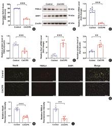
图1
CA/CPR诱导大鼠神经功能损伤及脑组织FBXL4、Drp1的表达变化注:A,各组大鼠自主循环恢复(ROSC)后24 h的神经功能缺损评分(NDS)比较;B,Western blot检测各组脑组织FBXL4和Drp1蛋白表达的代表性印记条带;C—F,各组大鼠脑组织中FBXL4和Drp1的mRNA相对表达量(C、E)及蛋白相对表达量(D、F)的统计学分析;G,各组大鼠脑组织皮层区域NeuN(绿色)、FBXL4(红色)及DAPI(蓝色)的代表性免疫荧光双标染色图(标尺 = 50 μm);H—I,各组神经元NeuN(H)及FBXL4(I)相对荧光强度的定量分析(n = 6)。结果以均数±标准差表示,n = 6。与 Control组比较,*P < 0.05,**P < 0.01,***P < 0.001,***P < 0.0001"
图2
脑组织过表达 FBXL4 对 CA/CPR 大鼠生存率及神经功能的影响注:A,侧脑室注射AAV9病毒及CA/CPR建模的体内实验流程图;B—D,各组大鼠脑组织中FBXL4蛋白表达(B、C)及mRNA水平(D)的验证;E,各组大鼠(每组6只)ROSC后7 d生存率曲线比较;F,各组大鼠ROSC后神经功能缺损(NDS)评分统计;G—H,Western blot检测CA/CPR建模后各组脑组织FBXL4蛋白的表达情况及定量分析;I—L,各组大鼠基础生理参数统计,包括体质量(I)、心率(J)、平均动脉压(K)及体温(L)。结果以均数±标准差表示,n = 6。ns,差异无统计学意义;与Control组比较,*P < 0.05,**P < 0.01,***P < 0.001"
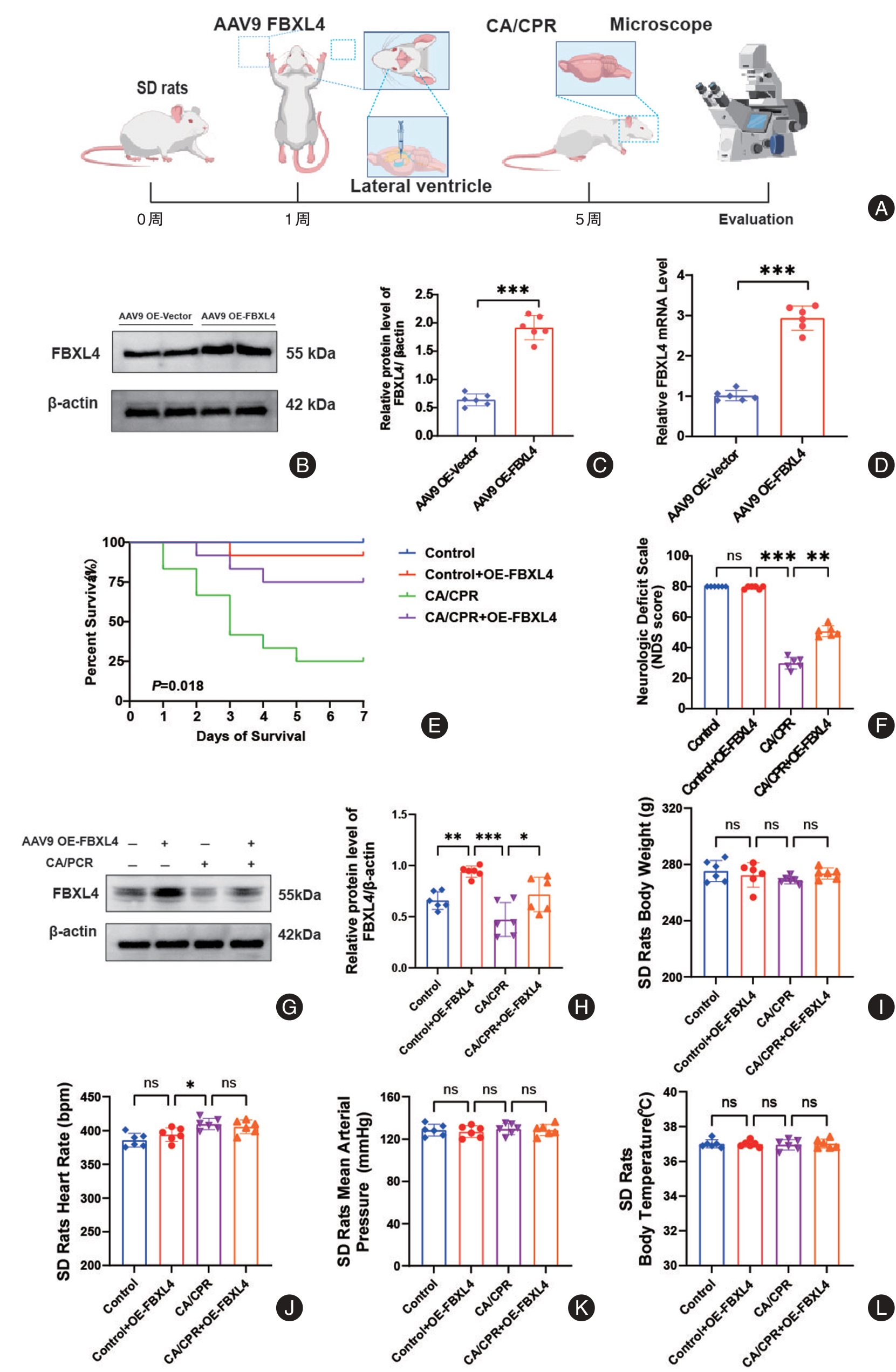
图3
过表达FBXL4减轻CA/CPR大鼠脑组织神经元病理损伤(HE染色)注:A,脑组织HE染色代表性图片。图中展示了4 ×低倍视野下的脑组织全貌(比例尺 = 250 μm),以及20 ×高倍视野下皮层、海马CA1区及CA3区的局部图(比例尺 = 50 μm)。可见CA/CPR组神经元损伤明显,而过表达FBXL4组损伤减轻。B—D,分别为皮层、海马CA1区及CA3区病变(损伤)神经元数量的定量分析。结果以均数±标准差表示,n = 6。ns,差异无统计学意义;与Control组比较,*P < 0.05,**P < 0.01,***P < 0.001"
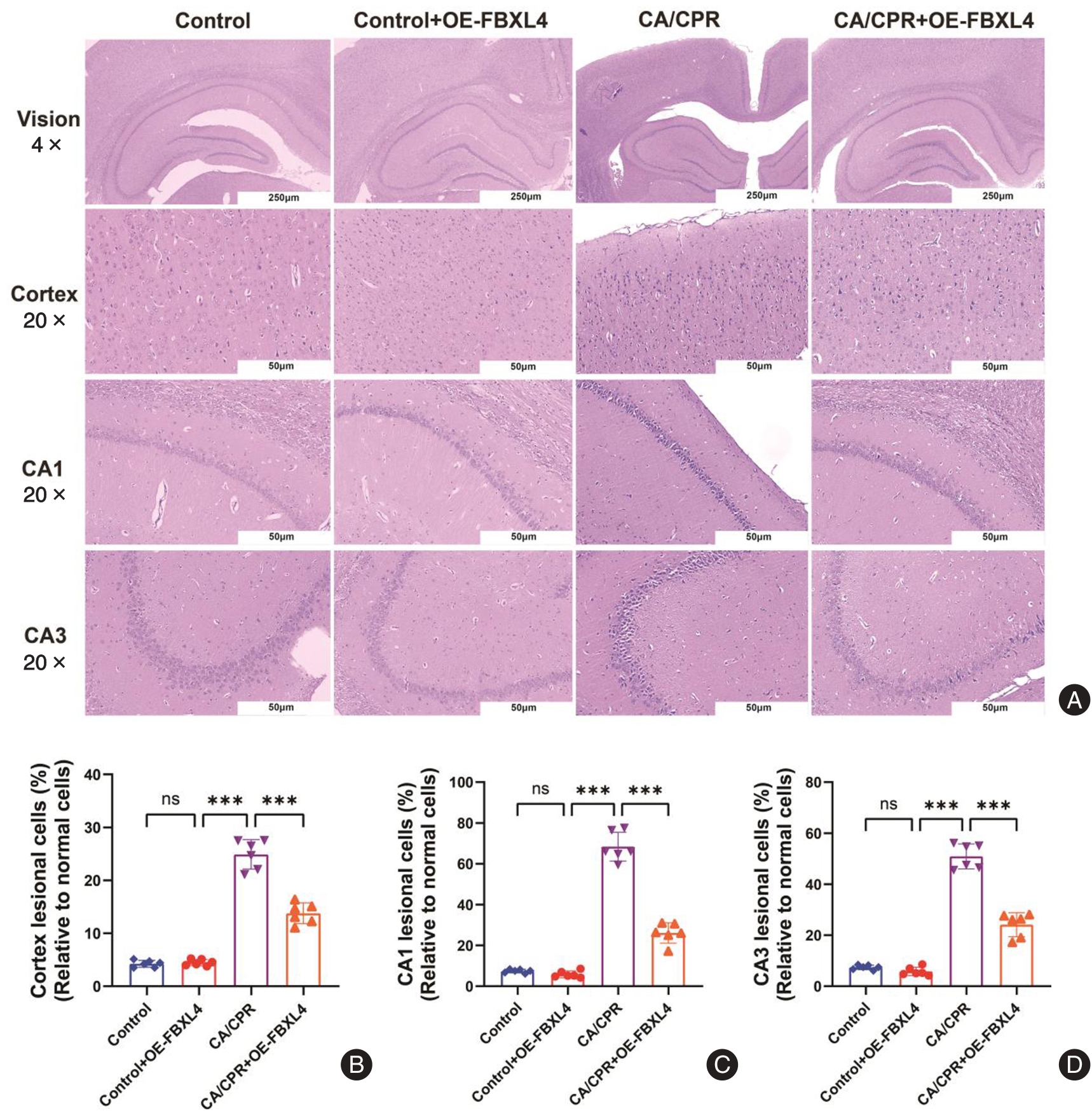
图4
过表达FBXL4减轻CA/CPR大鼠脑组织神经元病理损伤(Nissl染色)注:A,脑组织Nissl染色代表性图片。图中展示了4 ×低倍视野下的脑组织全貌(比例尺 = 250 μm),以及20 ×高倍视野下皮层、海马CA1区及CA3区的局部图(比例尺 = 50 μm)。可见CA/CPR组神经元损伤明显,而过表达FBXL4组损伤减轻。B—D,分别为皮层、海马CA1区及CA3区病变(损伤)神经元数量的定量分析。结果以均数±标准差表示,n = 6。ns,差异无统计学意义;与Control组比较,*P < 0.05,**P < 0.01,***P < 0.001"
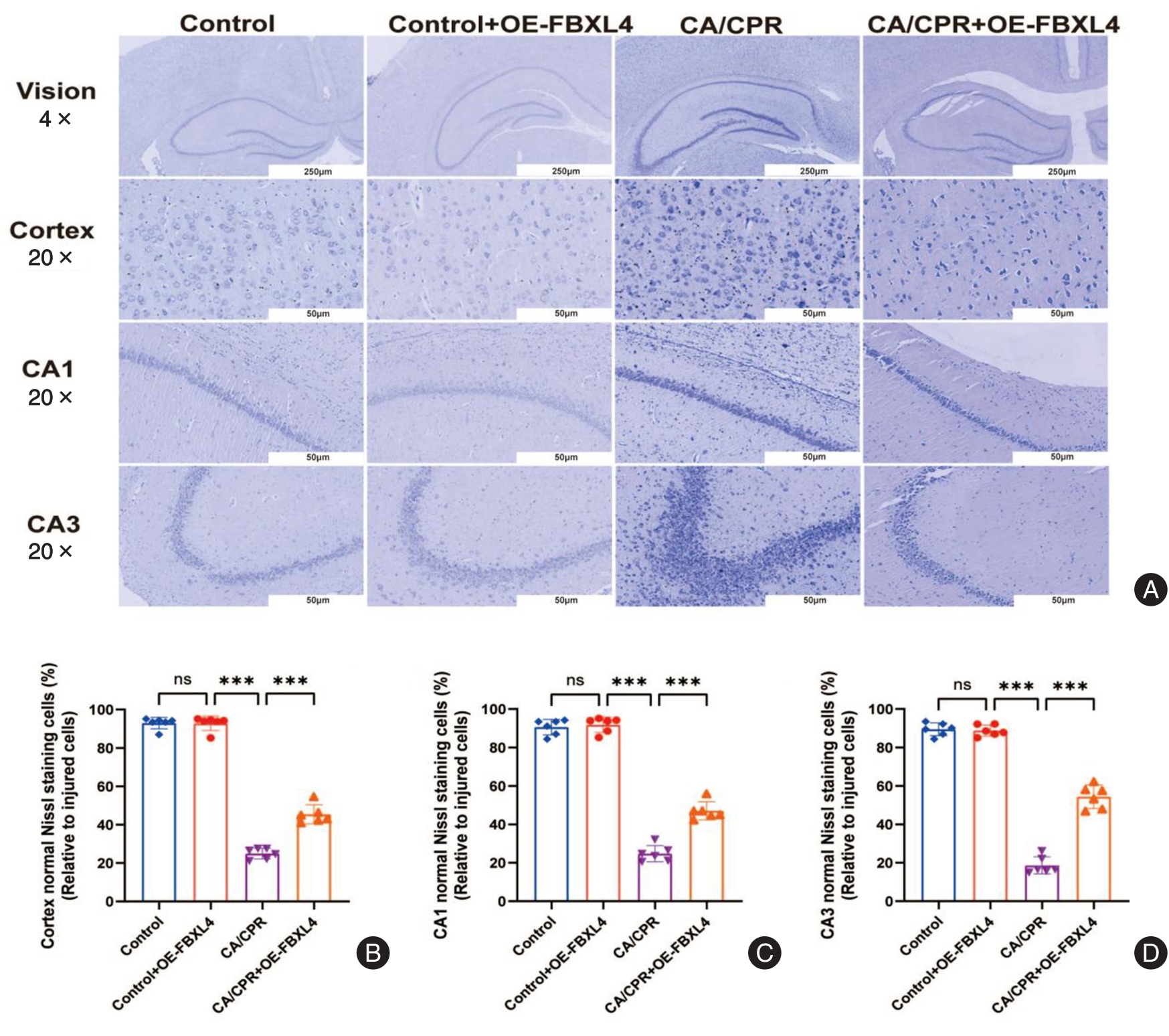
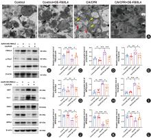
图5
过表达FBXL4通过调控Drp1/Fis1/MFF轴重塑线粒体动力学平衡注:A,大鼠皮层脑组织透射电镜(TEM)代表性图像(标尺 = 0.5μm)。黑色箭头指示结构正常的线粒体,黄色/红色箭头指示肿胀、嵴断裂及空泡化的受损线粒体。B,FBXL4、p-Drp1及Drp1蛋白表达的Western Blot代表性条带。C—E,FBXL4、p-Drp1及Drp1相对蛋白表达量的定量分析。F,线粒体分裂蛋白MFF、Fis1及融合蛋白OPA1、MFN1、MFN2的Western blot代表性条带。G,p-Drp1/Drp1比值的定量分析。H—L,MFF、Fis1、MFN1、MFN2及OPA1相对蛋白表达量的定量分析。结果以均数±标准差表示,n = 6。ns,差异无统计学意义;与Control组比较,*P < 0.05,**P < 0.01,***P < 0.001"
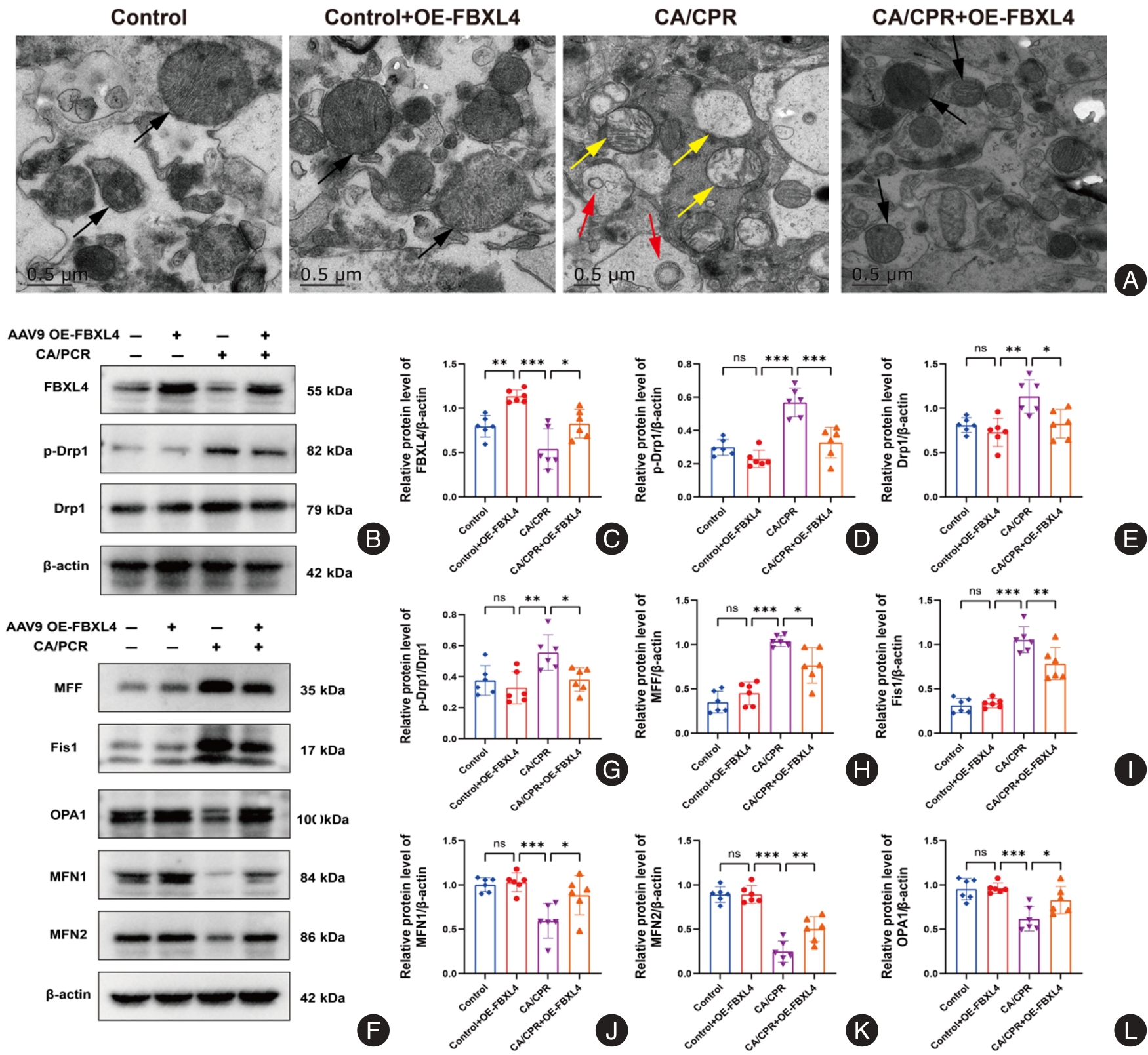
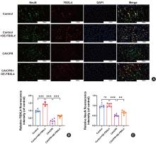
图6
FBXL4在皮层神经元中的定位表达及过表达后的神经保护作用注:A,大鼠脑皮层免疫荧光双标染色代表性图像(标尺 = 100 μm)。NeuN(绿色)标记神经元胞体,FBXL4(红色)标记目的蛋白,DAPI(蓝色)标记细胞核。Merge图像显示FBXL4主要定位于NeuN阳性神经元胞浆中。与Control组相比,CA/CPR组NeuN及FBXL4荧光强度均显著减弱;而OE-FBXL4干预显著恢复了FBXL4表达并改善了神经元形态。B,各组大鼠皮层内FBXL4相对荧光强度的定量分析。C,各组大鼠皮层内NeuN相对荧光强度的定量分析。结果以均数±标准差表示,n = 6。ns,差异无统计学意义;与Control组比较,*P < 0.05,**P < 0.01,***P < 0.001"
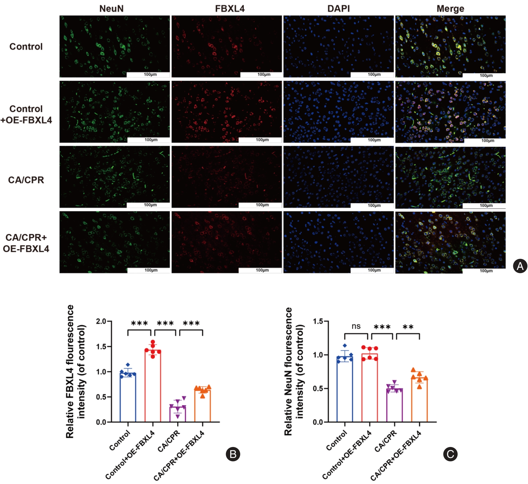
图7
过表达FBXL4对CA/CPR大鼠脑组织凋亡相关蛋白表达的影响注:A,各组大鼠脑组织样本中凋亡相关蛋白Caspase-3、Caspase-8、Bax及Bcl-2表达的代表性Western blot图像;B—E,各组大鼠脑组织样本中Caspase-3、Caspase-8、Bax及Bcl-2蛋白相对表达量的定量分析结果。结果以均数±标准差表示,n = 6。ns,差异无统计学意义;与Control组比较,*P < 0.05,**P < 0.01,***P < 0.001"
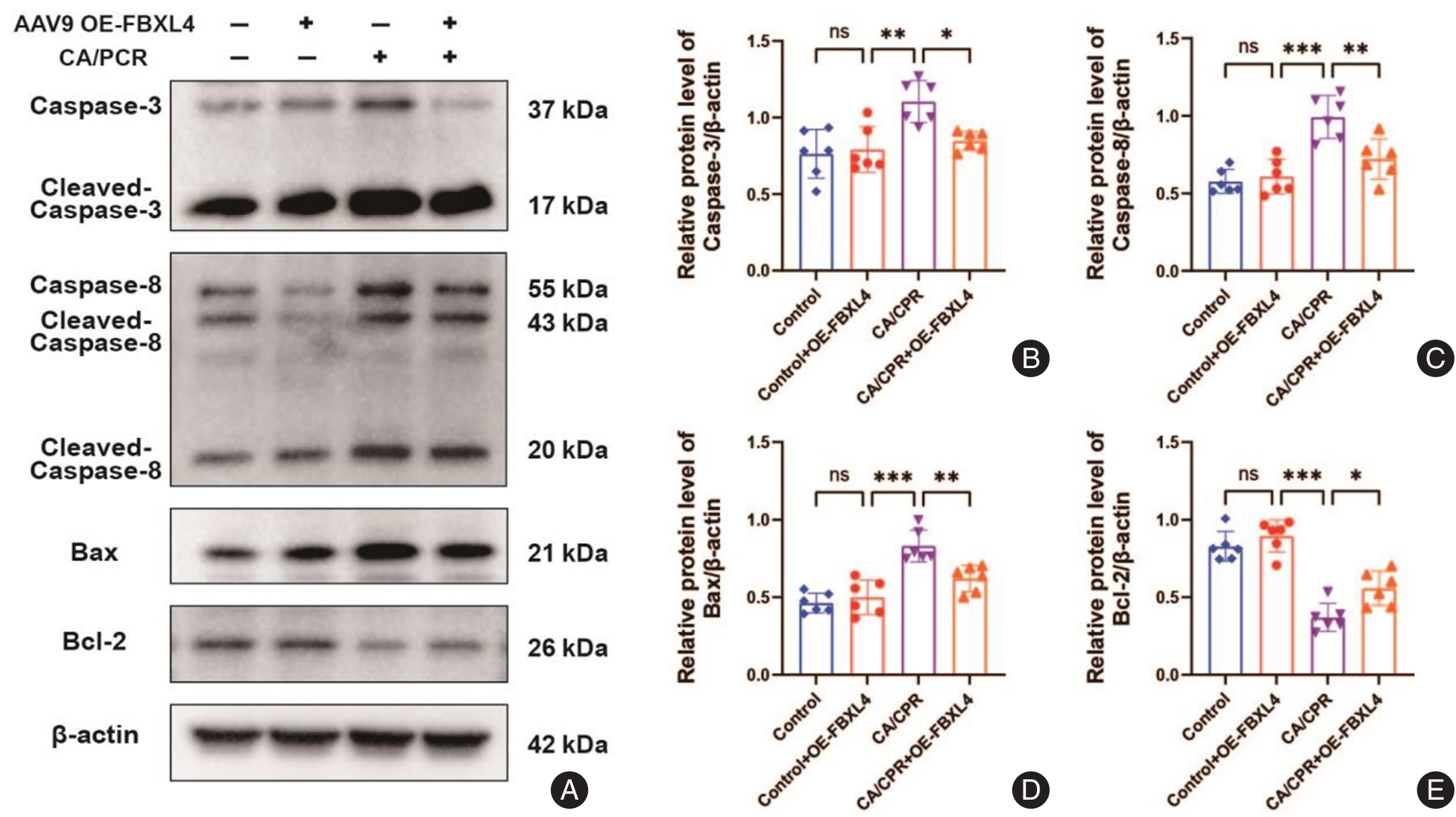
图8
过表达FBXL4对CA/CPR大鼠脑组织氧化应激指标及Nrf2/HO-1通路的影响注:A,各组大鼠脑组织样本中氧化应激蛋白Nrf2、HO-1表达的代表性Western blot图像;B、C,各组大鼠脑组织样本中氧化应激蛋白Nrf2、HO-1蛋白相对表达量的定量统计分析;D—G,各组大鼠脑组织样本中氧化应激酶MDA、GSH、CAT和SOD的活性的检测分析。结果以均数±标准差表示,n = 6。ns,差异无统计学意义;与Control组比较,*P < 0.05,**P < 0.01,***P < 0.001"
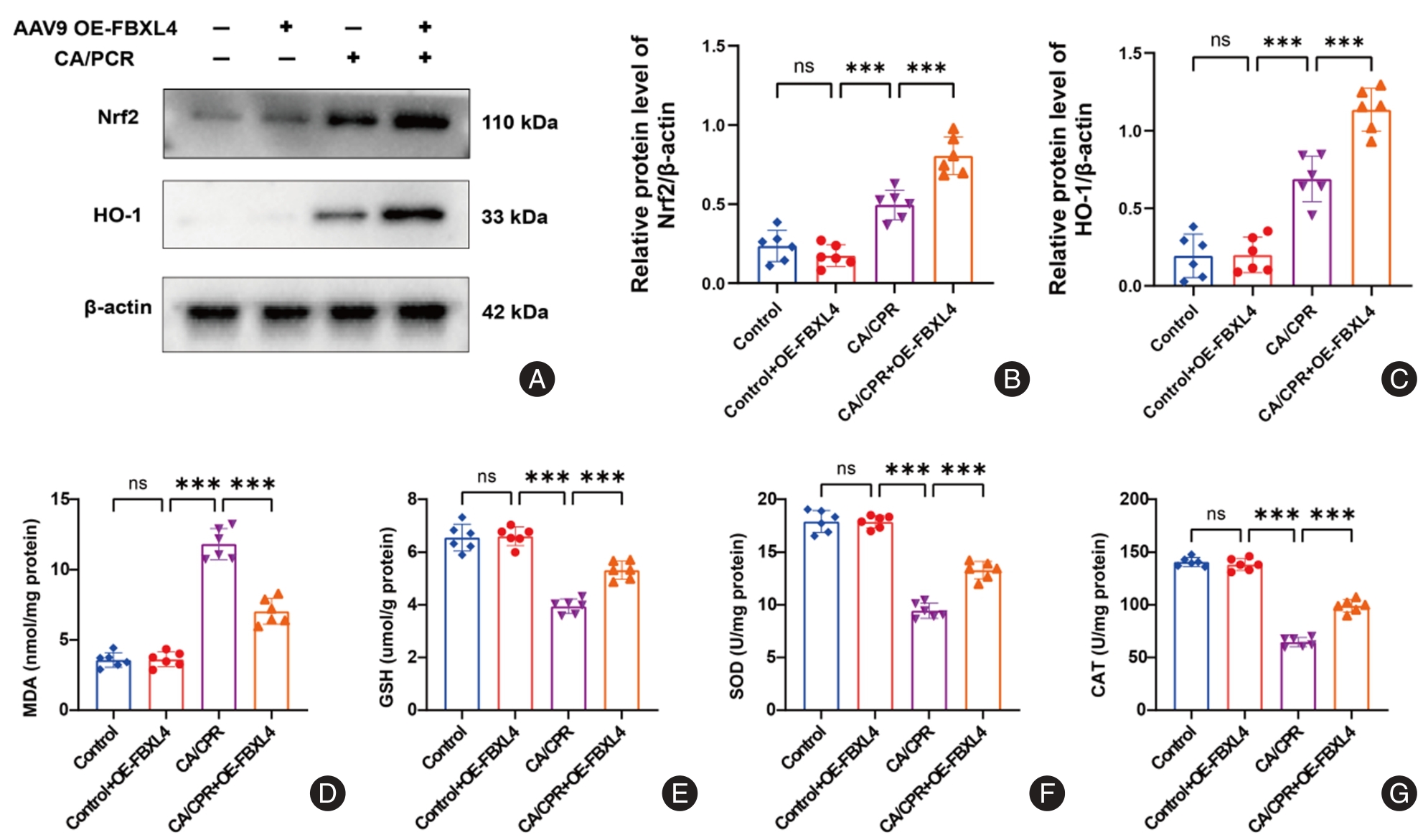
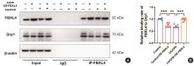
图9
Co-IP实验证实FBXL4与Drp1在大鼠脑组织中的存在相互作用注:A,各组大鼠脑组织Co-IP实验代表性Western blot图像。以FBXL4抗体免疫沉淀(IP:FBXL4),Western blot检测沉淀物中Drp1蛋白(IB:Drp1)。IgG组作为阴性对照,Input为总蛋白入样对照;B,各组FBXL4与Drp1相对结合量的定量分析结果。结果以均数±标准差表示,n = 6。ns,差异无统计学意义;与Control组比较,*P < 0.05,**P < 0.01,***P < 0.001"
| [1] |
ANDERSEN L W, HOLMBERG M J, BERG K M,et al. In-Hospital Cardiac Arrest: A Review[J]. JAMA,2019,321(12):1200-1210. doi:10.1001 /jama.2019.1696 .
doi: 10.1001 /jama.2019.1696 |
| [2] |
SANDRONI C, D'ARRIGO S, NOLAN J P. Prognostication after cardiac arrest[J]. Crit Care,2018,22(1):150. doi:10.1186/s13054-018-2060-7 .
doi: 10.1186/s13054-018-2060-7 |
| [3] |
MEHAFFEY J H, MONEY D, CHARLES E J,et al. Adenosine 2A Receptor Activation Attenuates Ischemia Reperfusion Injury During Extracorporeal Cardiopulmonary Resuscitation[J].Ann Surg,2019,269(6):1176-1183. doi:10.1097/sla. 0000000000002685 .
doi: 10.1097/sla. 0000000000002685 |
| [4] |
TUO Q Z, ZHANG S T, LEI P. Mechanisms of neuronal cell death in ischemic stroke and their therapeutic implications[J].Med Res Rev,2022,42(1):259-305. doi:10.1002/med.21817 .
doi: 10.1002/med.21817 |
| [5] |
MAO R, ZONG N, HU Y,et al. Neuronal Death Mechanisms and Therapeutic Strategy in Ischemic Stroke[J].Neurosci Bull,2022,38(10):1229-1247. doi:10.1007/s12264-022-00859-0 .
doi: 10.1007/s12264-022-00859-0 |
| [6] |
GUAN R, ZOU W, DAI X,et al. Mitophagy, a potential therapeutic target for stroke[J]. J Biomed Sci,2018,25(1):87. doi:10.1186/s12929-018-0487-4 .
doi: 10.1186/s12929-018-0487-4 |
| [7] |
LIANG W, HUANG X, CHEN W. The Effects of Baicalin and Baicalein on Cerebral Ischemia: A Review[J]. Aging Dis,2017,8(6):850-867. doi:10.14336/ad.2017.0829 .
doi: 10.14336/ad.2017.0829 |
| [8] |
QIU Z, WEI Y, SONG Q,et al. The Role of Myocardial Mitochondrial Quality Control in Heart Failure[J].Front Pharmacol,2019,10(1):1404. doi:10.3389/ fphar.2019.01404 .
doi: 10.3389/ fphar.2019.01404 |
| [9] |
DUAN X, LIU R, LAN W,et al. The Essential Role of Mitochondrial Dynamics in Viral Infections[J]. Int J Mol Sci,2025,26(5):1955. doi:10.3390/ijms 26051955 .
doi: 10.3390/ijms 26051955 |
| [10] |
WOODS L C, BERBUSSE G W, NAYLOR K. Microtubules Are Essential for Mitochondrial Dynamics-Fission, Fusion, and Motility-in Dictyostelium discoideum[J].Front Cell Dev Biol,2016,4(1):19. doi:10.3389/fcell. 2016.00019 .
doi: 10.3389/fcell. 2016.00019 |
| [11] |
SUÁREZ-RIVERO J M, VILLANUEVA-PAZ M, DE LA CRUZ-OJEDA P,et al. Mitochondrial Dynamics in Mitochondrial Diseases[J].Dig Dis,2016,5(1):1. doi:10.3390/diseases5010001 .
doi: 10.3390/diseases5010001 |
| [12] |
WANG N, WANG X, LAN B,et al. DRP1, fission and apoptosis[J].Cell Death Discov,2025,11(1):150. doi:10.1038/s41420-025-02458-0 .
doi: 10.1038/s41420-025-02458-0 |
| [13] |
ISHIHARA N, NOMURA M, JOFUKU A,et al. Mitochondrial fission factor Drp1 is essential for embryonic development and synapse formation in mice[J]. Nat Cell Biol,2009,11(8):958-966. doi:10.1038/ncb1907 .
doi: 10.1038/ncb1907 |
| [14] |
YU R, JIN S B, ANKARCRONA M,et al. The Molecular Assembly State of Drp1 Controls its Association With the Mitochondrial Recruitment Receptors Mff and MIEF1/2[J]. Front Cell Dev Biol,2021,9(1):706687. doi:10.3389/fcell. 2021.706687 .
doi: 10.3389/fcell. 2021.706687 |
| [15] |
SATO H, HOSHI M, IKEDA F,et al. Downregulation of mitochondrial biogenesis by virus infection triggers antiviral responses by cyclic GMP-AMP synthase[J].PLoS Pathog,2021,17(10):e1009841.doi:10.1371/journal.ppat. 1009841 .
doi: 10.1371/journal.ppat. 1009841 |
| [16] |
MURATA D, YAMADA T, TOKUYAMA T,et al. Mitochondrial Safeguard: A stress response that offsets extreme fusion and protects respiratory function via flickering-induced Oma1 activation[J]. EMBO J,2020,39(24):e105074. doi:10.15252/embj. 2020105074 .
doi: 10.15252/embj. 2020105074 |
| [17] |
CATANESE L, TARSIA J, FISHER M. Acute Ischemic Stroke Therapy Overview[J]. Circ Res,2017,120(3):541-558. doi:10.1161/circresaha. 116.309278 .
doi: 10.1161/circresaha. 116.309278 |
| [18] |
ZHAO Y X, CUI M, CHEN S F,et al. Amelioration of ischemic mitochondrial injury and Bax-dependent outer membrane permeabilization by Mdivi-1[J]. CNS Neurosci Ther,2014,20(6):528-538. doi:10.1111/cns.12266 .
doi: 10.1111/cns.12266 |
| [19] |
FLIPPO K H, LIN Z, DICKEY A S,et al. Deletion of a Neuronal Drp1 Activator Protects against Cerebral Ischemia[J]. J Neurosci,2020,40(15):3119-3129. doi:10.1523/jneurosci.1926-19.2020 .
doi: 10.1523/jneurosci.1926-19.2020 |
| [20] |
SHIELDS L Y, KIM H, ZHU L,et al. Dynamin-related protein 1 is required for normal mitochondrial bioenergetic and synaptic function in CA1 hippocampal neurons[J]. Cell Death Dis,2015,6(4):e1725. doi:10.1038/cddis.2015.94 .
doi: 10.1038/cddis.2015.94 |
| [21] |
PU Z X, WANG J L, LI Y Y,et al. A Bacterial Platform for Studying Ubiquitination Cascades Anchored by SCF-Type E3 Ubiquitin Ligases[J]. Biomolecules,2024,14(10):1209. doi:10.3390/biom14101209 .
doi: 10.3390/biom14101209 |
| [22] |
HWANG J, LAUINGER L, KAISER P. Distinct Stress Regulators in the CRL Family: Emerging Roles of F-Box Proteins: Cullin-RING Ligases and Stress-Sensing[J]. BioEssays,2025,47(5):e202400249. doi:10.1002/bies. 202400249 .
doi: 10.1002/bies. 202400249 |
| [23] |
CHEN Y, JIAO D, LIU Y,et al. FBXL4 mutations cause excessive mitophagy via BNIP3/BNIP3L accumulation leading to mitochondrial DNA depletion syndrome[J]. Cell Death Differ,2023,30(10):2351-2363. doi:10.1038/s41418- 023-01205-1 .
doi: 10.1038/s41418- 023-01205-1 |
| [24] |
PRASUN P. FBXL4-Related Mitochondrial Depletion Syndrome Underscores the role of Mitophagy in Stem Cell Differentiation during Embryogenesis[J]. Stem Cell Rep,2025,21(3):897-899. doi:10.1007/s12015 -025-10854-3 .
doi: 10.1007/s12015 -025-10854-3 |
| [25] |
BONNEN P E, YARHAM J W, BESSE A,et al. Mutations in FBXL4 cause mitochondrial encephalopathy and a disorder of mitochondrial DNA maintenance[J]. Am J Hum Genet,2013,93(3):471-481. doi:10.1016/j.ajhg. 2013.07.017 .
doi: 10.1016/j.ajhg. 2013.07.017 |
| [26] |
GAI X, GHEZZI D, JOHNSON M A,et al. Mutations in FBXL4, encoding a mitochondrial protein, cause early-onset mitochondrial encephalomyopathy[J]. Am J Hum Genet,2013,93(3):482-495. doi:10.1016/j.ajhg.2013.07.016 .
doi: 10.1016/j.ajhg.2013.07.016 |
| [27] |
ABUDUREYIMU M, LUO X, JIANG L,et al. FBXL4 protects against HFpEF through Drp1-Mediated regulation of mitochondrial dynamics and the downstream SERCA2a[J]. Redox Biol,2024,70(1):103081. doi:10.1016/j.redox. 2024.103081 .
doi: 10.1016/j.redox. 2024.103081 |
| [28] |
YU S, WU C, ZHU Y,et al. Rat model of asphyxia-induced cardiac arrest and resuscitation[J]. Front Neurosci,2022,16(1):1087725. doi:10.3389/fnins. 2022.1087725 .
doi: 10.3389/fnins. 2022.1087725 |
| [29] |
KATZ L, EBMEYER U, SAFAR P,et al. Outcome model of asphyxial cardiac arrest in rats[J]. J Cereb Blood Flow Metab,1995,15(6):1032-1039. doi:10.1038/jcbfm.1995.129 .
doi: 10.1038/jcbfm.1995.129 |
| [30] |
PERKINS G D, JACOBS I G, NADKARNI V M,et al. Cardiac arrest and cardiopulmonary resuscitation outcome reports: update of the Utstein Resuscitation Registry Templates for Out-of-Hospital Cardiac Arrest: a statement for healthcare professionals from a task force of the International Liaison Committee on Resuscitation (American Heart Association, European Resuscitation Council, Australian and New Zealand Council on Resuscitation, Heart and Stroke Foundation of Canada, InterAmerican Heart Foundation, Resuscitation Council of Southern Africa, Resuscitation Council of Asia); and the American Heart Association Emergency Cardiovascular Care Committee and the Council on Cardiopulmonary, Critical Care, Perioperative and Resuscitation[J]. Circulation,2015,132(13):1286-1300. doi:10.1161/cir. 0000000000000144 .
doi: 10.1161/cir. 0000000000000144 |
| [31] |
ADHIKARI A, LERNER T N, FINKELSTEIN J,et al. Basomedial amygdala mediates top-down control of anxiety and fear[J]. Nature,2015,527(7577): 179-185. doi:10.1038/nature15698 .
doi: 10.1038/nature15698 |
| [32] |
WANG P, YAO L, ZHOU L L,et al. Carbon Monoxide Improves Neurologic Outcomes by Mitochondrial Biogenesis after Global Cerebral Ischemia Induced by Cardiac Arrest in Rats[J]. Int J Biol Sci,2016,12(8):1000-1009. doi:10.7150/ijbs.13222 .
doi: 10.7150/ijbs.13222 |
| [33] |
CHEN M, LIU J, WU W,et al. SIRT1 restores mitochondrial structure and function in rats by activating SIRT3 after cerebral ischemia/reperfusion injury[J]. Cell Biol Toxicol,2024,40(1):31. doi:10.1007/s10565-024-09869-2 .
doi: 10.1007/s10565-024-09869-2 |
| [34] |
VONGSFAK J, PRATCHAYASAKUL W, APAIJAI N,et al. The Alterations in Mitochondrial Dynamics Following Cerebral Ischemia/Reperfusion Injury[J]. Antioxidants (Basel),2021,10(9):1384. doi:10.3390/antiox10091384 .
doi: 10.3390/antiox10091384 |
| [35] |
FLIPPO K H, STRACK S. Mitochondrial dynamics in neuronal injury, development and plasticity[J]. J Cell Sci,2017,130(4):671-681. doi:10.1242 /jcs.171017 .
doi: 10.1242 /jcs.171017 |
| [36] |
CHEN Y, GUO S, TANG Y,et al. Mitochondrial Fusion and Fission in Neuronal Death Induced by Cerebral Ischemia-Reperfusion and Its Clinical Application: A Mini-Review[J]. Med Sci Monit,2020,26(1):e928651. doi:10.12659/msm. 928651 .
doi: 10.12659/msm. 928651 |
| [37] |
HUANG J, CHEN L, YAO Z M,et al. The role of mitochondrial dynamics in cerebral ischemia-reperfusion injury[J].Biomed Pharmacother,2023,162(1): 114671. doi:10.1016/j.biopha.2023. 114671 .
doi: 10.1016/j.biopha.2023. 114671 |
| [38] |
TAN Y, ZHANG J, GE Q,et al. Ketone Body Improves Neurological Outcomes after Cardiac Arrest by Inhibiting Mitochondrial Fission in Rats[J]. Oxid Med Cell Longev,2022,2022(1):7736416. doi:10.1155/2022/7736416 .
doi: 10.1155/2022/7736416 |
| [39] |
CAI P, LI W, XU Y,et al. Drp1 and neuroinflammation: Deciphering the interplay between mitochondrial dynamics imbalance and inflammation in neurodegenerative diseases[J]. Neurobiol Dis, 2024,198(1):106561. doi:10.1016/j.nbd.2024.106561 .
doi: 10.1016/j.nbd.2024.106561 |
| [40] |
ZHANG J, WANG S, ZHANG H,et al. Drp1 acetylation mediated by CDK5-AMPK-GCN5L1 axis promotes cerebral ischemic injury via facilitating mitochondrial fission[J]. Mol Med, 2024,30(1):173. doi:10.1186/ s10020-024-00948-y .
doi: 10.1186/ s10020-024-00948-y |
| [41] |
GIACOMELLO M, PYAKUREL A, GLYTSOU C,et al. The cell biology of mitochondrial membrane dynamics[J]. Nat Rev Mol Cell Biol,2020,21(4): 204-224. doi:10.1038/s41580-020-0210-7 .
doi: 10.1038/s41580-020-0210-7 |
| [42] |
TILOKANI L, NAGASHIMA S, PAUPE V,et al. Mitochondrial dynamics: overview of molecular mechanisms[J]. Essays in biochemistry,2018, 62(3):341-360. doi:10.1042/ebc20170104 .
doi: 10.1042/ebc20170104 |
| [43] |
HAO S, HUANG H, MA R Y,et al. Multifaceted functions of Drp1 in hypoxia/ischemia-induced mitochondrial quality imbalance: from regulatory mechanism to targeted therapeutic strategy[J]. Mil Med Res,2023,10(1):46. doi:10.1186/s40779-023-00482-8 .
doi: 10.1186/s40779-023-00482-8 |
| [44] |
LIU M, YIN Z, LI B,et al. Targeting Drp1 in Cerebral Ischemia-Reperfusion Injury: Mechanisms and Therapeutic Implications[J]. CNS Neurosci Ther,2025,31(8):e70590. doi:10.1111/cns.70590 .
doi: 10.1111/cns.70590 |
| [45] |
SANDERSON T H, REYNOLDS C A, KUMAR R,et al. Molecular mechanisms of ischemia-reperfusion injury in brain: pivotal role of the mitochondrial membrane potential in reactive oxygen species generation[J]. Cell Mol Neurobiol,2013,47(1):9-23. doi:10.1007/s12035- 012-8344-z .
doi: 10.1007/s12035- 012-8344-z |
| [46] |
WANG P, LI Y, YANG Z,et al. Inhibition of dynamin-related protein 1 has neuroprotective effect comparable with therapeutic hypothermia in a rat model of cardiac arrest[J]. Transl Res,2018,194(1):68-78. doi:10.1016/j.trsl. 2018.01.002 .
doi: 10.1016/j.trsl. 2018.01.002 |
| [47] |
CHO B, CHO H M, KIM H J,et al. CDK5-dependent inhibitory phosphorylation of Drp1 during neuronal maturation[J]. Exp Mol Med,2014, 46(7):e105. doi:10.1038/emm.2014.36 .
doi: 10.1038/emm.2014.36 |
| [48] |
FLIPPO K H, GNANASEKARAN A, PERKINS G A,et al. AKAP1 Protects from Cerebral Ischemic Stroke by Inhibiting Drp1-Dependent Mitochondrial Fission[J]. J Neurosci,2018,38(38):8233-8242.doi:10.1523/jneurosci.0649-18. 2018 .
doi: 10.1523/jneurosci.0649-18. 2018 |
| [49] |
NHU N T, LI Q, LIU Y,et al. Effects of Mdivi-1 on Neural Mitochondrial Dysfunction and Mitochondria-Mediated Apoptosis in Ischemia-Reperfusion Injury After Stroke: A Systematic Review of Preclinical Studies[J]. Front Mol Neurosci,2021,14(1):778569. doi:10.3389/fnmol.2021.778569 .
doi: 10.3389/fnmol.2021.778569 |
| [50] |
TACCONE F S, CRIPPA I A, DELL'ANNA A M,et al. Neuroprotective strategies and neuroprognostication after cardiac arrest[J].Best Pract Res Clin Anaesthesiol,2015,29(4):451-464. doi:10.1016/j.bpa.2015.08.005 .
doi: 10.1016/j.bpa.2015.08.005 |
| [51] |
NOLAN J P, SANDRONI C, BÖTTIGER B W,et al. European Resuscitation Council and European Society of Intensive Care Medicine guidelines 2021: post-resuscitation care[J]. Intensive Care Med,2021,47(4):369-421. doi:10.1007/s00134-021-06368-4 .
doi: 10.1007/s00134-021-06368-4 |
| [1] | 周勇君,杨洪涛,许永劼,陈迪,李兴,潘卫,朱丽英. 基于hCMEC/D3人脑微血管内皮细胞探讨血糖波动促进铜稳态失衡致血脑屏障损伤的作用[J]. 实用医学杂志, 2026, 42(7): 1215-1224. |
| [2] | 臧梦雪,韩天奇,戴颖,陈海燕,彭立志,蒋勇明,杨桂新,梁挥,李雪斌,黄建敏. 抑制miR-203a-3p表达在脑缺血再灌注损伤大鼠侧支循环重建中的作用及机制[J]. 实用医学杂志, 2026, 42(6): 999-1007. |
| [3] | 赵亚洲,吴乐文,张喆,李宏涛,宋渊. 基于AMPK/SIRT1/NF-κB信号通路探讨消肿止痛合剂对糖尿病溃疡大鼠氧化应激和炎症损伤的影响[J]. 实用医学杂志, 2026, 42(5): 792-800. |
| [4] | 戴艳园,王燕,罗康,徐兵,廖鹏,王德明. 瑞马唑仑与右美托咪定对老年良性前列腺增生患者电切术后痛觉过敏及氧化应激的影响比较[J]. 实用医学杂志, 2026, 42(5): 846-853. |
| [5] | 张丽霞,范学菲,陈苏环,张梦妍,邵玉宝,周剑,陈晓宇. PINK1/Parkin通路介导的白藜芦醇对H2O2处理的骨关节炎成纤维样滑膜细胞的影响[J]. 实用医学杂志, 2026, 42(3): 363-370. |
| [6] | 刘佳欣,石梦琪,郭腾飞,赵雪,银多,张文龙,戈娜,袁雅婧,葛舒昱. 熊果酸通过Nrf2/GPX4信号通路对稀土氧化钕所致肝损伤的改善作用[J]. 实用医学杂志, 2026, 42(3): 387-394. |
| [7] | 李然,王璐瑶,陆带果,王贵红. 类风湿关节炎患者血清PYCARD、RANKL、CHI3L1表达及其与疾病活动度的关系[J]. 实用医学杂志, 2026, 42(11): 1981-1986. |
| [8] | 魏业煌,谭伟强,龚晓倩,王蓉蓉,刘旭东,刘容. 祛湿活血方调控NF-κB/Bcl-2通路对代谢功能障碍相关脂肪性肝病小鼠的影响[J]. 实用医学杂志, 2026, 42(10): 1787-1797. |
| [9] | 杨金山,贾本忠,钟思文,李涛,李登宝. 植物乳杆菌LB12对大鼠肾草酸钙结石的影响[J]. 实用医学杂志, 2025, 41(8): 1130-1138. |
| [10] | 孔蔺莎,何改改,李婷,方坤,何维,韦力. 磁性载药凝胶珠包裹阿霉素治疗结直肠癌的实验研究[J]. 实用医学杂志, 2025, 41(7): 953-959. |
| [11] | 甘美玉,吴春交,覃婧怡,罗佐杰. 双乙酰丙酮氧钒对人肾上腺皮质癌细胞增殖和侵袭的影响[J]. 实用医学杂志, 2025, 41(6): 781-789. |
| [12] | 王伟,王敏,成敏敏,张婷婷. 红花多糖对结直肠癌小鼠肿瘤生长及磷脂酰肌醇3-激酶/蛋白激酶B/雷帕霉素靶蛋白信号通路的影响[J]. 实用医学杂志, 2025, 41(5): 670-675. |
| [13] | 边红艳,张舒,孟珊珊,魏盈. 石榴皮多酚调控miR-138-5p/HIF-1α通路对结肠癌细胞恶性生物学行为的影响[J]. 实用医学杂志, 2025, 41(5): 676-682. |
| [14] | 杨慧霞,丁宁,马润秋,李桂忠,郝银菊,马胜超,姜怡邓,白志刚. 环状RNA mmu_circ_0000818在地塞米松导致的MC3T3-E1细胞凋亡中的作用及机制[J]. 实用医学杂志, 2025, 41(4): 478-489. |
| [15] | 李宗林,冯春林,刘欣,舒星铭,宋敏. CCCTC结合因子通过抵抗凋亡促进奥沙利铂相关胃癌药物耐受细胞形成[J]. 实用医学杂志, 2025, 41(4): 490-499. |
| 阅读次数 | ||||||
|
全文 |
|
|||||
|
摘要 |
|
|||||
