实用医学杂志 ›› 2024, Vol. 40 ›› Issue (1): 13-18.doi: 10.3969/j.issn.1006-5725.2024.01.003

• 专题报道:肺癌 • 上一篇    下一篇

p62/SQSTM1在非小细胞肺癌细胞增殖和侵袭转移中的作用

马雪,周世辉()   

  1. 锦州医科大学附属第一医院急诊科 (辽宁 锦州 121001 )
  • 收稿日期:2023-10-01 出版日期:2024-01-10 发布日期:2024-01-24
  • 通讯作者: 周世辉 E-mail:1060402586@qq.com
  • 基金资助:
    辽宁省科技计划项目(2022-MS-21)

Role and potential mechanisms of p62/SQSTM1 on migration and metastasis of non⁃small cell lung cancer

Xue MA,Shihui. ZHOU()   

  1. Department of Emergency,the First Affiliated Hospital of Jinzhou Medical University,Jinzhou 121001,China
  • Received:2023-10-01 Online:2024-01-10 Published:2024-01-24
  • Contact: Shihui. ZHOU E-mail:1060402586@qq.com

摘要:

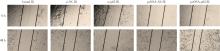
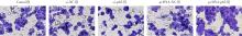
目的 探讨自噬调控多功能蛋白p62/SQSTM1对非小细胞肺癌生物学行为的影响和调控机制。 方法 RT-qPCR检测p62在正常人支气管上皮细胞和非小细胞肺癌细胞中的表达情况;CCK-8、划痕和Transwell实验分别检测抑制和促进p62表达后对非小细胞肺癌细胞增殖、迁移和侵袭能力的影响;Western blot实验检测抑制和促进p62表达后对非小细胞肺癌细胞凋亡相关蛋白(Bcl-2和Bax)和自噬相关蛋白(ATG5和Becline1)表达水平的影响;裸鼠皮下成瘤实验检测抑制p62表达后对非小细胞肺癌细胞体内肿瘤质量的影响。 结果 癌旁组织p62表达(1.01 ± 0.08)低于肺癌组织(2.81 ± 0.17)(P < 0.05);p62在si-NC、si-p62、pcDNA-NC、pcDNA-p62组的表达水平分别为1.02 ± 0.03、0.62 ± 0.07、1.03 ± 0.01、2.69 ± 0.12。与si-NC组细胞比较,si-p62组细胞中的p62表达显著降低(P < 0.05);与pcDNA-NC组细胞比较,pcDNA-p62组细胞中的p62表达显著升高(P < 0.05);转染si-p62后细胞增殖、迁移和侵袭能力显著降低(P < 0.05),而转染pcDNA-p62后细胞增殖、迁移和侵袭能力显著升高(P < 0.05);转染si-p62后,抗凋亡蛋白Bcl-2(0.31 ± 0.04)、自噬相关蛋白ATG5(0.48 ± 0.02)和Becline1表达水平(0.37 ± 0.03)明显降低,而促凋亡蛋白Bax表达水平(0.59 ± 0.07)明显升高(P < 0.05);而转染pcDNA-p62后,Bax蛋白水平(0.12 ± 0.02)降低,而Bcl-2(0.79 ± 0.07)、ATG5(0.93 ± 0.07)和Becline1表达水平(0.96 ± 0.04)明显升高(P < 0.05);与sh-NC组(2.50 ± 0.12)比较,sh-p62组(1.12 ± 0.08)小鼠的移植瘤质量明显减小(P < 0.05)。 结论 p62可以通过调控自噬促进非小细胞肺癌细胞A549的体内外生长。

关键词: 非小细胞肺癌, p62, 自噬, 增殖, 迁移, 侵袭

Abstract:

Objective To investigate the effect and regulatory mechanism of autophagy related multifunctional protein p62/SQSTM1 on biological behavior in non?small cell lung cancer (NSCLC). Methods RT?qPCR was used to detect the expression of p62 in normal lung cells and NSCLC cells. CCK?8, wound?healing and Transwell assays were used to detect the effects of inhibition and promotion of p62 expression on the proliferation, migration and invasion in NSCLC cells. Western blotting was used to detect the effects of inhibition and promotion of p62 expression on the expression of apoptosis?related proteins (Bcl?2 and Bax) and autophagy?related proteins (ATG5 and Becline1) in NSCLC cells. A nude mouse transplantation tumor experiment was used to detect the effect of inhibiting p62 expression on the tumor volume and mass of NSCLC cells in vivo. Results Compared with that in normal lung cells, the expression level of p62 in A549 cells was the highest. Cell function experiments in vitro showed that inhibition of p62 expression reduced the abilities of proliferation, migration and invasion in A549 cells, and suppressed autophagy and induced apoptosis. Consistently, p62 overexpression has the opposite effects. In addition, animal experiments in vivo showed that inhibition of p62 expression decreased the tumor volume and mass of tumor?bearing mice. Conclusion p62 could promote the growth of NSCLC A549 cell in vivo and in vitro by modulating autophagy.

Key words: non?small cell lung cancer, autophagy, proliferation, migration, invasion

中图分类号: