实用医学杂志 ›› 2024, Vol. 40 ›› Issue (20): 2923-2928.doi: 10.3969/j.issn.1006-5725.2024.20.017

• 医学检查与临床诊断 • 上一篇    下一篇

CD3+/CD4+T淋巴细胞水平及中性粒细胞与淋巴细胞比值在放射性肺炎中的预测价值

李鑫盼1,方懿2,邱俊1()   

  1. 1.湖南省人民医院(湖南师范大学附属第一医院)肿瘤二病区 (湖南 长沙 410016 )
    2.南华大学附属长沙中心医院麻醉科 (湖南 长沙 410006 )
  • 收稿日期:2024-06-24 出版日期:2024-10-25 发布日期:2024-11-05
  • 通讯作者: 邱俊 E-mail:68494976@qq.com
  • 基金资助:
    湖南省自然科学基金项目(2024JJ9299);湖南省卫生健康委科研立项课题(202204112874)

Predictive value of CD3+/CD4+T lymphocyte level and neutrophil to lymphocyte ratio in radiation pneumonia

Xinpan LI1,Yi FANG2,Jun QIU1()   

  1. *.Department of Tumor Ward 2,the First Affiliated Hospital of Hu′nan Normal University(Hu′nan Provincial People's Hospital),Changsha 410016,Hu′nan,China
  • Received:2024-06-24 Online:2024-10-25 Published:2024-11-05
  • Contact: Jun QIU E-mail:68494976@qq.com

摘要:

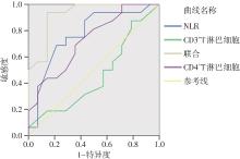
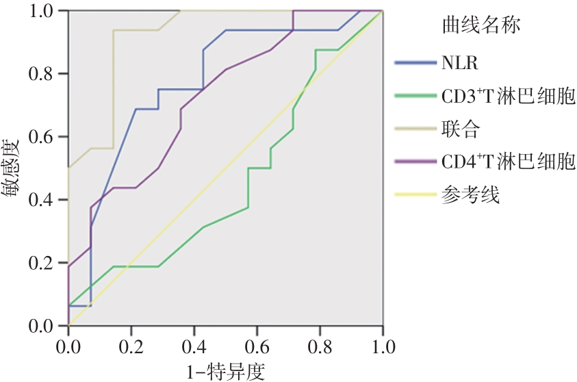
目的 探讨CD3+/CD4+T淋巴细胞水平及中性粒细胞与淋巴细胞比值(neutrophil-to-lymphocyte ratio, NLR)在放射性肺炎中的预测价值。 方法 回顾性分析2018年3月至2024年3月本院收治的局部晚期非小细胞肺癌者87例相关资料,其中发生放射性肺炎者43例,未发生放射性肺炎者44例,比较两组放射治疗前后T淋巴细胞亚群、NLR水平变化,统计T淋巴细胞亚群、NLR在肺癌放疗后放射性肺炎中的诊断价值,分析T淋巴细胞亚群、NLR与放射线肺炎分级严重程度的相关性,绘制T淋巴细胞亚群、NLR预测肺癌放疗后放射性肺炎的ROC曲线,并计算其AUC面积。 结果 放疗后发生组CD3+T淋巴细胞、CD4+T淋巴细胞水平显著低于放疗前且低于放疗后未发生组(P < 0.05),放疗后发生组NLR显著高于放疗前且显著高于放疗后未发生组(P < 0.05),CD3+T淋巴细胞、CD4+T淋巴细胞与NLR联合诊断的灵敏度、特异度、准确度以及阴性预测值、阳性预测值均高于NLR,且高于CD4+T淋巴细胞、CD3+T淋巴细胞,CD3+T淋巴细胞和CD4+T淋巴细胞水平变化与放射性肺炎分级严重程度之间呈负相关(P < 0.05),NLR变化与放射性肺炎分级严重程度之间呈正相关(P < 0.05),CD3+T淋巴细胞、CD4+T淋巴细胞与NLR联合检测预测肺癌放疗后放射性肺炎的AUC面积为0.924,大于CD3+T淋巴细胞、CD4+T淋巴细胞与NLR单独检测者。 结论 CD3+T淋巴细胞、CD4+T淋巴细胞和NLR联合检测,对局部晚期非小细胞肺癌接受放射治疗后出现放射性肺炎中具有较理想的诊断和预测价值,可作为临床早期诊断和预防放射性肺炎的指标,而得以应用推广。

关键词: CD3+T淋巴细胞, CD4+T淋巴细胞, 中性粒细胞与淋巴细胞比值, 放射性肺炎, 非小细胞肺癌

Abstract:

Objective To investigate the predictive value of CD3+/CD4+T lymphocyte level and neutrophil to lymphocyte ratio (NLR) in radiation pneumonia. Methods The data of 87 patients with locally advanced non-operative non-small cell lung cancer admitted to our hospital from March 2018 to October 2023 were retrospectively analyzed. Among them, 43 cases had radiation pneumonia and 44 cases did not have radiation pneumonia. The changes in T lymphocyte subsets and NLR before and after radiotherapy were compared between the two groups. The diagnostic value of T lymphocyte subsets and NLR in lung cancer radiation pneumonia after radiotherapy was analyzed, the correlation between T lymphocyte subsets and NLR and the grade severity of radiation pneumonia was analyzed, and the ROC curve of T lymphocyte subsets and NLR in predicting lung cancer radiation pneumonia after radiotherapy was drawn, and the AUC area was calculated. Results The levels of CD3+T lymphocytes and CD4+T lymphocytes in the post-radiotherapy group were significantly lower than those before radiotherapy and those without radiotherapy (P < 0.05). The NLR in the post-radiotherapy group was significantly higher than that before radiotherapy and those without radiotherapy (P < 0.05). The sensitivity, specificity, accuracy, negative predictive value, and positive predictive value of the combined diagnosis of CD3+T lymphocytes, CD4+T lymphocytes, and NLR were higher than those of NLR and higher than those of CD4+T lymphocytes and CD3+T lymphocytes. The changes of CD3+T lymphocyte and CD4+T lymphocyte levels were negatively correlated with grade severity of radiopneumonia (P < 0.05), while the changes of NLR were positively correlated with grade severity of radiopneumonia (P < 0.05). Combined detection of CD3+T lymphocytes, CD4+T lymphocytes, and NLR predicted that the AUC area of lung cancer after radiotherapy was 0.924, which was larger than that of CD3+T lymphocytes, CD4+T lymphocytes, and NLR alone. Conclusion The combined detection of CD3+T lymphocytes, CD4+T lymphocytes, and NLR has an ideal value in the diagnosis and prediction of radiation pneumonia after radiotherapy for locally advanced non-small cell lung cancer, and can be used as an indicator for early diagnosis and prevention of radiation pneumonia.

Key words: CD3+T lymphocytes, CD4+T lymphocytes, neutrophil to lymphocyte ratio, radiation pneumonia, non-small cell lung cancer

中图分类号: