实用医学杂志 ›› 2023, Vol. 39 ›› Issue (19): 2434-2439.doi: 10.3969/j.issn.1006-5725.2023.19.004

• 专题报道:结核专题 • 上一篇    下一篇

肺结核治疗过程中IL-6血清表达水平变化分析

谢贝1,2,许婉华3,龚兰1,2,杨瑜1,2,董海平2,4,吴玲1,2,孟繁荣1,2,王楠1,2,刘志辉1,2,李华1,2()   

  1. 1.广州市胸科医院肺部疾病研究所
    2.呼吸疾病国家重点实验室 (广州 510095 )
    3.广州市结核病防治所二分所 (广州 510095 )
    4.重症结核科 (广州 510095 )
    2.呼吸疾病国家重点实验室 (广州 510095 )
  • 收稿日期:2023-07-17 出版日期:2023-10-10 发布日期:2023-11-22
  • 通讯作者: 李华 E-mail:lh861218@163.com
  • 基金资助:
    广州市科技计划-重点研发项目(202206010134);广州市卫生健康科技项目(20231A011051);广州市科技计划(202002030152);广州市医学重点学科(2021-2023┫结核病学);国家重点研发计划“病原学与防疫技术体系研究”重点专项(2022YFC2304800)

Analysis of changes in serum expression levels of IL-6 during pulmonary tuberculosis treatment

Bei XIE1,2,WanHua XU3,Lan GONG1,2,Yu YANG1,2,Haiping DONG2,4,ling WU1,2,Fanrong MENG1,2,Nan WANG1,2,Zhihui LIU1,2,Hua. LI1,2()   

  1. *.Institute of Pulmonary Diseases,Guangzhou Chest Hospital Guangzhou 510095,China
    *.ICU of tuberculosis,Guangzhou Chest Hospital,Guangzhou 510095,China
  • Received:2023-07-17 Online:2023-10-10 Published:2023-11-22
  • Contact: Hua. LI E-mail:lh861218@163.com

摘要:

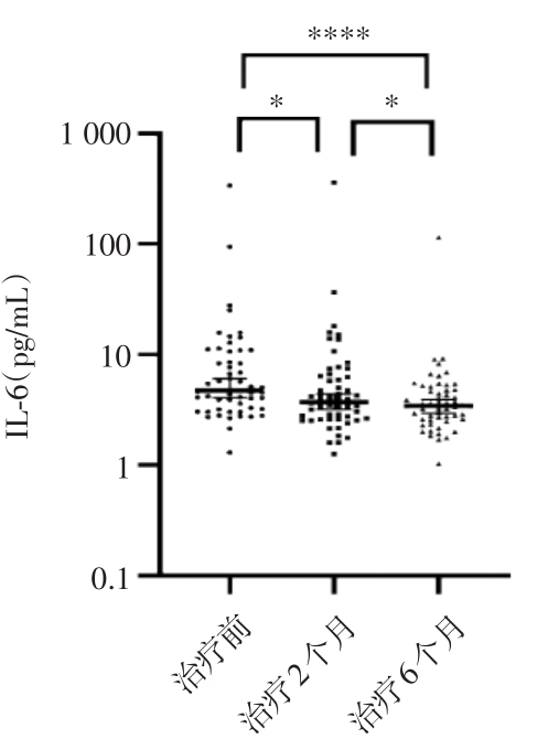
目的 探究血清中白介素-6(IL-6)水平在肺结核治疗过程中的变化趋势及影响因素。 方法 分别收集56位肺结核患者治疗前、治疗2个月(强化期结束)和治疗6个月时血清,通过流式细胞术测量其中IL-6的表达水平。 结果 肺结核患者治疗期间血清中IL-6的表达水平呈降低趋势,且其表达水平差异具有统计学意义(P = 0.000)。ROC曲线分析显示,治疗前和治疗2个月时,血清中IL-6表达水平差异的线下面积为0.627,界值为3.840,鉴别的灵敏度为71.40%,特异度为51.80%。二元logistic回归显示治疗2个月后血清中IL-6表达水平是否降低至3.840 pg/mL以下与治疗周期、年龄有关。治疗前IL-6的血清表达水平在有无空洞组、治疗周期为6个月组和12个月组、治疗2个月时对比未治疗前影像学病灶吸收组和无明显变化组、治疗前病原学检查结果阴阳性组之间的差异具有统计学意义。治疗2个月时,IL-6的血清表达水平在不同年龄组、不同治疗周期组、治疗前及治疗2个月时病原学检查结果阴阳性组之间的差异具有统计学意义。治疗6个月时,IL-6的血清表达水平在是否有糖尿病组、不同治疗周期组、治疗6个月时病原学检查结果阴阳性组之间的差异具有统计学意义。ROC曲线分析显示,将入组病例限制到治疗前病原学阳性时,治疗6个月和治疗2个月血清中IL-6表达水平差异的线下面积为0.805,界值为5.450,鉴别的灵敏度为81.25%,特异度为75.00%;当将入组病例年龄限制到44岁及以下时,治疗2个月和治疗前、治疗6个月和治疗2个月血清中IL-6表达水平差异的线下面积分别为0.726、0.721,界值分别为3.840、3.755,鉴别的灵敏度均为70.59%,特异度分别为70.59%、67.65%。 结论 肺结核治疗过程中IL-6在血清中的表达水平呈下降趋势,其水平与患者肺部有无空洞、病原学结果、年龄、病情进展、是否合并糖尿病等有关。

关键词: 肺结核, 白介素-6, 表达水平变化

Abstract:

Objective To explore the trend and influencing factors of serum IL-6 level in the treatment of pulmonary tuberculosis. Methods The serum of 56 patients with pulmonary tuberculosis was collected before treatment, 2 months of treatment (the end of the intensive period) and 6 months of treatment, and the expression level of IL-6 was measured by flow cytometry. Results The expression level of IL-6 in serum during treatment in tuberculosis patients tended to decrease, and the expression level was statistically significant (P = 0.000). ROC curve analysis showed that before treatment and 2 months of treatment, the subline area of IL-6 concentration difference in serum was 0.627, the cut-off value was 3.840, the sensitivity for discrimination was 71.40%, and the specificity was 51.80%. Binary logistic regression showed that whether the serum IL-6 concentration decreased to less than 3.840 pg/mL after 2 months of treatment was related to treatment cycle and age. The difference between the serum concentration of IL-6 before treatment was statistically significant between the group with or without cavitation, the treatment period group with 6 months and 12 months, and the group with no significant change in imaging lesions before treatment at 2 months, and the negative group with positive etiological test results before treatment. At 2 months of treatment, the difference between serum concentrations of IL-6 was statistically significant between different age groups, different treatment cycle groups, and the group with negative etiological test results before treatment and at 2 months of treatment. At 6 months of treatment, the difference between the serum concentration of IL-6 was statistically significant in the group with diabetes, in the group with different treatment cycles, and in the group with negative etiological test results at 6 months of treatment. ROC curve analysis showed that when the enrolled cases were limited to positive etiology before treatment, the off-line area of the difference in serum IL-6 concentration between 6 months of treatment and 2 months of treatment was 0.805, the cut-off value was 5.450, the sensitivity for discrimination was 81.25%, and the specificity was 75.00%. When the age of the enrolled cases was limited to 44 years and below, the off-line areas of IL-6 concentration differences in serum after 2 months of treatment and before treatment, 6 months of treatment and 2 months of treatment were 0.726 and 0.721, the cut-off values were 3.840 and 3.755, and the sensitivity and specificity were 70.59% and 67.65%, respectively. Conclusion During the treatment of pulmonary tuberculosis, the concentration of IL-6 in serum show a downward trend, and its level is related to whether the patient's lungs have cavities, etiological results, age, disease progression, and whether diabetes is present.

Key words: tuberculosis, IL-6, changes in expression level