实用医学杂志 ›› 2023, Vol. 39 ›› Issue (19): 2517-2523.doi: 10.3969/j.issn.1006-5725.2023.19.018

• 临床研究 • 上一篇    下一篇

糖尿病视网膜病变发展中的免疫图谱变化

吴晓君1,卜诗淼1,陈湘婷2,俞军1,刘勇1,张跃红1,2,辛松青3,刘雨丰1()   

  1. 1.华南理工大学医学院附属第二医院 (广州 510006 )
    2.广州市第一人民医院眼科
    1.广州 510180 ),广东省东莞市常安医院检验科,(广东东莞 523000
  • 收稿日期:2023-07-12 出版日期:2023-10-10 发布日期:2023-11-22
  • 通讯作者: 刘雨丰 E-mail:eyyufengliu@scut.edu.cn
  • 基金资助:
    国家自然科学基金面上项目(8217060280);广东省自然科学基金面上项目(2114050002084);广州市基础与应用基础研究专题(优秀博士“续航”项目)(SL2024A04J00240);东莞市社会科技发展(重点)项目(20221800904962)

Immune mapping changes in the development of diabetic retinopathy

Xiaojun WU1,Shimiao BU1,Xiangting CHEN2,Jun YU1,Yong LIU1,Yuehong ZHANG1,2,Songqing XIN3,Yufeng. LIU1()   

  1. *.The Second Affiliated Hospital,School of Medicine,South China University of Technology,Guangzhou 510006,China
  • Received:2023-07-12 Online:2023-10-10 Published:2023-11-22
  • Contact: Yufeng. LIU E-mail:eyyufengliu@scut.edu.cn

摘要:

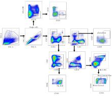
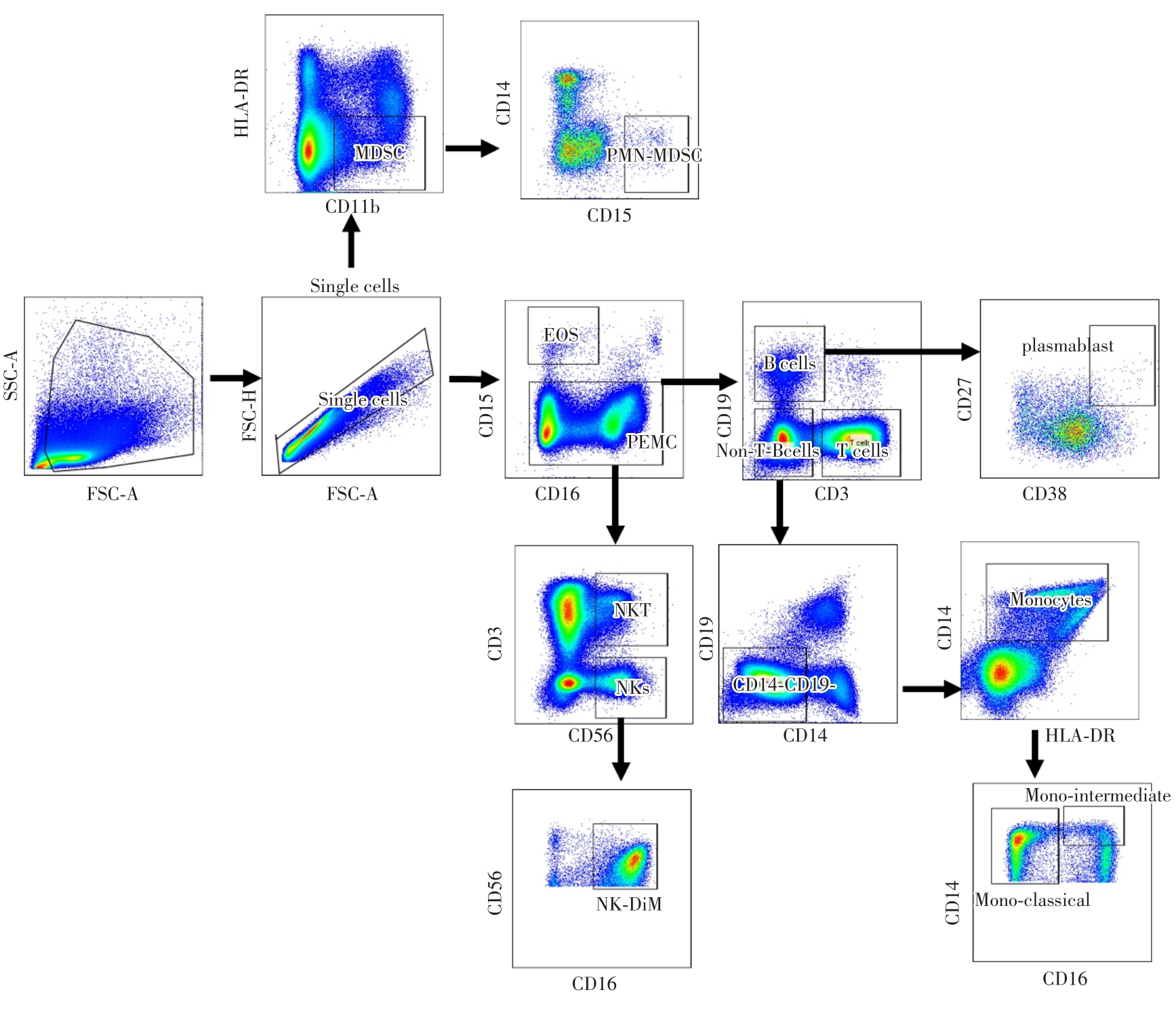
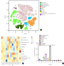
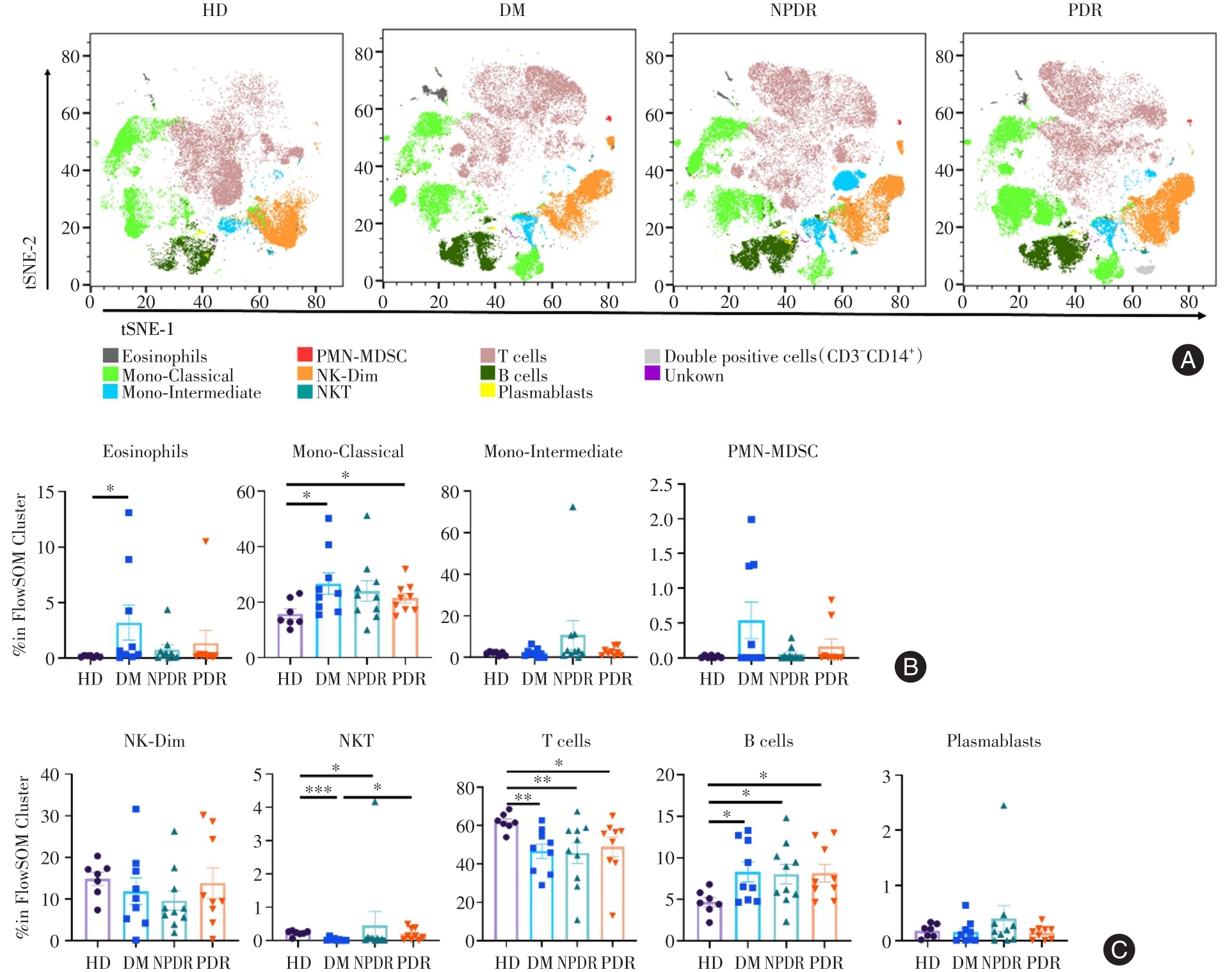
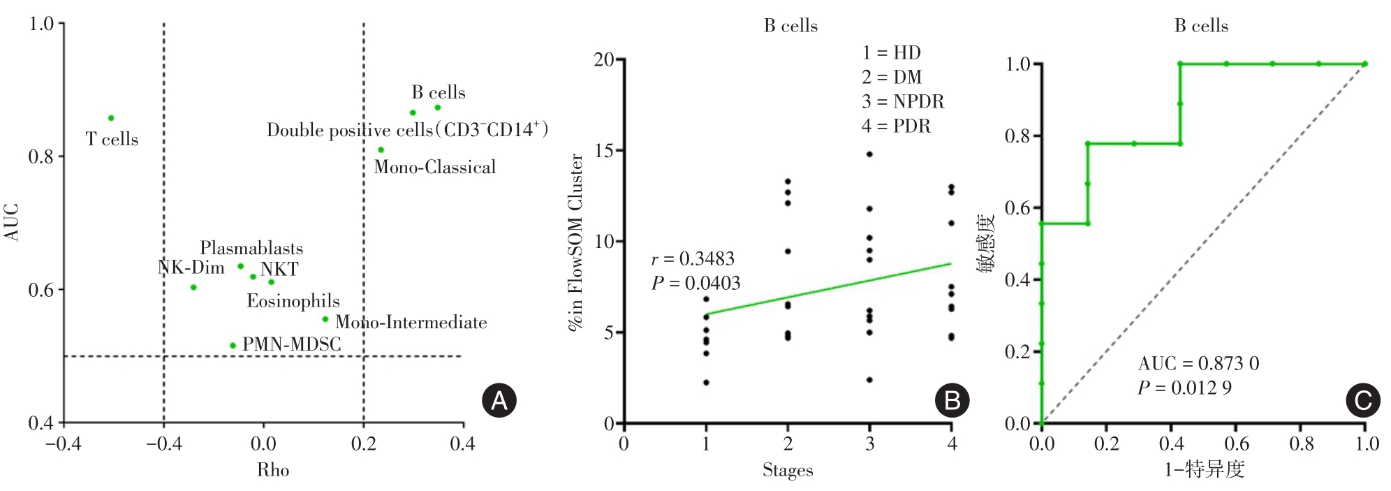
目的 为解析糖尿病向糖尿病视网膜病变进展的免疫学调控机制,流式细胞术分析健康人(health donor,HD)、糖尿病患者(diabetes mellitus,DM)、非增殖型糖尿病视网膜病变患者(non-proliferative diabetic retinopathy, NPDR)以及增殖型糖尿病视网膜病变患者(proliferative diabetic retinopathy, PDR)外周血中的免疫细胞图谱之间的差异。 方法 收集HD、DM、NPDR以及PDR患者的外周血,采用密度梯度离心法分离获得外周血免疫细胞。13色流式抗体共同染色并上流式细胞仪进行检测。结合Flowjo及其插件对细胞进行降维及聚类处理分析,最后采用Spearman相关性分析及受试者工作特征(receiver operating characteristic,ROC)曲线评估外周血免疫细胞预测DM到DR阶段进展的诊断价值。 结果 将外周血免疫细胞分为10大群,在固有免疫细胞中,DM和PDR中mono-classical细胞群较健康人升高(P < 0.05);适应性免疫细胞中,DM和NPDR中的B细胞较HD组升高(P < 0.05),NKT、T细胞则较HD组下降(P < 0.05);PDR中的B细胞和健康人对比则均升高(P < 0.05),但T细胞下降(P < 0.05)。此外,B细胞和各阶段呈正相关,并且在PDR阶段中,ROC曲线分析B细胞预测DM到DR阶段进展的效能大于其他免疫细胞。 结论 健康人、DM和DR患者的3个阶段的外周血免疫细胞图谱之间存在差异,此外B细胞和各阶段正相关,有着预测疾病进展的诊断价值的潜力。

关键词: 糖尿病, 糖尿病视网膜病变, 免疫细胞, 流式细胞术

Abstract:

Objective To analyze the immunological regulation mechanism of the progression of diabetes to diabetic retinopathy. We used Flow cytometry to compare the differences of immune cell profiles in peripheral blood of healthy donors (HD), diabetic patients (DM), non-proliferative diabetic retinopathy patients (NPDR) and proliferative diabetic retinopathy patients (PDR). Methods Peripheral blood was collected from HD, DM, NPDR and PDR, and peripheral blood immune cells were isolated by density gradient centrifugation. The 13-color flow cytometry antibody was co-stained and detected by flow cytometry. Combined with Flowjo and its plug-in, the cells were analyzed by dimensionality reduction and clustering. Finally, Spearman correlation analysis and receiver operating characteristic (ROC) curve were used to evaluate the diagnostic value of peripheral blood immune cells in predicting the progression from DM to DR stage. Results Peripheral blood immune cells were divided into 10 groups. Among the innate immune cells, the number of mono-classical cells in DM and PDR was higher than that in healthy people (P < 0.05). In adaptive immune cells, B cells in DM and NPDR were higher than those in HD group (P < 0.05), while NKT and T cells were lower than those in HD group (P < 0.05). Compared with healthy people, B cells in PDR were increased (P < 0.05), but T cells were decreased (P < 0.05). In addition, B cells were positively correlated with each stage. And in the PDR stage, ROC curve analysis showed that the efficiency of B cells in predicting the progression of DM to DR was greater than that of other immune cells. Conclusion Differences were noted between the three stages of peripheral blood immune cell profiles in HD, DM and DR. In addition, B cells were positively correlated with each stage, and had the potential to predict the diagnostic value of disease progression.

Key words: diabetes mellitus, diabetic retinopathy, immune cells, flow cytometry

中图分类号: