实用医学杂志 ›› 2023, Vol. 39 ›› Issue (18): 2395-2400.doi: 10.3969/j.issn.1006-5725.2023.18.019

• 医学检查与临床诊断 • 上一篇    下一篇

18F-FDG PET/CT联合血清CA19-9、CEA、NSE鉴别胰腺导管腺癌与胰腺神经内分泌肿瘤

郑立春1,张欢2,顾程1,申新宇1,张晓明1,欧阳向柳3()   

  1. 1.唐山市工人医院,核医学科,(河北 唐山 063000 )
    3.唐山市工人医院,超声医学科,(河北 唐山 063000 )
    2.华北理工大学研究生学院 (河北 唐山 063000 )
  • 收稿日期:2023-04-14 出版日期:2023-09-25 发布日期:2023-10-10
  • 通讯作者: 欧阳向柳 E-mail:usouyangxl@163.com
  • 基金资助:
    河北省省级科技计划项目基金资助项目(182777145);唐山市科学技术研究与发展计划项目基金资助项目(19150220E)

The differential diagnosis of pancreatic duct a ladenocarcinoma and pancreatic neuroendocrine tumors using 18F⁃FDG PET/CT combined with serum CA19⁃9, CEA and NSE

Lichun ZHENG1,Huan ZHANG2,Cheng GU1,Xinyu SHEN1,Xiaoming ZHANG1,Xiangliu. OUYANG3()   

  1. 1.Department of Nuclear Medicine,Tangshan Gongren Hospital,Tangshan 063000,China
  • Received:2023-04-14 Online:2023-09-25 Published:2023-10-10
  • Contact: Xiangliu. OUYANG E-mail:usouyangxl@163.com

摘要:

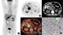
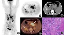
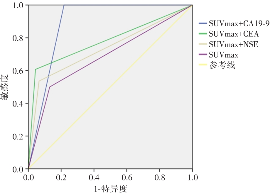
目的 探讨18F-FDG PET/CT联合血清CA19-9、CEA、NSE在胰腺导管腺癌(pDAC)与胰腺神经内分泌肿瘤(pNETs)鉴别诊断的临床价值。 方法 回顾性分析经病理确诊的pDAC和pNETs患者临床资料、18F-FDG PET/CT影像表现和血清肿瘤标志物CA19-9、CEA、NSE结果,对比两组患者年龄、性别、病灶大小、位置、胰管受累情况及18F-FDG PET/CT显像中代谢形态、最大标准化摄取值(SUVmax)、周围淋巴结及肝转移情况、血清CA19-9、CEA、NSE结果,分析两者的有效鉴别诊断指标,对比18F-FDG PET/CT显像中SUVmax联合血清CA19-9、CEA、NSE的诊断效能。 结果 共入组74例患者,男42例、女32例,pDAC 46例,年龄46 ~ 82岁,平均(66.48 ± 8.84)岁,pNETs 28例,年龄47 ~ 73岁,平均(58.64 ± 6.50)岁,pDAC组SUVmax均值(5.55 ± 2.01),pNETs组SUVmax均值(4.62 ± 2.10)。两组在性别、18F-FDG PET/CT影像中代谢形态、SUVmax、周围淋巴结转移情况方面差异均无统计学意义(P > 0.05),而在年龄、病灶大小、位置、胰管受累情况及肝转移情况、血清CA19-9、CEA、NSE结果方面差异均有统计学意义(P < 0.05)。SUVmax联合血清CA19-9、CEA、NSE较单独依据SUVmax的诊断效能均有所提高,其中以SUVmax联合CA19-9诊断效能最高,ROC曲线下AUC为0.891(95%CI:0.816 ~ 0.967,P < 0.001)。 结论 18F-FDG PET/CT联合CA19-9、CEA、NSE对胰腺导管腺癌与胰腺神经内分泌肿瘤的鉴别有较高临床价值,弥补了两者在18F-FDG PET/CT显像中鉴别困难的不足,以SUVmax联合CA19-9诊断效能最高。

关键词: 胰腺导管腺癌, 胰腺神经内分泌肿瘤, 正电子发射型体层摄影术, 体层摄影术,X线计算机, 18F氟脱氧葡萄糖, 肿瘤标志物

Abstract:

Objective To discuss the value of 18F?FDG PET/CT combined with serum CA19?9, CEA and NSE in the differential diagnosis pancreatic ductal adenocarcinoma(pDAC) and pancreatic neuroendocrine tumors(pNETs). Methods Retrospective analys is of patients who were diagnosed as pDAC or pNETs by pathology, including the clinical data, 18F?FDG PET/CT images and the level of tumor markers CA19?9, CEA and NSE, and compared age, sex, size and location of the lesions, the involvement of pancreatic ductal, the morphology of the lesionson PET image, SUVmax, surrounding lymphatic metastasis and hepatic metastasis and the level of serum CA19?9, CEA, NSE between the two groups, and figure out the effective indicators in differential diagnosis. The diagnostic efficacy of 18F?FDG PET/CT imaging SUVmax combined with serum CA19?9, CEA and NSE was compared. Results There were 74 patients were included. The age were between 46 and 82(66.48 ± 8.84), and 32 were male, 32 were female. 46 patients were pDAC, 28 were pNETs. The mean value of SUVmax were (5.55 ± 2.01) in pDAC group, (4.62 ± 2.01) in pNETs group. It was not statistically significant between the two groups in sex, morphology of the lesions in PET image, SUVmax, surrounding lymphatic metastasis(P > 0.05), and was statistically significant in age, size of the lesions, location of the lesions, the involvement of pancreatic ductal hepatic metastasis and serum CA19?9, CEA, NSE(P < 0.05). The diagnostic efficacy of SUVmax combined with serum CA19?9, CEA and NSE was improved compared with SUVmax, which SUVmax combined with CA19?9 had the best diagnostic efficacy with the AUC of ROC curve was 0.891 (95%CI: 0.816~0.967, P < 0.001). Conclusion The using of 18F?FDG PET/CT combined with serum CA19?9, CEA, NSE has a high differential diagnosis value in pancreatic ductal adenocarcinoma and pancreatic neuroendocrine tumors, that make up the shortage of using 18F?FDG PET/CT alone, which the diagnostic efficiency of SUVmax combined with CA19?9 was the best.

Key words: pancreatic ductal adenocarcinoma, pancreatic neuroendocrine tumors, positron emission tomography, X ray computed tomography, 18F?deoxyglucose, tumor marker

中图分类号: