实用医学杂志 ›› 2023, Vol. 39 ›› Issue (18): 2401-2404.doi: 10.3969/j.issn.1006-5725.2023.18.020

• 医学检查与临床诊断 • 上一篇    下一篇

超声纹理分析在直肠癌T分期中的诊断价值

谢亚宁1,温德惠2(),王立坤2,王琪1,武雪亮3   

  1. 1.河北北方学院 (河北 张家口 075000 )
    2.河北北方学院附属第一医院2 超声医学科
    3.普外科;肿瘤研究所 (河北 张家口 075000 )
  • 收稿日期:2023-07-20 出版日期:2023-09-25 发布日期:2023-10-10
  • 通讯作者: 温德惠 E-mail:15530396808@163.com
  • 基金资助:
    2022年度河北省医学科学研究课题计划(20220028);河北省临床医学优秀人才培养项目

Diagnostic value of ultrasound texture analysis in T stage of rectal cancer

Yaning XIE1,Dehui WEN2(),Likun WANG2,Qi WANG1,Xueliang. WU3   

  1. 1.Hebei North University,Zhangjiakou 075000,China
  • Received:2023-07-20 Online:2023-09-25 Published:2023-10-10
  • Contact: Dehui WEN E-mail:15530396808@163.com

摘要:

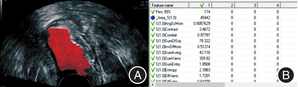
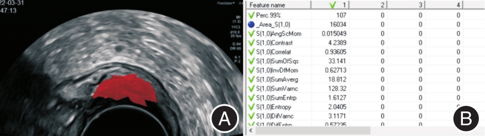
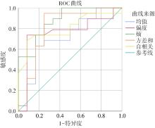
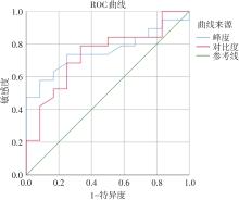
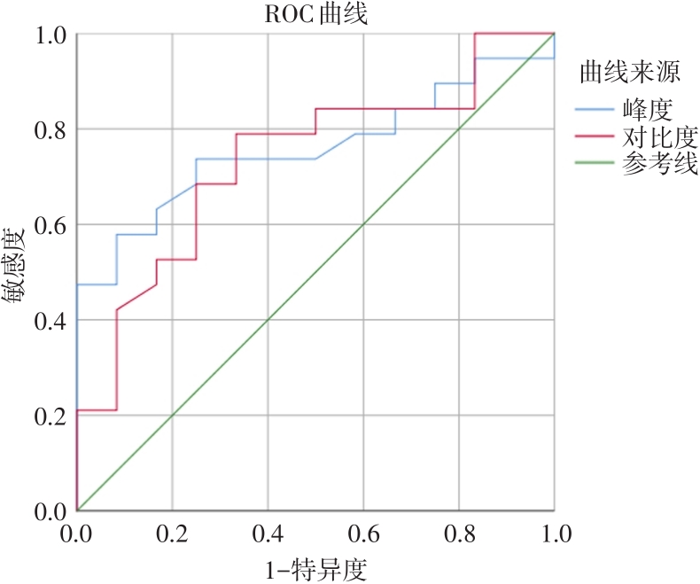
目的 研究超声纹理分析在直肠癌T分期中的诊断价值。 方法 选取经手术或病理证实的31例直肠癌患者术前超声检查图像,采用mazda软件勾画感兴趣区域(ROI),软件自动计算出均值、峰度、偏度、对比度、熵、方差和、自相关、能量、逆差距纹理特征参数,比较不同分期直肠癌各纹理参数的差异,并对差异有统计学意义的参数绘制受试者工作特征曲线(ROC),比较曲线下面积(AUC),评估其诊断效能。 结果 统计得到,峰度、偏度、对比度、熵、方差和、自相关均是直肠癌T分期的影响因素,晚期直肠癌(pT3-4期)较早期直肠癌(pT1-2期)偏度、熵、方差和、自相关偏高(P < 0.05),而峰度、对比度较早期直肠癌偏低(P < 0.05)。其中,熵具有较高的诊断效能,AUC值为0.88。 结论 超声纹理分析可为直肠癌T分期提供更客观的影像学依据,与早期直肠癌相比,晚期直肠癌的纹理特征更为复杂。

关键词: 直肠癌, 超声, 纹理分析, T分期

Abstract:

Objective To investigate the diagnostic value of ultrasound texture analysis in T stage of rectal cancer. Methods The preoperative ultrasound images of 31 patients with rectal cancer confirmed by surgery or pathology were selected, and the region of interest (ROI) was delineated by mazda software. The software automatically calculated the texture characteristic parameters of mean, kurtosis, skewness, contrast, entropy, variance sum, autocorrelation, energy and deficit distance, compared the differences of texture parameters of rectal cancer in different stages, and drew the subject's working characteristic curve (ROC) for the statistically significant parameters, compared the area under the curve (AUC), and evaluated its diagnostic efficiency. Results It was statistically shown that kurtosis, skewness, contrast, entropy, variance sum and autocorrelation were all influencing factors of T stage of rectal cancer, and the skewness, entropy, variance sum and autocorrelation of advanced rectal cancer (pT3-4 stage) were higher than those of early rectal cancer (pT1-2 stage) (P < 0.05), while the kurtosis and contrast were lower than those of early rectal cancer (P < 0.05). Among them, entropy has a higher diagnostic efficiency, and the AUC value is 0.88. Conclusion Ultrasound texture analysis can provide a more objective imaging basis for T staging of rectal cancer, and the texture characteristics of advanced rectal cancer are more complex than those of early rectal cancer.

Key words: rectal cancer, ultrasound, texture analysis, T stage

中图分类号: