

The Journal of Practical Medicine ›› 2026, Vol. 42 ›› Issue (13): 2404-2418.doi: 10.3969/j.issn.1006-5725.2026.13.017
• Treatise:Mechanism and Practice • Previous Articles
Xuefei XIANG1,Heyou GAO1,2,Zhiyun CHEN1,Qingchuang HU1,Liu HE1,Yu WANG1,2( )
)
Received:2026-02-10
Online:2026-07-10
Published:2026-07-14
Contact:
Yu WANG
E-mail:wangyuscu@foxmail.com
CLC Number:
Xuefei XIANG,Heyou GAO,Zhiyun CHEN,Qingchuang HU,Liu HE,Yu WANG. Study on the role of the PI3K/AKT pathway in the regulation of osteogenesis on the surface of calcium phosphate ceramics by erythropoietin[J]. The Journal of Practical Medicine, 2026, 42(13): 2404-2418.
Tab.1
Primer sequences for each gene"
| 基因 | 引物序列(5′-3′) |
|---|---|
| Runx2 | 正义链:AAG GAG CAG ACC GTC AAA GG 反义链:CCC AGA GTC CAC CTC TCT GA |
| Osterix | 正义链:GCA TCT GAA AGC CCA CTT GC 反义链:AGT GGT CGC TTC GGG TAA AG |
| OPN | 正义链:GAT GAC GAC GAC GAT GAC GA 反义链:GCT GGC AGT GAA GGA CTC AT |
| CYTL1 | 正义链:CAC CGT CCT CCA GAG TGA AC 反义链:CGG ACC CAG TTT GGA TGG AA |
| MTSS1 | 正义链;GCG GTT TTC TCT GAC CCA AG 反义链:CCC CAA GTC CCA ATC CGA AA |
| GAPDH | 正义链:GAC ATG CCG CCT GGA GAA AC 反义链:AGC CCA GGA TGC CCT TTA GT |

Fig.1
Gross observation results of the EPO/pDA/BCP composite scaffold"
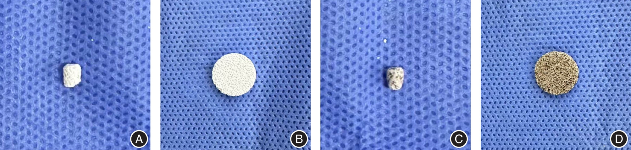
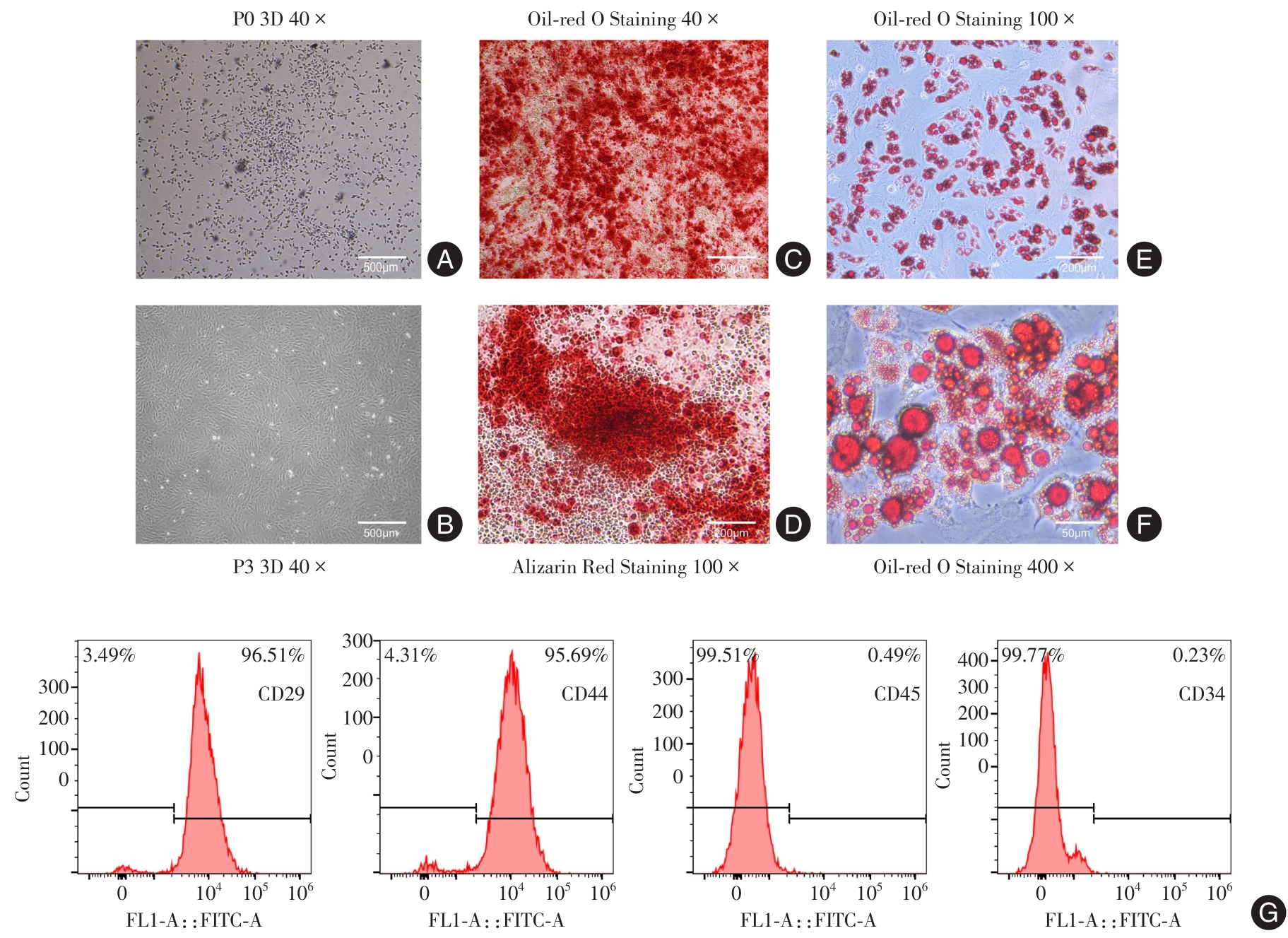
Fig.2
Identification results of rat bone marrow mesenchymal stem cells"
Tab.2
CCK-8 assay results of bone marrow mesenchymal stem cells in each group(n = 3)"
| 组别 | 第1天 | 第3天 | 第7天 |
|---|---|---|---|
| 空白组 | 0.28 ± 0.03 | 0.47 ± 0.06 | 1.02 ± 0.07 |
| BCP组 | 0.32 ± 0.01 | 0.47 ± 0.06 | 0.99 ± 0.06 |
| EPO/pDA/BCP组 | 0.27 ± 0.01 | 0.41 ± 0.01 | 1.04 ± 0.04 |
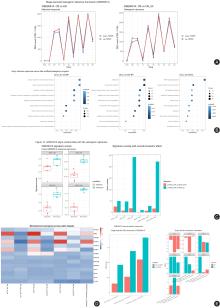
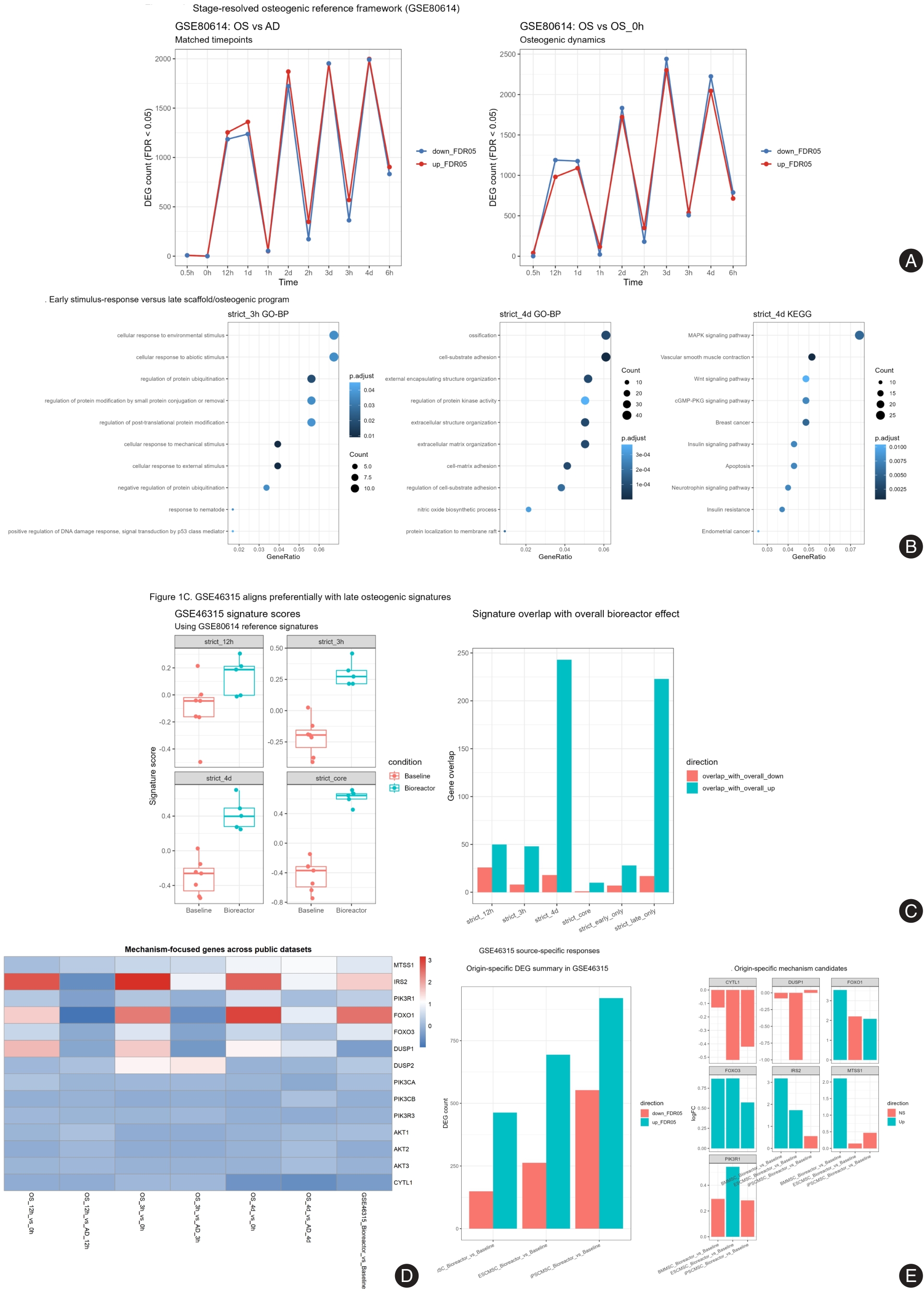
Fig.3
Analysis results of osteogenic stage characteristics and PI3K/AKT response network based on bioinformatics mining"
Tab.3
Results of alkaline phosphatase activity detection in bone marrow mesenchymal stem cells of each group after osteogenic induction(n = 3)"
| 组别 | 第3天 | 第7天 |
|---|---|---|
| 空白组 | 0.02 ± 0.00 | 0.05 ± 0.00 |
| BCP组 | 0.04 ± 0.01* | 0.07 ± 0.00* |
| EPO/pDA/BCP组 | 0.06 ± 0.01*# | 0.08 ± 0.00*# |
| EPO/pDA/BCP+LY组 | 0.04 ± 0.00△ | 0.05 ± 0.01△ |
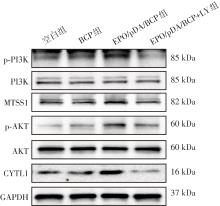
Fig.4
Western blot bands showing expression of PI3K/AKT pathway-related proteins in bone marrow mesenchymal stem cells of each group"
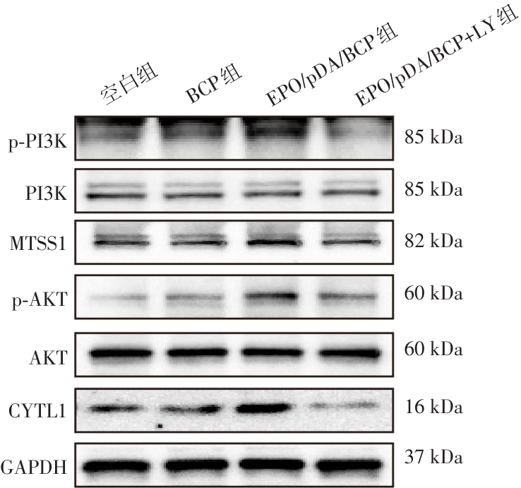
Tab.4
Quantitative analysis results of Western blot for expression of PI3K/AKT pathway-related proteins in bone marrow mesenchymal stem cells of each group(n = 3)"
| 组别 | p-PI3K/PI3K | p-AKT/AKT | CYTL1 | MTSS1 |
|---|---|---|---|---|
| 空白组 | 0.29 ± 0.13 | 0.24 ± 0.07 | 0.46 ± 0.03 | 0.41 ± 0.08 |
| BCP组 | 0.47 ± 0.07 | 0.48 ± 0.06 | 0.48 ± 0.04 | 0.51 ± 0.10 |
| EPO/pDA/BCP组 | 0.85 ± 0.09*# | 0.80 ± 0.03*# | 0.61 ± 0.05*# | 0.71 ± 0.05*# |
| EPO/pDA/BCP+LY组 | 0.49 ± 0.13△ | 0.39 ± 0.12△ | 0.39 ± 0.05△ | 0.40 ± 0.06△ |
Tab.5
Quantitative RT-qPCR analysis results of MTSS1 and CYTL1 in bone marrow mesenchymal stem cells of each group(n = 3)"
| 组别 | CYTL1 | MTSS1 |
|---|---|---|
| 空白组 | 1.01 ± 0.17 | 1.00 ± 0.15 |
| BCP组 | 2.50 ± 0.55 | 2.56 ± 0.41 |
| EPO/pDA/BCP组 | 4.10 ± 0.75*# | 3.34 ± 0.12*# |
| EPO/pDA/BCP+LY组 | 2.11 ± 0.12△ | 2.21 ± 0.11△ |
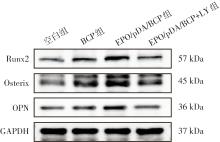
Fig.5
Western blot bands showing expression of PI3K/AKT pathway-related proteins in bone marrow mesenchymal stem cells of each group"
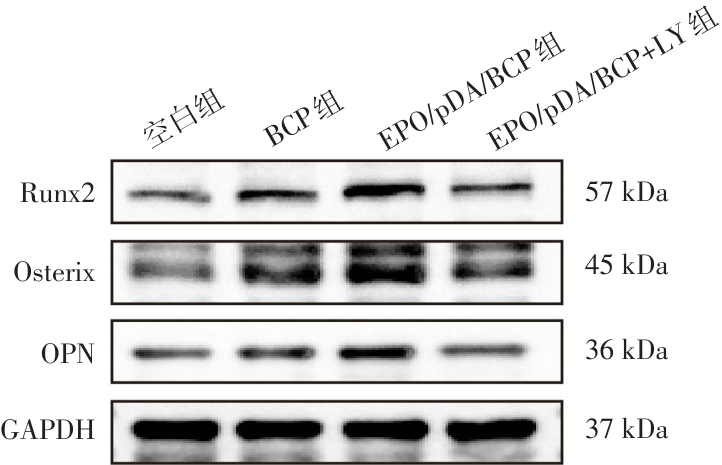
Tab.6
Western blot band images of osteogenesis-related protein expression in bone marrow mesenchymal stem cells of each group(n = 3)"
| 组别 | Runx2 | Osterix | OPN |
|---|---|---|---|
| 空白组 | 0.27 ± 0.07 | 0.38 ± 0.01 | 0.41 ± 0.05 |
| BCP组 | 0.49 ± 0.08* | 0.58 ± 0.06* | 0.51 ± 0.02 |
| EPO/pDA/BCP组 | 0.83 ± 0.12*# | 0.80 ± 0.11*# | 0.75 ± 0.08*# |
| EPO/pDA/BCP+LY组 | 0.45 ± 0.02△ | 0.54 ± 0.07△ | 0.49 ± 0.13△ |
Tab.7
Quantitative RT-qPCR analysis results of osteogenesis-related genes in bone marrow mesenchymal stem cells of each group(n = 3)"
| 组别 | Runx2 | Osterix | OPN |
|---|---|---|---|
| 空白组 | 1.01 ± 0.18 | 1.00 ± 0.09 | 1.01 ± 0.15 |
| BCP组 | 3.36 ± 0.26* | 3.05 ± 0.11* | 1.35 ± 0.11* |
| EPO/pDA/BCP组 | 4.87 ± 1.02*# | 4.15 ± 0.33*# | 1.84 ± 0.16*# |
| EPO/pDA/BCP+LY组 | 2.34 ± 0.38△ | 1.34 ± 0.19△ | 1.05 ± 0.03△ |
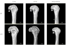
Fig.6
Micro-CT reconstruction images of the femoral defects in SD rats of each group at 4 weeks and 12 weeks after surgery"
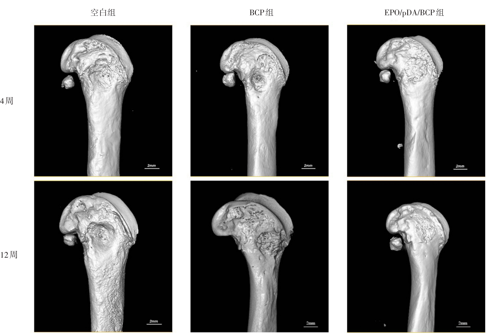
Tab.8
Semi-quantitative analysis results of bone volume fraction, trabecular bone thickness, bone mineral density, and trabecular bone number at 4 weeks and 12 weeks after surgery(n = 6)"
| 组别 | 骨体积分数/% | 骨小梁厚度/mm | 骨密度/(mg/cm3) | 骨小梁数量/(1/mm) |
|---|---|---|---|---|
| 空白组(4周) | 25.15 ± 8.02 | 0.30 ± 0.06 | 162.0 ± 60.67 | 0.42 ± 0.03 |
| BCP组(4周) | 45.90 ± 1.06* | 0.49 ± 0.03* | 312.72 ± 25.24* | 0.59 ± 0.04* |
| EPO/pDA/BCP组(4周) | 56.09 ± 3.55*# | 0.69 ± 0.07*# | 423.73 ± 11.54*# | 0.75 ± 0.09*# |
| 空白组(12周) | 30.20 ± 3.31 | 0.40 ± 0.06 | 238.85 ± 29.61 | 0.54 ± 0.03 |
| BCP组(12周) | 53.31 ± 1.57* | 0.55 ± 0.02* | 389.68 ± 28.29* | 0.80 ± 0.06* |
| EPO/pDA/BCP组(12周) | 72.83 ± 1.70*# | 0.80 ± 0.07*# | 631.11 ± 51.07*# | 0.96 ± 0.06*# |
Fig.7
Results of histological analysis of the femoral defect sites in rats of each group at 4 and 12 weeks after surgery"
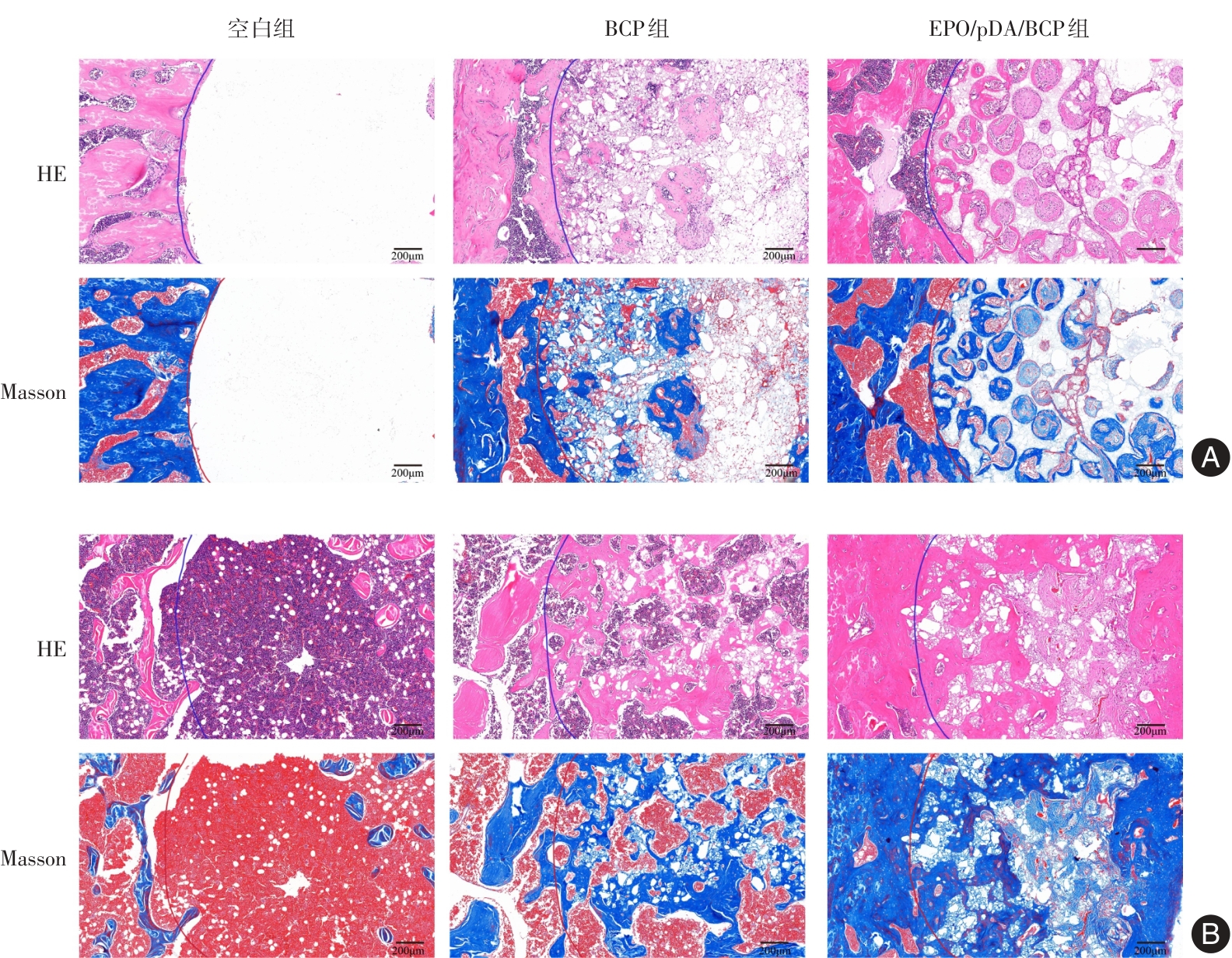
Fig.8
Results of immunohistochemical staining analysis of the femoral defect sites in rats of each group at 4 and 12 weeks after surgery"
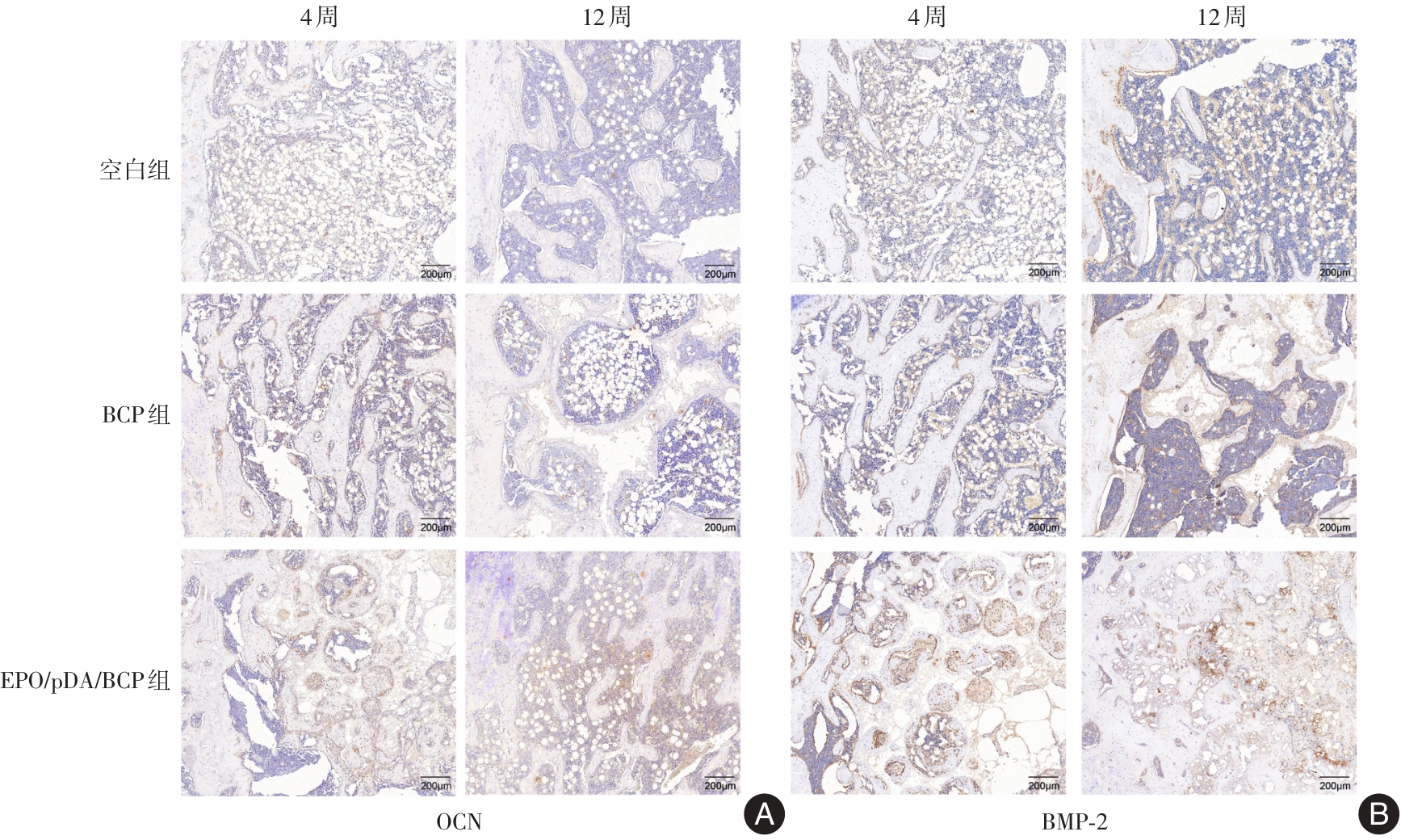
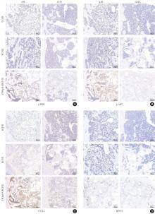
Fig.9
Results of immunohistochemical staining analysis of the femoral defect sites in rats of each group at 4 and 12 weeks after surgery"

| [1] |
LIU J, MENG Z, SONG J, et al. Yoda1-Loaded Microfibrous Scaffolds Accelerate Osteogenesis through Piezo1-F-Actin Pathway-Mediated YAP Nuclear Localization and Functionalization[J]. ACS Appl Mater Interfaces, 2025, 17(21): 30559-30572. doi: 10.1021/acsami.5c03093 .
doi: 10.1021/acsami.5c03093 |
| [2] |
QIU S, CAO L, XIANG D, et al. Enhanced osteogenic differentiation in 3D hydrogel scaffold via macrophage mitochondrial transfer[J]. J Nanobiotechnology, 2024, 22(1): 540. doi: 10.1186/s12951-024-02757-1 .
doi: 10.1186/s12951-024-02757-1 |
| [3] |
XUE N, DING X, HUANG R, et al. Bone tissue engineering in the treatment of bone defects[J]. Pharmaceuticals, 2022, 15(7): 879. doi: 10.3390/ph15070879 .
doi: 10.3390/ph15070879 |
| [4] |
宋李治, 陈慧坤, 张奕纯, 等. 二甲双胍在骨缺损修复中的研究进展和临床应用[J]. 实用医学杂志, 2022, 38(12): 1443-1448. doi: 10.3969/j.issn.1006-5725.2022.12.001 .
doi: 10.3969/j.issn.1006-5725.2022.12.001 |
| [5] |
栾非凡, 陈嘉怡, 郑臣校, 等. 伤科九味健骨片通过调控基质细胞衍生因子-1α/C-X-C趋化因子受体4型轴对骨髓间充质干细胞成骨分化及牵张成骨的影响[J]. 实用医学杂志, 2026, 42(2): 285-294. doi: 10.3969/j.issn.1006-5725.2026.02.014 .
doi: 10.3969/j.issn.1006-5725.2026.02.014 |
| [6] |
DELYANEE M, TABATABAEE S, SAMANIPOUR R, et al. Effects of gamma radiation as terminal sterilization on mechanical properties and organic composition of bone and skin allograft[J]. J Mater Res, 2025, 40(9): 1374-1389. doi: 10.1557/s43578-025-01584-w .
doi: 10.1557/s43578-025-01584-w |
| [7] |
吴妍廷, 李宇, 廖金凤. 氧化镁纳米粒调控成骨与血管生成相关基因表达促进骨缺损愈合[J]. 中国组织工程研究, 2026, 30(8): 1885-1895. doi: 10.12307/2026.030 .
doi: 10.12307/2026.030 |
| [8] |
HOU X, ZHANG L, ZHOU Z, et al. Calcium phosphate-based biomaterials for bone repair[J]. J Funct Biomater, 2022, 13(4): 187. doi: 10.3390/jfb13040187 .
doi: 10.3390/jfb13040187 |
| [9] |
王宁宁, 徐志民, 郑叶, 等. 聚多巴胺涂层3D打印双相磷酸钙组织工程支架的制备及其评价[J]. 吉林大学学报(医学版), 2019, 45(4): 955-959. doi: 10.13481/j.1671-587x.20190438 .
doi: 10.13481/j.1671-587x.20190438 |
| [10] |
YANG Z, XIE L, ZHANG B, et al. Preparation of BMP-2/PDA-BCP bioceramic scaffold by DLP 3D printing and its ability for inducing continuous bone formation[J]. Front Bioeng Biotechnol, 2022, 10: 854693. doi: 10.3389/fbioe.2022.854693 .
doi: 10.3389/fbioe.2022.854693 |
| [11] |
杨玉芳, 杨芷姗, 段棉棉, 等. 促红细胞生成素在骨组织工程中的应用及前景[J]. 中国组织工程研究, 2024, 28(9): 1443-1449. doi: 10.12307/2024.014 .
doi: 10.12307/2024.014 |
| [12] |
TSIFTSOGLOU A S. Erythropoietin (EPO) as a key regulator of erythropoiesis, bone remodeling and endothelial transdifferentiation of multipotent mesenchymal stem cells (MSCs): Implications in regenerative medicine[J]. Cells, 2021, 10(8): 2140. doi: 10.3390/cells10082140 .
doi: 10.3390/cells10082140 |
| [13] |
ZHANG X, LIU J, LI Y, et al. Erythropoietin Functionalized Hierarchical Scaffolds Enable Immunomodulated and Vascularized Bone Repair in Periodontal Defect[J]. ACS Appl Mater Interfaces, 2026, 18(9): 14256-14273. doi: 10.1021/acsami. 5c24484 .
doi: 10.1021/acsami. 5c24484 |
| [14] |
WU X, ZHU Y, QI Y, et al. Erythropoietin, as a biological macromolecule in modification of tissue engineered constructs: A review[J]. Int J Biol Macromol, 2021, 193: 2332-2342. doi: 10.1016/j.ijbiomac.2021.11.065 .
doi: 10.1016/j.ijbiomac.2021.11.065 |
| [15] |
SENATOV F, AMANBEK G, ORLOVA P, et al. Biomimetic UHMWPE/HA scaffolds with rhBMP-2 and erythropoietin for reconstructive surgery[J]. Mater Sci Eng C, 2020, 111: 110750. doi: 10.1016/j.msec.2020.110750 .
doi: 10.1016/j.msec.2020.110750 |
| [16] |
SENATOV F, ZIMINA A, CHUBRIK A, et al. Effect of recombinant BMP-2 and erythropoietin on osteogenic properties of biomimetic PLA/PCL/HA and PHB/HA scaffolds in critical-size cranial defects model[J]. Mater Sci Eng C, 2022, 135: 112680. doi: 10.1016/j.msec.2022.112680 .
doi: 10.1016/j.msec.2022.112680 |
| [17] |
WANG Y, WANG P, WU Q, et al. Loading of erythropoietin on biphasic calcium phosphate bioceramics promotes osteogenesis and angiogenesis by regulating EphB4/EphrinB2 molecules[J]. J Mater Sci Mater Med, 2022, 33(2): 19. doi: 10.1007/s10856-022-06644-9 .
doi: 10.1007/s10856-022-06644-9 |
| [18] |
YU Y, MA L, ZHANG H, et al. EPO could be regulated by HIF-1 and promote osteogenesis and accelerate bone repair[J]. Artif Cells Nanomed Biotechnol, 2020, 48(1): 206-217. doi: 10.1080/21691401.2019.1699827 .
doi: 10.1080/21691401.2019.1699827 |
| [19] |
QIN Q, LIU Y, YANG Z, et al. Hypoxia-inducible factors signaling in osteogenesis and skeletal repair[J]. Int J Mol Sci, 2022, 23(19): 11201. doi: 10.3390/ijms231911201 .
doi: 10.3390/ijms231911201 |
| [20] |
卢燕勤, 易芳, 鞠巍, 等. Ca-P涂层镁合金支架负载缓释微球修复股骨缺损[J]. 中国组织工程研究, 2019, 23(2): 232-238. doi: 10.3969/j.issn.2095-4344.0689 .
doi: 10.3969/j.issn.2095-4344.0689 |
| [21] |
MA S, LIU X, XIAO B, et al. An Artificial Intelligence‐Selected Multifunctional Oligopeptide Coated Biphasic Calcium Phosphate (BCP) Scaffold Promotes Bone Defect Repair[J]. Adv Healthc Mater, 2026, 15(16): e05458. doi: 10.1002/adhm.202505458 .
doi: 10.1002/adhm.202505458 |
| [22] |
XIANG M, ZHU F, HAO Z, et al. Vat-Photopolymerized Biphasic Calcium Phosphate Bioceramics with Cortical Bone-Like Properties via Microwave Sintering[J]. Ceram Int, 2026. doi: 10.1016/j.ceramint.2026.03.379 .
doi: 10.1016/j.ceramint.2026.03.379 |
| [23] |
KIM S E, PARK K. Recent advances of biphasic calcium phosphate bioceramics for bone tissue regeneration[J]. Adv Exp Med Biol, 2020, 1250: 177-188. doi: 10.1007/978-981-15-3262-7_12 .
doi: 10.1007/978-981-15-3262-7_12 |
| [24] |
YASUOKA Y, IZUMI Y, SANDS J M, et al. Progress in the Detection of Erythropoietin in Blood, Urine, and Tissue[J]. Molecules, 2023, 28(11): 4446. doi: 10.3390/molecules28114446 .
doi: 10.3390/molecules28114446 |
| [25] |
RÖLFING J H D, BAATRUP A, STIEHLER M, et al. The osteogenic effect of erythropoietin on human mesenchymal stromal cells is dose-dependent and involves non-hematopoietic receptors and multiple intracellular signaling pathways[J]. Stem Cell Rev Rep, 2014, 10(1): 69-78. doi: 10.1007/s12015-013-9476-x .
doi: 10.1007/s12015-013-9476-x |
| [26] |
LIU S, WANG Z, LI Y, et al. Erythropoietin‐stimulated macrophage‐derived extracellular vesicles in chitosan hydrogel rescue BMSCs fate by targeting EGFR to alleviate inflammatory bone loss in periodontitis[J]. Adv Sci, 2025, 12(23): 2500554. doi: 10.1002/advs.202500554 .
doi: 10.1002/advs.202500554 |
| [27] |
KIM J, JUNG Y, SUN H, et al. Erythropoietin mediated bone formation is regulated by mTOR signaling[J]. J Cell Biochem, 2012, 113(1): 220-228. doi: 10.1002/jcb.23347 .
doi: 10.1002/jcb.23347 |
| [28] |
VASILEVA R, CHAPRAZOV T. Evaluation of recombinant human erythropoietin as a promoter of bone healing in cats with femoral fractures[J]. Open Vet J, 2024, 14(4): 1012. doi: 10.5455/OVJ.2024.v14.i4.8 .
doi: 10.5455/OVJ.2024.v14.i4.8 |
| [29] |
HOLSTEIN J H, ORTH M, SCHEUER C, et al. Erythropoietin stimulates bone formation, cell proliferation, and angiogenesis in a femoral segmental defect model in mice[J]. Bone, 2011, 49(5): 1037-1045. doi: 10.1016/j.bone.2011.08.004 .
doi: 10.1016/j.bone.2011.08.004 |
| [30] |
CHAPRAZOV T, VASILEVA R, ATLIEV K, et al. Diagnostic imaging studies on local and systemic erythropoietin application for promoting bone regeneration in rat calvarial defects[J]. Vet Sci, 2022, 9(10): 578. doi: 10.3390/vetsci9100578 .
doi: 10.3390/vetsci9100578 |
| [31] |
LI X, TANG J, GUO W, et al. Polydopamine Nanocomposite Hydrogel for Drug Slow-Release in Bone Defect Repair: A Review of Research Advances[J]. Gels, 2025, 11(3): 190. doi: 10.3390/gels11030190 .
doi: 10.3390/gels11030190 |
| [32] |
BEDHIAFI T, IDOUDI S, ALHAMS A A, et al. Applications of polydopaminic nanomaterials in mucosal drug delivery[J]. J Control Release, 2023, 353: 842-849. doi: 10.1016/j.jconrel.2022. 12.037 .
doi: 10.1016/j.jconrel.2022. 12.037 |
| [33] |
GHORBANI F, KIM M, GHALANDARI B, et al. Architecture of β-lactoglobulin coating modulates bioinspired alginate dialdehyde-gelatine/polydopamine scaffolds for subchondral bone regeneration[J]. Acta Biomater, 2024, 181: 188-201. doi: 10.1016/j.actbio.2024.04.028 .
doi: 10.1016/j.actbio.2024.04.028 |
| [34] |
HUANG S, LIANG N, HU Y, et al. Polydopamine-assisted surface modification for bone biosubstitutes[J]. Biomed Res Int, 2016, 2016(1): 2389895. doi: 10.1155/2016/2389895 .
doi: 10.1155/2016/2389895 |
| [35] |
LI D, LI Q, XU T, et al. Pro-vasculogenic fibers by PDA-mediated surface functionalization using cell-free fat extract (CEFFE)[J]. Biomacromolecules, 2024, 25(3): 1550-1562. doi: 10.1021/acs.biomac.3c01124 .
doi: 10.1021/acs.biomac.3c01124 |
| [36] |
SUN X, JIAO X, WANG Z, et al. Polydopamine-coated 3D-printed β-tricalcium phosphate scaffolds to promote the adhesion and osteogenesis of BMSCs for bone-defect repair: mRNA transcriptomic sequencing analysis[J]. J Mater Chem B, 2023, 11(8): 1725-1738. doi: 10.1039/d2tb02280j .
doi: 10.1039/d2tb02280j |
| [37] |
WANG P, YIN H M, LI X, et al. Simultaneously constructing nanotopographical and chemical cues in 3D-printed polylactic acid scaffolds to promote bone regeneration[J]. Mater Sci Eng C, 2021, 118: 111457. doi: 10.1016/j.msec.2020.111457 .
doi: 10.1016/j.msec.2020.111457 |
| [38] |
SWETHA S, RAO G N, MAHALAKSHMI V, et al. Alkaline phosphatase and acid phosphatase in health and disease-A systematic review[J]. J Oral Maxillofac Pathol, 2025, 29(2): 324-334. doi: 10.4103/jomfp.jomfp_304_24 .
doi: 10.4103/jomfp.jomfp_304_24 |
| [39] |
VIMALRAJ S. Alkaline phosphatase: Structure, expression and its function in bone mineralization[J]. Gene, 2020, 754: 144855. doi: 10.1016/j.gene.2020.144855 .
doi: 10.1016/j.gene.2020.144855 |
| [40] |
WANG Y, PAN J, ZHANG Y, et al. Wnt and Notch signaling pathways in calcium phosphate‐enhanced osteogenic differentiation: A pilot study[J]. J Biomed Mater Res B Appl Biomater, 2019, 107(1): 149-160. doi: 10.1002/jbm.b.34105 .
doi: 10.1002/jbm.b.34105 |
| [41] |
胡俊, 谈荣珍, 袁忠, 等. 锁阳多糖通过激活PI3K/Akt/GSK3β/β-catenin信号通路促进MC3T3-E1细胞成骨分化[J]. 实用医学杂志, 2022, 38(22): 2774-2779. doi: 10.3969/j.issn.1006-5725.2022.22.003 .
doi: 10.3969/j.issn.1006-5725.2022.22.003 |
| [42] |
LI J, HUANG Z, LI B, et al. Mobilization of transplanted bone marrow mesenchymal stem cells by erythropoietin facilitates the reconstruction of segmental bone defect[J]. Stem Cells Int, 2019, 2019(1): 5750967. doi: 10.1155/2019/5750967 .
doi: 10.1155/2019/5750967 |
| [43] |
刘岚, 魏松, 张丞, 等. 小檗碱通过PI3K/AKT信号通路调控人牙周膜干细胞的增殖和成骨分化[J]. 中国细胞生物学学报, 2024, 46(3): 494-501. doi: 10.11844/cjcb.2024.03.0012 .
doi: 10.11844/cjcb.2024.03.0012 |
| [44] |
陶恩东, 夏伟杰, 王福林, 等.骨髓间充质干细胞来源的外泌体通过PI3K/AKT信号通路逆转高糖诱导的骨髓间充质干细胞成骨功能障碍[J]. 温州医科大学学报, 2024, 54(8): 641-649. doi: 10.3969/j.issn.2095-9400.2024.08.005 .
doi: 10.3969/j.issn.2095-9400.2024.08.005 |
| [45] |
LI B, ZHANG S, YUN X, et al. NEDD4's effect on osteoblastogenesis potential of bone mesenchymal stem cells in rats concerned with PI3K/Akt pathway[J]. Differentiation, 2025, 141: 100830. doi: 10.1016/j.diff.2024.100830 .
doi: 10.1016/j.diff.2024.100830 |
| [46] |
CHEN M, SHAN L, GAN Y, et al. Metastasis suppressor 1 controls osteoblast differentiation and bone homeostasis through regulating Src-Wnt/β-catenin signaling[J]. Cell Mol Life Sci, 2022, 79(2): 107. doi: 10.1007/s00018-022-04147-y .
doi: 10.1007/s00018-022-04147-y |
| [47] |
SHIN Y, WON Y, YANG J I, et al. CYTL1 regulates bone homeostasis in mice by modulating osteogenesis of mesenchymal stem cells and osteoclastogenesis of bone marrow-derived macrophages[J]. Cell Death Dis, 2019, 10(2): 47. doi: 10.1038/s41419-018-1284-4 .
doi: 10.1038/s41419-018-1284-4 |
| Viewed | ||||||
|
Full text |
|
|||||
|
Abstract |
|
|||||
