

The Journal of Practical Medicine ›› 2025, Vol. 41 ›› Issue (21): 3428-3434.doi: 10.3969/j.issn.1006-5725.2025.21.019
• Medical Examination and Clinical Diagnosis • Previous Articles
Linmei HAN,Yingli REN,Yiman LI,Fen HUANG,Taoming. DU( )
)
Received:2025-08-08
Online:2025-11-10
Published:2025-11-13
Contact:
Taoming. DU
E-mail:dtm0528@sina.com
CLC Number:
Linmei HAN,Yingli REN,Yiman LI,Fen HUANG,Taoming. DU. Feasibility study of low⁃dose chest CT with deep learning reconstruction algorithm combined with axial scan in children with mycoplasma pneumoniae pneumonia[J]. The Journal of Practical Medicine, 2025, 41(21): 3428-3434.
Tab.1
Comparison of objective image quality between DLIR and ASIR-V"
| 项目 | SD(HU) | SNR | CNR |
|---|---|---|---|
| ASIR-V20% | 33.75 ± 5.02 | 10.81 ± 2.40 | 12.06 ± 3.49 |
| ASIR-V50% | 26.40 ± 4.81 | 12.96 ± 2.72 | 16.44 ± 3.65 |
| ASIR-V80% | 19.77 ± 4.53 | 16.67 ± 3.58 | 23.70 ± 4.22 |
| F值 | 340.901 | 162.392 | 383.056 |
| P值 | < 0.001 | < 0.001 | < 0.001 |
| DLIR-L | 28.64 ± 4.04 | 12.11 ± 2.82 | 13.62 ± 3.74 |
| DLIR-M | 21.89 ± 3.50 | 16.45 ± 3.75 | 18.81 ± 3.92 |
| DLIR-H | 14.81 ± 4.15 | 20.95 ± 4.40 | 28.73 ± 4.31 |
| F值 | 501.301 | 226.671 | 590.284 |
| P值 | < 0.001 | < 0.001 | < 0.001 |
Tab.2
Comparison of subjective image quality between DLIR and ASIR-V"
| 项目 | 1分 | 2分 | 3分 | 4分 | 5分 |
|---|---|---|---|---|---|
| ASIR-V20% | 0(0.00) | 11(6.88) | 19(11.88) | 99(61.88) | 31(19.38) |
| ASIR-V50% | 0(0.00) | 6(3.75) | 13(8.13) | 95(59.38) | 46(28.75) |
| ASIR-V80% | 0(0.00) | 2(1.25) | 7(4.38) | 82(51.25) | 69(43.13) |
| χ2值 | 21.972 | ||||
| P值 | < 0.001 | ||||
| DLIR-L | 0(0.00) | 0(0.00) | 6(3.75) | 116(72.50) | 38(23.75) |
| DLIR-M | 0(0.00) | 0(0.00) | 1(0.63) | 99(61.88) | 60(37.50) |
| DLIR-H | 0(0.00) | 0(0.00) | 0(0.00) | 64(40.00) | 96(60.00) |
| χ2值 | 34.982 | ||||
| P值 | < 0.001 | ||||
Tab.3
The detection rate of CT signs of DLIR and ASIR-V"
| CT征象 | DLIR-H | ASIR-V80% | χ2值 | P值 |
|---|---|---|---|---|
| 支气管壁增厚 | 146(91.25) | 137(85.63) | 2.475 | 0.116 |
| 空气支气管征 | 111(69.38) | 80(50.00) | 12.481 | < 0.001 |
| 淋巴结肿大 | 53(33.13) | 48(30.00) | 0.362 | 0.548 |
| 间质增厚 | 31(19.38) | 31(19.38) | 0.000 | 1.000 |
| 肺实变影 | 139(86.88) | 115(71.88) | 10.995 | 0.001 |
| 间质浸润 | 33(20.63) | 12(7.50) | 11.404 | 0.001 |
| 胸腔积液 | 40(25.00) | 34(21.25) | 0.633 | 0.426 |
Tab.4
The consistency between the severity of MPP diagnosed by DLIR and ASIR-V and the clinical diagnosis"
| 重建条件 | 临床诊断/例 | 合计 | Kappa值 | P值 | 95%CI | |
|---|---|---|---|---|---|---|
| 轻度 | 重度 | |||||
| DLIR-H | 0.856 | < 0.001 | 0.711 ~ 0.996 | |||
| 轻度 | 113 | 5 | 118 | |||
| 重度 | 4 | 38 | 42 | |||
| 合计 | 117 | 43 | 160 | |||
| ASIR-V80% | 0.498 | < 0.001 | 0.346 ~ 0.650 | |||
| 轻度 | 108 | 20 | 128 | |||
| 重度 | 9 | 23 | 32 | |||
| 合计 | 117 | 43 | 160 | |||
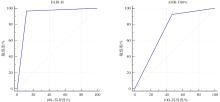
Fig.1
ROC curves of DLIR and ASIR-V for diagnosing the severity of MPP"
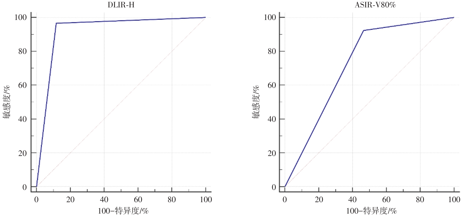
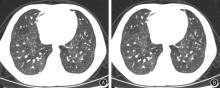
Fig.2
Typical case 1"
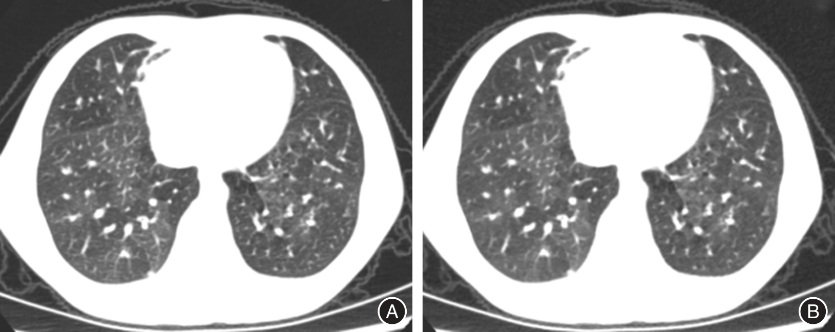
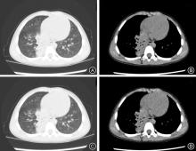
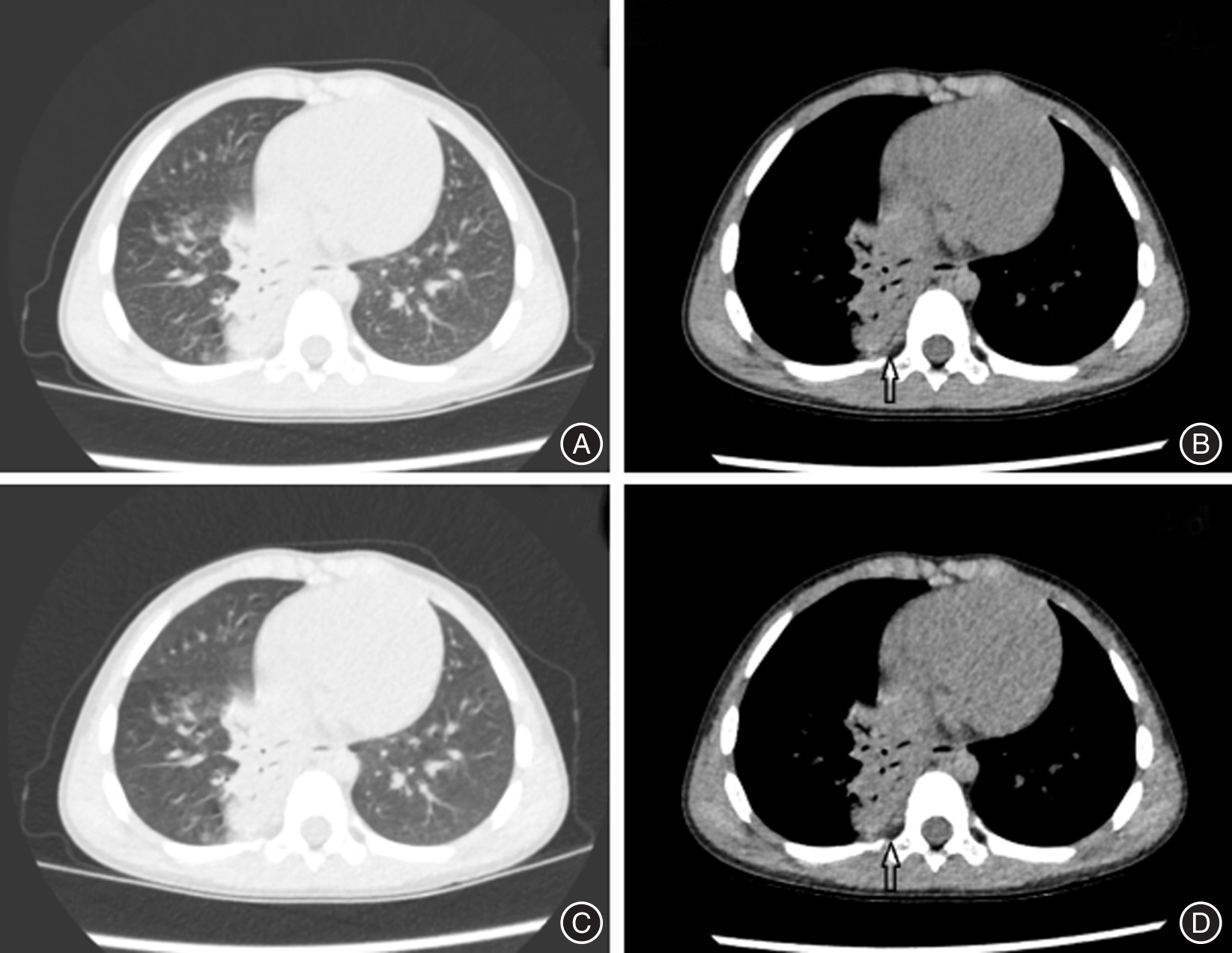
Fig.3
Typical case 2"
| [1] | 陈茜茜,林秋玉,张湘云,等. 大环内酯类耐药肺炎支原体感染与儿童难治性肺炎支原体肺炎的关系[J]. 实用医学杂志,2024,40(22):3190-3195. |
| [2] |
XU M, LI Y, SHI Y,et al. Molecular epidemiology of Mycoplasma pneumoniae pneumonia in children,Wuhan,2020-2022[J]. BMC Microbiol,2024,24(1):23-30. doi:10.1186/s12866-024-03180-0
doi: 10.1186/s12866-024-03180-0 |
| [3] | 陈海静,杨亚英,赵卫,等. 增强CT和MRI在鉴别鼻腔鼻窦鳞状细胞癌与淋巴瘤中的应用[J]. 实用医学杂志,2024,40(3):394-399. |
| [4] |
YANG S H, PU Q, LEI C T,et al. Low-dose CT denoising with a high-level feature refinement and dynamic convolution network[J]. Med Phys,2023,50(6):3597-3611. doi:10.1002/mp.16175
doi: 10.1002/mp.16175 |
| [5] |
DENG J T, MA T, YAN J,et al. Effect of Low Tube Voltage (100 kV) Combined with ASIR-V on the Visualization and Image Quality of the Adamkiewicz Artery:A Comparison with 120 kV Protocol[J]. Diagnostics (Basel),2023,13(15):2495-2499. doi:10.3390/diagnostics13152495
doi: 10.3390/diagnostics13152495 |
| [6] |
CAO J J, MROUEH N, MERCALDO N,et al. Detectability of Hypoattenuating Liver Lesions with Deep Learning CT Reconstruction:A Phantom and Patient Study[J]. Radiology,2024,313(1):e232749. doi:10.1148/radiol.232749
doi: 10.1148/radiol.232749 |
| [7] |
ROSSI A, GENNARI A G, ETTER D,et al. Impact of deep learning image reconstructions (DLIR) on coronary artery calcium quantification[J]. Eur Radiol,2023,33(6):3832-3838. doi:10.1007/s00330-022-09287-0
doi: 10.1007/s00330-022-09287-0 |
| [8] |
ZHONG JY, SHEN H L, CHEN Y,et al. Evaluation of Image Quality and Detectability of Deep Learning Image Reconstruction (DLIR) Algorithm in Single- and Dual-energy CT[J]. J Digit Imaging,2023,36(4):1390-1407. doi:10.1007/s10278-023-00806-z
doi: 10.1007/s10278-023-00806-z |
| [9] | 中华医学会儿科学分会呼吸学组,《中华实用儿科临床杂志》编辑委员会. 儿童肺炎支原体肺炎诊治专家共识(2015年版)[J]. 中华实用儿科临床杂志,2015,30(17):1304-1308. |
| [10] |
DING G D, ZHANG X B, VINTURACHE A, et al. Challenges in the treatment of pediatric Mycoplasma pneumoniae pneumonia[J]. Eur J Pediatr,2024,183(7):3001-3011. doi:10.1007/s00431-024-05519-1
doi: 10.1007/s00431-024-05519-1 |
| [11] | 袁静,陈庆仪,杨好贤,等. 含凝血功能五项Nomogram预测模型预测重症肺炎支原体肺炎患儿预后的效果研究[J]. 临床误诊误治,2023,36(3):90-94. |
| [12] |
YANG L X, SUN J H, LI J Y,et al. Dual-energy spectral CT imaging of pulmonary embolism with Mycoplasma pneumoniae pneumonia in children[J]. Radiol Med,2022,127(2):154-161. doi:10.1007/s11547-021-01442-9
doi: 10.1007/s11547-021-01442-9 |
| [13] | 苏布德格日乐,刘伟民,斯琴格日勒,等. 儿童肺炎支原体肺炎急性期高分辨率CT特征与血清炎症因子、病情严重程度及预后的相关性[J]. 放射学实践,2023,38(9):1173-1177. |
| [14] |
DE SANTIS D, POLIDORI T, TREMAMUNNO G,et al. Deep learning image reconstruction algorithm: Impact on image quality in coronary computed tomography angiography[J]. Radiol Med,2023,128(4):434-444. doi:10.1007/s11547-023-01607-8
doi: 10.1007/s11547-023-01607-8 |
| [15] |
KIM C H, CHUNG M J, CHA Y K,et al. The impact of deep learning reconstruction in low dose computed tomography on the evaluation of interstitial lung disease[J]. PLoS One,2023,18(9):e0291745. doi:10.1371/journal.pone.0291745
doi: 10.1371/journal.pone.0291745 |
| [16] | 陈依林,刘元芬,王莉莉,等. 深度学习重建算法对低kV逆血流扫描下肢动脉CT血管成像图像质量的影响[J]. 中华放射学杂志,2022,56(11):1188-1194. |
| [17] |
YANG K, CAO J J, PISUCHPEN N,et al. CT image quality evaluation in the age of deep learning: Trade-off between functionality and fidelity[J]. Eur Radiol,2023,33(4):2439-2449. doi:10.1007/s00330-022-09233-0
doi: 10.1007/s00330-022-09233-0 |
| [18] |
KANAN A, PEREIRA B, HORDONNEAU C,et al. Deep learning CT reconstruction improves liver metastases detection[J]. Insights Imaging,2024,15(1):167-172. doi:10.1186/s13244-024-01753-1
doi: 10.1186/s13244-024-01753-1 |
| [19] |
TOIA G V, ZAMORA D A, SINGLETON M,et al. Detectability of Small Low-Attenuation Lesions With Deep Learning CT Image Reconstruction:A 24-Reader Phantom Study[J]. AJR Am J Roentgenol,2023,220(2):283-295. doi:10.2214/ajr.22.28407
doi: 10.2214/ajr.22.28407 |
| [20] |
SVALKVIST A, FAGMAN E, VIKGREN J,et al. Evaluation of deep-learning image reconstruction for chest CT examinations at two different dose levels[J]. J Appl Clin Med Phys,2023,24(3):e13871. doi:10.1002/acm2.13871
doi: 10.1002/acm2.13871 |
| [21] |
LEI L M, ZHOU Y H, GUO X X,et al. The value of a deep learning image reconstruction algorithm in whole-brain computed tomography perfusion in patients with acute ischemic stroke[J].Quant Imaging Med Surg,2023,13(12):8173-8189. doi:10.21037/qims-23-547
doi: 10.21037/qims-23-547 |
| [22] |
WANG H, LI X Y, WANG T Z,et al. The value of using a deep learning image reconstruction algorithm of thinner slice thickness to balance the image noise and spatial resolution in low-dose abdominal CT[J]. Quant Imaging Med Surg,2023,13(3):1814-1824. doi:10.21037/qims-22-353
doi: 10.21037/qims-22-353 |
| [23] |
ZHENG Z, AI Z, LIANG Y,et al. Clinical value of deep learning image reconstruction on the diagnosis of pulmonary nodule for ultra-low-dose chest CT imaging[J]. Clin Radiol,2024,79(8):628-636. doi:10.1016/j.crad.2024.04.008
doi: 10.1016/j.crad.2024.04.008 |
| [24] |
KOH S, LEE N K, KIM S,et al. The efficacy of low-dose CT with deep learning image reconstruction in the surveillance of incidentally detected pancreatic cystic lesions[J]. Abdom Radiol (NY),2023,48(8):2585-2595. doi:10.1007/s00261-023-03958-2
doi: 10.1007/s00261-023-03958-2 |
| [25] |
ZHU H Y, HUANG Z K, CHEN Q H,et al. Feasibility of Sub-milliSievert Low-dose Computed Tomography with Deep Learning Image Reconstruction in Evaluating Pulmonary Subsolid Nodules:A Prospective Intra-individual Comparison Study[J]. Acad Radiol,2025,32(4):2309-2319. doi:10.1016/j.acra.2024.11.042
doi: 10.1016/j.acra.2024.11.042 |
| [26] |
KLEMENZ A C, ALBRECHT L, MANZKE M,et al. Improved image quality in CT pulmonary angiography using deep learning-based image reconstruction[J]. Sci Rep,2024,14(1):2494-2498. doi:10.1038/s41598-024-52517-2
doi: 10.1038/s41598-024-52517-2 |
| [1] | Pao YU,Feng ZHU,Zheng GE,Bi ZHOU,Lixia ZHANG. Phenotypic characteristics of early lymphocyte subsets and bronchoscopy findings in children with severe Mycoplasma pneumoniae pneumonia [J]. The Journal of Practical Medicine, 2025, 41(7): 1062-1069. |
| [2] | Dongli LIU,Zilin QUAN,Lingxiu ZHONG,Qiqi CHEN,Wenqiao CAI,Senpei ZHUANG,Ying WEI,Huiyi PAN,Yawen. LIN. Construction and validation of a predictive model for antibiotic-associated diarrhea after surgery in children with congenital heart disease [J]. The Journal of Practical Medicine, 2025, 41(5): 683-690. |
| [3] | Peng XU,Yun ZHOU,Rong JIA,Can QI,Linmeng SHI,Jingda GAO,Dengwei CHU,Xu. GAO. Clinical efficacy of testicular fascial compartment decompression in the treatment of testicular torsion in children [J]. The Journal of Practical Medicine, 2025, 41(2): 220-224. |
| [4] | Zhiqing XIAO,Xue WU,Rui QIU,Jinghan CHI,Shaodong HUA,Bin ZHU,De CHANG. Construction and verification of multi⁃factor prediction model for refractory mycoplasma pneumoniae pneumonia in children [J]. The Journal of Practical Medicine, 2025, 41(13): 2004-2010. |
| [5] | Bingjie QUAN,Yijing LIU,Xiaoqin LI,Fang ZHOU. Clinical features of hepatitisassociatedaplastic anemia in children [J]. The Journal of Practical Medicine, 2025, 41(1): 84-89. |
| [6] | Qingwen WANG,Shuya ZHANG,Weilin XIONG,Xiaolei HU,Ziwei LI,Qingyin. GUO. Characteristics of oral flora and its metabolites in children with henoch⁃schonlein purpura [J]. The Journal of Practical Medicine, 2024, 40(9): 1244-1250. |
| [7] | Jun ZHENG,Qiye WU,Xia ZENG,Zhixian LEI,Dufei. ZHANG. Clinical characteristics and risk factors of the occurrence of hypoxic hepatitis in children with shock [J]. The Journal of Practical Medicine, 2024, 40(15): 2126-2132. |
| [8] |
HU Haiyu, ZHANG Kunlong, CHU Jinhua, WANG Ningling, CHENG Yan..
The distribution and drug resistance ofpathogens of bloodstream infection in pediatric hematology depart⁃ ment in 2016⁃2021 [J]. The Journal of Practical Medicine, 2023, 39(8): 991-996. |
| [9] |
JIN Bei, CHENG Cheng, ZHUANG Hongjie, OUYANG Xiaojun, LI Jie, CHEN Lizhi, JIANG Xiaoyun..
The clinicopathological features and long ⁃ term prognosis inlupus nephritis children of different gender [J]. The Journal of Practical Medicine, 2023, 39(8): 997-1001. |
| [10] |
HUA Qun, HUANG Liqu, ZHOU Xin, BIAN Menglu, CHEN Jun, ZHU Shanliang..
The value of clinical interpretation combined with ultrasonic measurement of rectal transverse diameter in the diagnosis of cystorectal dysfunction in children [J]. The Journal of Practical Medicine, 2023, 39(6): 752-756. |
| [11] | Shijun YOU,Xue LIANG,Chunlian WANG,Yuhan. SONG. The effect of modified ginseng and schisandra decoction on the efficacy and inflammatory indicators of children with lobar pneumonia (lung spleen deficiency syndrome) caused by mycoplasma pneumonia infection [J]. The Journal of Practical Medicine, 2023, 39(24): 3281-3285. |
| [12] | Ruhai YAN,Lihong SUN,Yingtong YE,Ming. ZHANG. Clinical characteristics of 88 infants or young children with allergic rhinitis and follow⁃up after drug treatment [J]. The Journal of Practical Medicine, 2023, 39(23): 3101-3105. |
| [13] | Chenyang CHANG,Shaowen HU,Guoping DENG,Huifang ZHU,Kaiyuan. LUO. Predictive value of neutrophil to lymphocyte and platelet ratio in prognosis of children with sepsis [J]. The Journal of Practical Medicine, 2023, 39(22): 2958-2963. |
| [14] | Yalan HU,Ting WANG,Qiang. FU. Comparison of therapeutic effects between single⁃dose CTX intravenous pulse therapy and IVIG pulse therapy for severe gastrointestinal involvement of Henoch Schönlein purpura in children [J]. The Journal of Practical Medicine, 2023, 39(22): 2974-2978. |
| [15] | Li PENG,Lili ZHONG,Lin LIN,Han HUANG,Xiaofang DING,Min CHEN,Xiaojuan. LIN. Expression and clinical significance of MUC5AC in airway of children with mycoplasma pneumoniae pneumonia [J]. The Journal of Practical Medicine, 2023, 39(20): 2618-2622. |
| Viewed | ||||||
|
Full text |
|
|||||
|
Abstract |
|
|||||