

The Journal of Practical Medicine ›› 2025, Vol. 41 ›› Issue (14): 2264-2268.doi: 10.3969/j.issn.1006-5725.2025.14.021
• Medical Examination and Clinical Diagnosis • Previous Articles
Yonggang MU1,Longyun CHEN1,Tongkuai CONG1,Xuxiu JING1,Suzhen. ZHANG2( )
)
Received:2025-04-09
Online:2025-07-25
Published:2025-07-29
Contact:
Suzhen. ZHANG
E-mail:zhangsuzhen_2013@126.com
CLC Number:
Yonggang MU,Longyun CHEN,Tongkuai CONG,Xuxiu JING,Suzhen. ZHANG. Clinical application of near⁃infrared spectroscopy in assisting the diagnosis of schizophrenia[J]. The Journal of Practical Medicine, 2025, 41(14): 2264-2268.
Tab.1
Comparison of demographic characteristics between two groups"
| 项目 | 精神分裂症组 | 健康对照组 | t/χ2值 | P值 |
|---|---|---|---|---|
| 年龄/岁 | 42.78 ± 6.88 | 40.57 ± 8.47 | 1.242 | 0.218 |
| 性别(男/女)/例 | 21/24 | 15/15 | 0.800 | 0.777 |
| 受教育年限 | 14.53 ± 2.62 | 15.23 ± 2.92 | -1.083 | 0.282 |
Tab.2
Comparison of word count and fNRIS measures between groups in VFT"
| 项目 | 精神分裂症组 | 健康对照组 | t/Z值 | P值 |
|---|---|---|---|---|
| 组词数/个 | 8.98 ± 3.22 | 11.97 ± 3.44 | 3.817 | < 0.001 |
| 额叶积分值[M(Q1,Q3)] | 25.40(-3.00,66.90) | 57.70(35.30,77.00) | 2.742 | 0.006 |
| 额叶重心值/s | 59.59 ± 15.33 | 56.43 ± 8.80 | -0.102 | 0.310 |
| 额叶斜率 | 0.000 5 ± 0.001 1 | 0.001 0 ± 0.001 1 | 2.214 | 0.027 |
| 颞叶积分值[M(Q1,Q3)] | 28.00(-1.55, 65.20) | 110.10(49.98,157.50) | 4.110 | < 0.001 |
| 颞叶重心值/s | 58.66 ± 15.21 | 59.67 ± 7.65 | 0.340 | 0.735 |
| 颞叶斜率 | 0.000 4 ± 0.000 1 | 0.000 6 ± 0.000 8 | 1.593 | 0.111 |
Tab.3
Association between fNRIS measures and schizophrenia: A logistic regression analysis"
| 项目 | β | SE | P值 | OR | 95%CI |
|---|---|---|---|---|---|
| 组词数 | -0.149 | 0.078 | 0.057 | 0.861 | 0.739 ~ 1.004 |
| 额叶积分值 | 0.005 | 0.008 | 0.562 | 1.005 | 0.989 ~ 1.021 |
| 额叶斜率a | -0.024 | 0.029 | 0.417 | 0.976 | 0.922 ~ 1.034 |
| 颞叶积分值 | -0.021 | 0.007 | 0.005 | 0.979 | 0.965 ~ 0.994 |
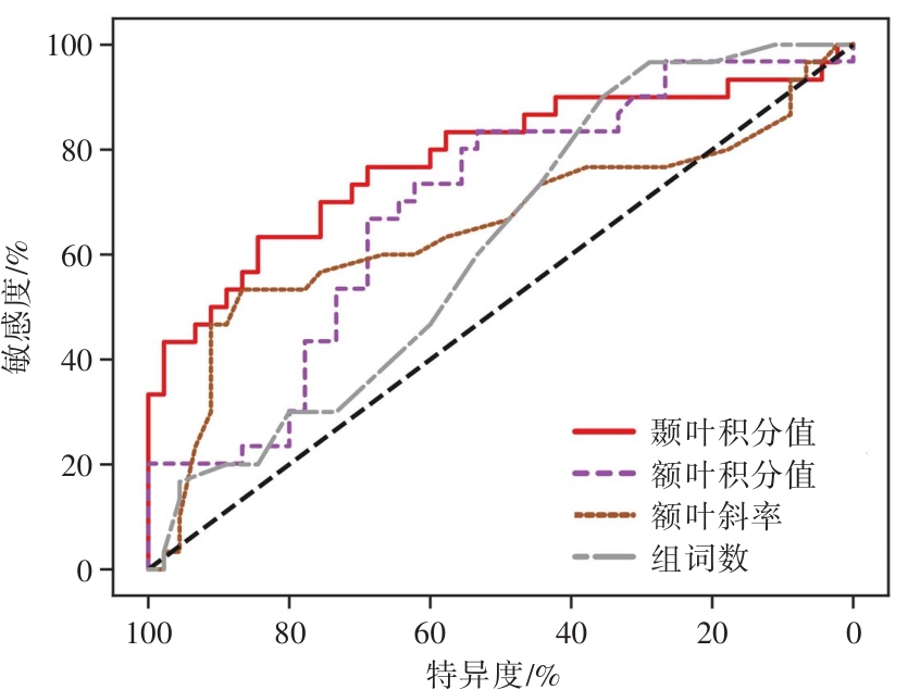
Fig.1
ROC curve of fNRIS measures for diagnosing schizophrenia"
Tab.4
fNRIS measures in assisting the diagnosis of schizophrenia"
| 项目 | 最佳截断值 | 灵敏度 | 特异度 | P值 | AUC | 95%CI |
|---|---|---|---|---|---|---|
| 组词数 | 8.5 | 0.900 | 0.364 | 0.086 | 0.618 | 0.491 ~ 0.745 |
| 额叶积分值 | 50.9 | 0.667 | 0.682 | 0.008 | 0.681 | 0.558 ~ 0.805 |
| 额叶斜率 | 0.011 5 | 0.533 | 0.864 | 0.036 | 0.644 | 0.504 ~ 0.783 |
| 颞叶积分值 | 72.6 | 0.633 | 0.844 | < 0.001 | 0.779 | 0.664 ~ 0.894 |
| [1] |
MARDER S R, CANNON T D. Schizophrenia [J]. N Engl J Med, 2019, 381(18): 1753-1761. doi:10.1056/nejmra1808803
doi: 10.1056/nejmra1808803 |
| [2] |
CHEN J, MULLER V I, DUKSRT J, et al. Intrinsic Connectivity Patterns of Task-Defined Brain Networks Allow Individual Prediction of Cognitive Symptom Dimension of Schizophrenia and Are Linked to Molecular Architecture [J]. Biol Psychiatry, 2021, 89(3): 308-319. doi:10.1016/j.biopsych.2020.09.024
doi: 10.1016/j.biopsych.2020.09.024 |
| [3] |
ZHANG S, LI W, XIANG Q, et al. Longitudinal alterations of modular functional-metabolic coupling in first-episode schizophrenia [J]. J Psychiatr Res, 2022, 156:705-712. doi:10.1016/j.jpsychires.2022.10.067
doi: 10.1016/j.jpsychires.2022.10.067 |
| [4] | 俞天悦, 郭茜, 胡昊, 等. 精神分裂症中氧化应激相关通路与诊断和预测价值的研究进展[J]. 实用医学杂志, 2024,40(20):2935-2940. |
| [5] |
AYANO G, DEMELASH S, YOHANNES Z, et al. Misdiagnosis, detection rate, and associated factors of severe psychiatric disorders in specialized psychiatry centers in Ethiopia [J]. Ann Gen Psychiatry, 2021, 20(1): 10. doi:10.1186/s12991-021-00333-7
doi: 10.1186/s12991-021-00333-7 |
| [6] | 郝飞, 范丰梅, 朱小林, 等. 精神分裂症近红外光谱脑功能成像研究进展 [J]. 中华精神科杂志, 2016, 49(2): 113-117. |
| [7] |
KOIKE S, SATMOURA Y, KAWASAKI S, et al. Application of functional near infrared spectroscopy as supplementary examination for diagnosis of clinical stages of psychosis spectrum [J]. Psychiatry Clin Neurosci, 2017, 71(12): 794-806. doi:10.1111/pcn.12551
doi: 10.1111/pcn.12551 |
| [8] |
WEI Y, CHEN Q, CURTIN A, et al. Functional near-infrared spectroscopy (fNIRS) as a tool to assist the diagnosis of major psychiatric disorders in a Chinese population [J]. Eur Arch Psychiatry Clin Neurosci, 2021, 271(4): 745-757. doi:10.1007/s00406-020-01125-y
doi: 10.1007/s00406-020-01125-y |
| [9] |
QIAO Y, SONG X, YAN J, et al. Neurological activation during verbal fluency task and resting-state functional connectivity abnormalities in obsessive-compulsive disorder: A functional near-infrared spectroscopy study [J]. Front Psychiatry, 2024, 15:151416810. doi:10.3389/fpsyt.2024.1416810
doi: 10.3389/fpsyt.2024.1416810 |
| [10] |
ROSENBAUM D, HAGEN K, DEPPERMANN S, et al. State-dependent altered connectivity in late-life depression: A functional near-infrared spectroscopy study[J].Neurobiol Aging, 2016, 39: 57-68. doi:10.1016/j.neurobiolaging.2015.11.022
doi: 10.1016/j.neurobiolaging.2015.11.022 |
| [11] | RIVHSRDS A L, PARDINAS A F, FRIZZATI A, et al. The Relationship Between Polygenic Risk Scores and Cognition in Schizophrenia [J]. Schizophr Bull, 2020, 46(2): 336-344. |
| [12] |
RAHMAN M A, SIDDIK A B, GHOSH T K, et al. A Narrative Review on Clinical Applications of fNIRS [J]. J Digit Imaging, 2020, 33(5): 1167-1184. doi:10.1007/s10278-020-00387-1
doi: 10.1007/s10278-020-00387-1 |
| [13] | 徐若愚, 李献云. 稳定期精神分裂症在不同情绪启动下反应抑制特点 [J]. 中国神经精神疾病杂志, 2022, 48(10): 614-618. |
| [14] | 管晓枫, 胡欣怡, 陆峥. 精神分裂症诊断标准更新与分类变化[J]. 重庆医科大学学报,2021,46(7):760-763. |
| [15] |
VITA A, GAEBEL W, MUCCI A, et al. European Psychiatric Association guidance on assessment of cognitive impairment in schizophrenia[J]. Eur Psychiatry, 2022, 65(1): e58. doi:10.1192/j.eurpsy.2022.2316
doi: 10.1192/j.eurpsy.2022.2316 |
| [16] | 李佳, 林荫, 吉训琦, 等. 载脂蛋白E和MTHFR C677T基因多态性与精神分裂症及患者认知功能的关联性[J]. 实用医学杂志, 2021,37(18):2391-2394. |
| [17] | RUND B R, BARDER H E, EVENSEN J, et al. Neurocognition and Duration of Psychosis: A 10-year Follow-up of First-Episode Patients [J]. Schizophr Bull, 2016, 42(1): 87-95. |
| [18] |
LIU D, JI C, ZHUO K, et al. Impaired cue identification and intention retrieval underlie prospective memory deficits in patients with first-episode schizophrenia [J]. Aust N Z J Psychiatry, 2017, 51(3): 270-277. doi:10.1177/0004867416640097
doi: 10.1177/0004867416640097 |
| [19] |
LIANG J, CHEN L, LI Y, et al. Unraveling the Prefrontal Cortex-Basolateral Amygdala Pathway's Role on Schizophrenia's Cognitive Impairments: A Multimodal Study in Patients and Mouse Models [J]. Schizophr Bull, 2024, 50(4):913-923. doi:10.1093/schbul/sbae063
doi: 10.1093/schbul/sbae063 |
| [20] |
HUSAIN S F, TANG T B, YU R, et al. Cortical haemodynamic response measured by functional near infrared spectroscopy during a verbal fluency task in patients with major depression and borderline personality disorder [J]. EBioMedicine, 2020, 51: 102586. doi:10.1016/j.ebiom.2019.11.047
doi: 10.1016/j.ebiom.2019.11.047 |
| [21] |
YAMAMURO K, KIMOTO S, IIDA J, et al. Distinct patterns of blood oxygenation in the prefrontal cortex in clinical phenotypes of schizophrenia and bipolar disorder [J]. J Affect Disord, 2018, 234:45-53. doi:10.1016/j.jad.2018.02.065
doi: 10.1016/j.jad.2018.02.065 |
| [22] |
PAUL T, SEE J W, VIJAKUMAR V, et al. Neurostructural changes in schizophrenia and treatment-resistance: A narrative review [J]. Psychoradiology, 2024, 4:kkae015. doi:10.1093/psyrad/kkae015
doi: 10.1093/psyrad/kkae015 |
| [23] |
YEUNG M K, LIN J. Probing depression, schizophrenia, and other psychiatric disorders using fNIRS and the verbal fluency test: A systematic review and meta-analysis [J]. J Psychiatr Res, 2021, 140:416-435. doi:10.1016/j.jpsychires.2021.06.015
doi: 10.1016/j.jpsychires.2021.06.015 |
| [24] | ZHANG K, JIN X, HE Y, et al. Atypical frontotemporal cortical activity in first-episode adolescent-onset schizophrenia during verbal fluency task: A functional near-infrared spectroscopy study [J]. Front Psychiatry, 2023, 14:1126131. |
| [25] | CHOU P H, LIU W C, LIN W H, et al. NIRS-aided differential diagnosis among patients with major depressive disorder, bipolar disorder, and schizophrenia [J]. J Affect Disord, 2023, 15(341): 366-373. |
| [1] | Lu ZHAO,Huiwen ZHI,Yafeng LI. The role of biomarkers in the diagnosis and prediction of disease progression of IgA nephropathy [J]. The Journal of Practical Medicine, 2025, 41(9): 1267-1272. |
| [2] | Rui GUO,Tao LIU,Xi. SU. Establishment of a nomogram model for comorbid cardiovascular disease in schizophrenia based on electrocardiogram parameters and kynurenine metabolites [J]. The Journal of Practical Medicine, 2025, 41(8): 1205-1211. |
| [3] | Guixian PAN,Sitao LI,Hu HAO,Wei LIU,Qiuping YANG,Xin XIAO,Yao. CAI. Pedigree analysis and prenatal diagnosis in a family with congenital ectopia lentis [J]. The Journal of Practical Medicine, 2025, 41(6): 824-828. |
| [4] | Jiaqi SHEN,Yu KANG,Xuhong NAN,Xiaoxi. SHA. The application value of color doppler ultrasound combined with real⁃time shear wave elastography in the diagnosis of vascular erectile dysfunction [J]. The Journal of Practical Medicine, 2025, 41(6): 877-881. |
| [5] | Gaokai HU,Ya'nan NIU,Yukang GONG,Yang HU,Ruixuan XU,Wenshan. GAO. Research progress on the application of deep learning in lumbar spine disease [J]. The Journal of Practical Medicine, 2025, 41(6): 921-928. |
| [6] | Yiyang ZHAI,Yunyi MA,Junying ZHAI,Hongli NIU,Ying. WANG. The relationship between tumor necrosis factor alpha inducible protein 8 family members 2, cell proliferation nuclear antigen expression levels, and clinical pathological parameters and prognosis in endometrial cancer tissue [J]. The Journal of Practical Medicine, 2025, 41(3): 379-384. |
| [7] | Danping WANG,Yufeng CAI,Dehua HU,Liang. ZHANG. The role of LncRNA RMST in gastric cancer: Expression levels, diagnostic value, and prognostic implicationssion [J]. The Journal of Practical Medicine, 2025, 41(3): 409-413. |
| [8] | Cong LIU,Fei ZHAI,Min LI,Xiaoli ZHANG,Han SHI,Ningning GUO,Changhong WANG. Association between smoking status, cognitive function, and personality traits in first⁃episode male patients with schizophrenia [J]. The Journal of Practical Medicine, 2025, 41(12): 1922-1928. |
| [9] | Long WANG,Yingmin WU,Sha SHANG,Lu WANG. The diagnostic value and clinical research of 5′⁃tRF⁃GlyGCC in the early stage of colorectal cancer [J]. The Journal of Practical Medicine, 2025, 41(11): 1730-1735. |
| [10] | Dandan SHANG,Ping LIU,Lizhen LIU,Yiwei PANG,Chao ZHOU. Diagnosis and advances in individualized management of resistant ovary syndrome and premature ovarian insufficiency [J]. The Journal of Practical Medicine, 2025, 41(1): 146-152. |
| [11] | Min ZHAO,Ping NI,Huiying ZHAI,Xiaoke JIN,Yuqiong. YANG. Analysis of the expression of lymphoid enhancer binding factor 1 in B cell chronic lymphoproliferative disorders [J]. The Journal of Practical Medicine, 2024, 40(7): 984-988. |
| [12] | Yuxin CHENG,Liang LIU,Shiyu DONG,Shengchao LI,Meng ZHANG. Research advances in exosomal proteins, mRNA and non⁃coding RNA regulation of Hepatocellular Carcinoma [J]. The Journal of Practical Medicine, 2024, 40(6): 748-755. |
| [13] | Juan TANG,Yi LI,Liqiong ZHAI,Shaowen LIU,Yong SHEN,Shuo CONG,Yongmei. LIU. The diagnostic value of miR⁃571 levels in blood from the peripherals in liver fibrosis [J]. The Journal of Practical Medicine, 2024, 40(5): 653-657. |
| [14] | Weifeng LIU,Zheng DAI,Yibin ZHOU,Kaiwen FENG,Kai WEI,Gule SUN,Dongrong YANG,Jin. ZHU. The value of urine protein kinase Y⁃linked gene promoter site methylation in early diagnosis of prostate cancer [J]. The Journal of Practical Medicine, 2024, 40(5): 688-694. |
| [15] | Haijing CHEN,Yaying YANG,Wei ZHAO,Jihong HU,Li WU,Linglin ZHENG,Yan WU,Qingqing LI. Contrast⁃enhanced CT and MRI in differentiating squamous cell carcinoma of the nasal cavity and sinuses from lymphoma [J]. The Journal of Practical Medicine, 2024, 40(3): 394-399. |
| Viewed | ||||||
|
Full text |
|
|||||
|
Abstract |
|
|||||