

The Journal of Practical Medicine ›› 2025, Vol. 41 ›› Issue (12): 1859-1866.doi: 10.3969/j.issn.1006-5725.2025.12.013
• Clinical Research • Previous Articles
Weiran SHI1,Ying HUANG2,Maji SUN1,Feng YUAN1( )
)
Received:2025-04-01
Online:2025-06-25
Published:2025-07-02
Contact:
Feng YUAN
E-mail:xzmuyf@163.com
CLC Number:
Weiran SHI,Ying HUANG,Maji SUN,Feng YUAN. Clinical efficacy analysis of transparent fully visualized working channel in percutaneous endoscopic interlaminar discectomy[J]. The Journal of Practical Medicine, 2025, 41(12): 1859-1866.

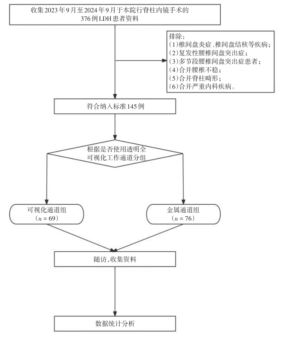
Fig.1
Research design flowchart"
Tab.1
Comparison of baseline data between the two groups"
| 项目 | 透明通道组(n = 69) | 金属通道组(n = 76) | χ2/t值 | P值 |
|---|---|---|---|---|
| 性别构成比(男/女)/例 | 33/36 | 36/40 | 0.003 | 0.956 |
| 年龄/岁 | 52.3 ± 14.2 | 51.6 ± 13.7 | 0.302 | 0.763 |
| BMI/(kg/m2) | 25.6 ± 4.7 | 25.3 ± 3.5 | 0.439 | 0.662 |
| 病程/月 | 12.8 ± 4.9 | 13.4 ± 5.3 | 0.706 | 0.482 |
| 病变节段/例 | 0.829 | 0.363 | ||
| L4/L5 | 37 | 35 | ||
| L5/S1 | 32 | 41 |
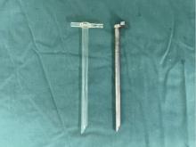
Fig.2
Different working channels"
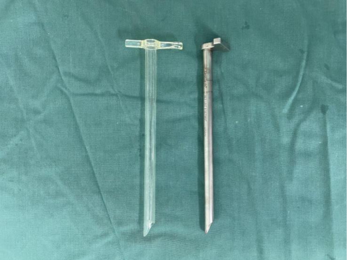
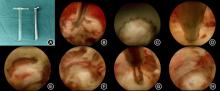
Fig.3
Transparent, fully visible working channel for use during surgery"

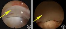
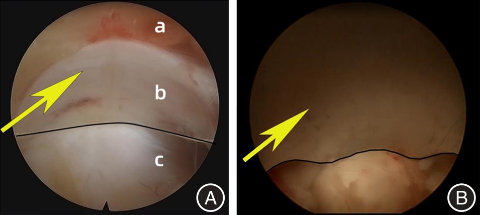
Fig.4
Improved surgical field of view with transparent, fully visible working channel compared to metal working channel"
Tab.2
Comparison of clinical data related to the two groups of operations"
| 组别 | 平均手术时间/min | 住院时间/d | 术中透视 次数 |
|---|---|---|---|
| 透明通道组(n = 69) | 65.4 ± 7.2 | 2.9 ± 1.3 | 6.5 ± 1.3 |
| 金属通道组(n = 76) | 68.3 ± 8.1 | 3.1 ± 1.7 | 6.6 ± 1.6 |
| t值 | 2.269 | 0.790 | 0.411 |
| P值 | 0.025 | 0.431 | 0.682 |
Tab.3
Comparison of the number of intraoperative electromyography neurophysiological alarms"
| 组别 | 放置工作通道时 | 术中减压时 | 探查神经时 |
|---|---|---|---|
| 透明通道组(n = 69) | 69 | 8 | 69 |
| 金属通道组(n = 76) | 76 | 29 | 76 |
| χ2值 | - | 13.427 | - |
| P 值 | - | < 0.001 | - |
Tab.4
Comparison of the incidence of surgical complications between the two groups of patients"
| 项目 | 透明通道组(n = 69) | 金属通道组(n = 76) | χ2值 | P值 |
|---|---|---|---|---|
| 感觉异常 | 1 | 7 | 2.823 | 0.093 |
| 术后复发 | 1 | 2 | 0.007 | 0.932 |
Tab.5
Comparison of VAS scores and ODI scores for low back pain before and after surgery in the two groups of patients x ± s"
| 项目 | 透明通道组 (n = 69) | 金属通道组 (n = 76) | t值 | P值 |
|---|---|---|---|---|
| VAS | ||||
| 术前 VAS | 7.6 ± 1.2 | 7.8 ± 1.4 | 0.919 | 0.359 |
| 术后1个月 | 2.6 ± 0.8 | 2.7 ± 1.1 | 0.621 | 0.536 |
| 术后3个月 | 2.0 ± 0.6 | 2.1 ± 0.7 | 0.803 | 0.424 |
| 术后6个月 | 1.6 ± 0.6 | 1.7 ± 0.5 | 1.094 | 0.276 |
| 末次随访 | 0.7 ± 0.4 | 0.8 ± 0.6 | 1.169 | 0.245 |
| ODI | ||||
| 术前ODI | 54.3 ± 9.0 | 55.6 ± 9.4 | 0.849 | 0.398 |
| 术后1个月 | 15.2 ± 5.4 | 15.8 ± 5.7 | 0.649 | 0.517 |
| 术后3个月 | 10.8 ± 2.9 | 11.1 ± 3.2 | 0.589 | 0.557 |
| 术后6个月 | 8.1 ± 2.5 | 8.5 ± 2.8 | 0.904 | 0.368 |
| 末次随访 | 6.7 ± 2.3 | 7.1 ± 2.7 | 0.955 | 0.341 |
Tab. 6
Assessment of the modified MacNab criteria in the two groups of patients"
| 组别 | 差 | 可 | 良 | 优 | 优良率/% |
|---|---|---|---|---|---|
| 透明通道组(n = 69) | 0 | 9 | 48 | 12 | 86.9 |
| 金属通道组(n = 76) | 0 | 13 | 47 | 16 | 82.9 |
| Z值 | 0.122 | ||||
| P 值 | 0.903 | ||||
| 1 |
JIANG H W, CHEN C D, ZHAN B S, et al. Unilateral biportal endoscopic discectomy versus percutaneous endoscopic lumbar discectomy in the treatment of lumbar disc herniation: A retrospective study[J]. J Orthop Surg Res,2022,17(1):30. doi:10.1186/s13018-022-02929-5
doi: 10.1186/s13018-022-02929-5 |
| 2 |
YUAN C, WEN B, LIN H. Clinical Analysis of Minimally Invasive Percutaneous Treatment of Severe Lumbar Disc Herniation with UBE Two-Channel Endoscopy and Foraminal Single-Channel Endoscopy Technique[J]. Oxid Med Cell Longev,2022,2022:9264852. doi:10.1155/2022/9264852
doi: 10.1155/2022/9264852 |
| 3 | KÖGL N, PETR O, LÖSCHER W, et al. Lumbar Disc Herniation—the Significance of Symptom Duration for the Indication for Surgery[J]. Dtsch Arztebl Int,2024,121(13):440-448. |
| 4 |
CHEN J, JING X, LI C, et al. Percutaneous Endoscopic Lumbar Discectomy for L5S1 Lumbar Disc Herniation Using a Transforaminal Approach Versus an Interlaminar Approach: A Systematic Review and Meta-Analysis[J]. World Neurosurg,2018,116:412-420.e412. doi:10.1016/j.wneu.2018.05.075
doi: 10.1016/j.wneu.2018.05.075 |
| 5 |
CHEN Z, ZHANG L, DONG J, et al. Percutaneous Transforaminal Endoscopic Discectomy Versus Microendoscopic Discectomy for Lumbar Disk Herniation: Five-year Results of a Randomized Controlled Trial[J]. Spine (Phila Pa 1976),2023,48(2):79-88. doi:10.1097/brs.0000000000004468
doi: 10.1097/brs.0000000000004468 |
| 6 | 廖烨晖, 叶入裴, 唐超, 等. 双通道与单通道椎板间入路髓核摘除术治疗腰椎间盘突出症的临床疗效比较[J]. 华西医学,2024,39(10):1543-1550. |
| 7 |
CHOI K C, KIM J S, LEE D C, et al. Percutaneous endoscopic lumbar discectomy: Minimally invasive technique for multiple episodes of lumbar disc herniation[J]. BMC Musculoskelet Disord,2017,18(1):329. doi:10.1186/s12891-017-1697-8
doi: 10.1186/s12891-017-1697-8 |
| 8 |
FAN N, YUAN S, DU P, et al. Complications and risk factors of percutaneous endoscopic transforaminal discectomy in the treatment of lumbar spinal stenosis[J]. BMC Musculoskelet Disord,2021,22(1):1041. doi:10.1186/s12891-021-04940-z
doi: 10.1186/s12891-021-04940-z |
| 9 | 韩秀丰, 路文超, 赵学航. PETD与PEID治疗钙化型腰椎间盘突出症的疗效及并发症分析[J]. 颈腰痛杂志,2024,45(5):972-976. |
| 10 |
CHENG Y P, CHENG X K, WU H. A comparative study of percutaneous endoscopic interlaminar discectomy and transforaminal discectomy for L5-S1 calcified lumbar disc herniation[J]. BMC Musculoskelet Disord,2022,23(1):244. doi:10.1186/s12891-022-05186-z
doi: 10.1186/s12891-022-05186-z |
| 11 |
MA H, SHEN M, TANG Z, et al. Percutaneous endoscopic interlaminar discectomy for high-grade migrated lumbar disc herniation: Clinical efficacy and safety assessment[J]. Int Orthop,2024,48(9):2455-2463. doi:10.1007/s00264-024-06246-w
doi: 10.1007/s00264-024-06246-w |
| 12 |
LI T, YANG G, ZHONG W, et al. Percutaneous endoscopic transforaminal vs. interlaminar discectomy for L5-S1 lumbar disc herniation: A retrospective propensity score matching study[J]. J Orthop Surg Res,2024,19(1):64. doi:10.1186/s13018-024-04543-z
doi: 10.1186/s13018-024-04543-z |
| 13 |
ZHOU Z, NI HJ, ZHAO W, et al. Percutaneous Endoscopic Lumbar Discectomy via Transforaminal Approach Combined with Interlaminar Approach for L4/5 and L5/S1 Two-Level Disc Herniation[J]. Orthop Surg,2021,13(3):979-988. doi:10.1111/os.12862
doi: 10.1111/os.12862 |
| 14 |
HUANG H, YANG M, FU Z, et al. Predicting and analysing of the unfavourable outcomes of early applicated percutaneous endoscopic interlaminar discectomy for lumbar disc herniation: development and validation based nomogram[J]. Eur Spine J,2024,33(3):906-914. doi:10.1007/s00586-024-08141-9
doi: 10.1007/s00586-024-08141-9 |
| 15 |
GAO A, YANG H, ZHU L, et al. Comparison of Interlaminar and Transforaminal Approaches for Treatment of L(5) /S(1) Disc Herniation by Percutaneous Endoscopic Discectomy[J]. Orthop Surg,2021,13(1):63-70. doi:10.1111/os.12831
doi: 10.1111/os.12831 |
| 16 |
HUANG H, HU H, LIN X, et al. Percutaneous endoscopic interlaminar discectomy via inner border of inferior pedicle approach for downmigrated disc herniation: A retrospective study[J]. J Orthop Surg Res,2022,17(1):359. doi:10.1186/s13018-022-03245-8
doi: 10.1186/s13018-022-03245-8 |
| 17 |
LI W, LIU Y C, ZHENG C F, et al. Diagnosis of Compressed Nerve Root in Lumbar Disc Herniation Patients by Surface Electromyography[J]. Orthop Surg,2018,10(1):47-55. doi:10.1111/os.12362
doi: 10.1111/os.12362 |
| 18 |
CHOI K C, SHIM H K, HWANG J S, et al. Comparison of Surgical Invasiveness Between Microdiscectomy and 3 Different Endoscopic Discectomy Techniques for Lumbar Disc Herniation[J]. World Neurosurg,2018,116:e750-e758. doi:10.1016/j.wneu.2018.05.085
doi: 10.1016/j.wneu.2018.05.085 |
| 19 |
王威威, 连鸿凯. 经皮椎间孔镜技术对巨大型腰椎间盘突出症患者功能恢复、创伤程度的前瞻性研究[J]. 实用医学杂志,2021,37(5):648-653. doi:10.3969/j.issn.1006-5725.2021.05.018
doi: 10.3969/j.issn.1006-5725.2021.05.018 |
| 20 | 李坚,李锦锦,何丽萍. 经皮椎间孔镜脊柱系统术治疗腰椎间盘突出症合并神经根管狭窄[J]. 中山大学学报(医学科学版),2023,44(2):342-347. |
| 21 |
LIU X, YUAN S, TIAN Y, et al. Comparison of percutaneous endoscopic transforaminal discectomy, microendoscopic discectomy, and microdiscectomy for symptomatic lumbar disc herniation: Minimum 2-year follow-up results[J]. J Neurosurg Spine,2018,28(3):317-325. doi:10.3171/2017.6.spine172
doi: 10.3171/2017.6.spine172 |
| 22 | 韩铁鹏, 虞攀峰, 黄鹏. 经皮脊柱内镜对比椎间盘镜治疗腰椎间盘突出症疗效的荟萃分析[J]. 中国疼痛医学杂志,2023,29(6):463-470. |
| 23 | 赵晓东, 邓立明, 王建波,等. 椎间孔镜与椎间盘镜治疗腰椎间盘突出症5年疗效比较[J]. 中国微创外科杂志,2019,19(8):684-687,701. |
| 24 | REN C, QIN R, LI Y, et al. Microendoscopic Discectomy Combined with Annular Suture Versus Percutaneous Transforaminal Endoscopic Discectomy for Lumbar Disc Herniation: A Prospective Observational Study[J]. Pain Physician,2020,23(6):E713-E721. |
| 25 |
CHANG H, XU J, YANG D, et al. Comparison of full-endoscopic foraminoplasty and lumbar discectomy (FEFLD), unilateral biportal endoscopic (UBE) discectomy, and microdiscectomy (MD) for symptomatic lumbar disc herniation[J]. Eur Spine J,2023,32(2):542-554. doi:10.1007/s00586-022-07510-6
doi: 10.1007/s00586-022-07510-6 |
| 26 |
KIM S K, KANG S S, HONG Y H, et al. Clinical comparison of unilateral biportal endoscopic technique versus open microdiscectomy for single-level lumbar discectomy: A multicenter, retrospective analysis[J]. J Orthop Surg Res,2018,13(1):22. doi:10.1186/s13018-018-0725-1
doi: 10.1186/s13018-018-0725-1 |
| 27 |
GU G, LIU T, GUO H, et al. Treatment of L5-S1 Floating Calcified Lumbar Disc Herniation with Percutaneous Endoscopic Interlaminar Discectomy[J]. Orthop Surg,2024,16(3):620-627. doi:10.1111/os.14007
doi: 10.1111/os.14007 |
| 28 |
HAN J, TANG W, LI G, et al. Comparison of Percutaneous Endoscopic Transforaminal and Interlaminar Approaches in Treating Adjacent Segment Disease Following Lumbar Decompression Surgery: A Clinical Retrospective Study[J]. Pain Physician,2023,26(7):E833-E842. doi:10.36076/ppj.2023.26.e833
doi: 10.36076/ppj.2023.26.e833 |
| 29 | PAN M, LI Q, LI S, et al. Percutaneous Endoscopic Lumbar Discectomy: Indications and Complications[J]. Pain Physician,2020,23(1):49-56. |
| 30 |
CHENG L, CAI H, LIU Z, et al. Modified Full-Endoscopic Interlaminar Discectomy via an Inferior Endplate Approach for Lumbar Disc Herniation: Retrospective 3-Year Results from 321 Patients[J]. World Neurosurg,2020,141:e537-e544. doi:10.1016/j.wneu.2020.05.234
doi: 10.1016/j.wneu.2020.05.234 |
| 31 |
SOLIMAN H, FRIDLEY J, TELFEIAN A, et al. Minimally Invasive, Far Lateral Lumbar Microdiscectomy with Intraoperative Computed Tomography Navigational Assistance and Electrophysiological Monitoring[J]. World Neurosurg,2019,122:e1228-e1239. doi:10.1016/j.wneu.2018.11.020
doi: 10.1016/j.wneu.2018.11.020 |
| 32 |
WU T M, CHOI D J, CHANG W S, et al. Exploring Physical Lumbar Microvascular Geometry Through Endoscopy and Illustrations: Implications for Clinical Interpretation[J]. Global Spine J,2025,15(2):969-980. doi:10.1177/21925682231218729
doi: 10.1177/21925682231218729 |
| 33 |
ZHOU S, A J, XU X, et al. Comparison of surgical invasiveness and hidden blood loss between unilateral double portal endoscopic lumbar disc extraction and percutaneous endoscopic interlaminar discectomy for lumbar spinal stenosis[J]. J Orthop Surg Res,2024,19(1):778. doi:10.1186/s13018-024-05274-x
doi: 10.1186/s13018-024-05274-x |
| 34 |
KIYOSUE H, MATSUMARU Y, NIIMI Y, et al. Angiographic and Clinical Characteristics of Thoracolumbar Spinal Epidural and Dural Arteriovenous Fistulas[J]. Stroke,2017,48(12):3215-3222. doi:10.1161/strokeaha.117.019131
doi: 10.1161/strokeaha.117.019131 |
| 35 |
SHLOBIN NA, RAZ E, SHAPIRO M, et al. Spinal neurovascular complications with anterior thoracolumbar spine surgery: A systematic review and review of thoracolumbar vascular anatomy[J]. Neurosurg Focus,2020,49(3):E9. doi:10.3171/2020.6.focus20373
doi: 10.3171/2020.6.focus20373 |
| 36 |
WU T L, YUAN J H, JIA J Y, et al. Percutaneous Endoscopic Interlaminar Discectomy via Laminoplasty Technique for L(5) -S(1) Lumbar Disc Herniation with a Narrow Interlaminar Window[J]. Orthop Surg,2021,13(3):825-832. doi:10.1111/os.12978
doi: 10.1111/os.12978 |
| 37 | 梁磊, 段晓晓, 刘兰涛. 两种内镜椎间盘切除术早期临床结果比较[J]. 中国矫形外科杂志,2023,31(22):2099-2103. |
| 38 | ZHOU C, ZHANG G, PANCHAL R R, et al. Unique Complications of Percutaneous Endoscopic Lumbar Discectomy and Percutaneous Endoscopic Interlaminar Discectomy[J]. Pain Physician,2018,21(2):E105-E112. |
| Viewed | ||||||
|
Full text |
|
|||||
|
Abstract |
|
|||||