

The Journal of Practical Medicine ›› 2025, Vol. 41 ›› Issue (4): 500-508.doi: 10.3969/j.issn.1006-5725.2025.04.006
• Basic Research • Previous Articles
Zhizhou XIAO1,Ying HUANG2,Huawei. BIAN1( )
)
Received:2024-11-25
Online:2025-02-25
Published:2025-02-28
Contact:
Huawei. BIAN
E-mail:southnutrition@sina.com
CLC Number:
Zhizhou XIAO,Ying HUANG,Huawei. BIAN. To investigate the effect of aloperine on bone metabolism in osteoporotic mice based on autophagy and apoptosis mediated by Wnt/β⁃catenin signaling pathway[J]. The Journal of Practical Medicine, 2025, 41(4): 500-508.
Tab.1
Comparison of bone mineral density and bone microstructure parameters among each group of mice(n = 10)"
| 组别 | BMD/(g/cm2) | 骨小梁厚度/μm | 骨小梁分离度/(pg/mL) | 骨表面积与体积比/(1/mm) |
|---|---|---|---|---|
| Sham组 | 0.46 ± 0.05 | 140.55 ± 14.94 | 322.33 ± 61.57 | 22.98 ± 3.26 |
| OP组 | 0.22 ± 0.03? | 93.23 ± 13.61? | 869.52 ± 89.29? | 38.52 ± 3.34? |
| L-ALO组 | 0.28 ± 0.04# | 106.97 ± 13.53# | 842.14 ± 86.99# | 32.91 ± 3.07# |
| M-ALO组 | 0.37 ± 0.05#△ | 120.26 ± 12.87#△ | 637.16 ± 73.85#△ | 28.28 ± 3.11#△ |
| H-ALO组 | 0.44 ± 0.04#▽ | 132.48 ± 12.01#▽ | 416.97 ± 71.55#▽ | 23.46 ± 3.03#▽ |
| EV组 | 0.38 ± 0.03# | 123.42 ± 14.04# | 716.28 ± 70.85# | 27.16 ± 3.24# |
| F值 | 975.960 | 951.862 | 914.697 | 982.343 |
| P值 | < 0.001 | < 0.001 | < 0.001 | < 0.001 |

Fig.1
Micro-CT scan images"
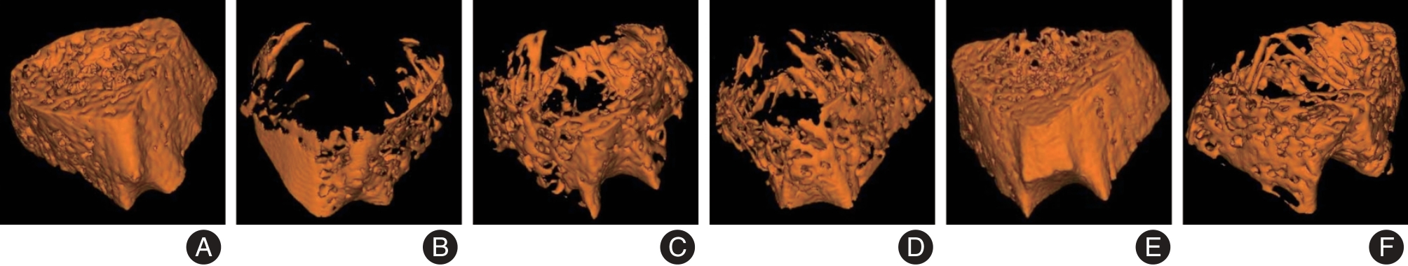

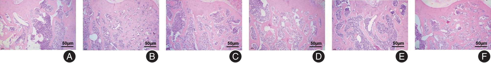
Fig.2
Morphology of tibial bone tissue in each group of mice (HE staining, × 400)"
Tab.2
Comparison of bone metabolism-related indexes among each group of mice (n = 10)"
| 组别 | OPG/(ng/L) | OCN/(ng/L) | ALP/(U/L) | Ca/(mmol/L) | P/(mmol/L) |
|---|---|---|---|---|---|
| Sham组 | 11.87 ± 1.33 | 17.24 ± 1.67 | 41.68 ± 5.27 | 2.64 ± 0.31 | 2.89 ± 0.39 |
| OP组 | 4.23 ± 0.71? | 7.28 ± 1.18? | 86.94 ± 8.97? | 1.13 ± 0.16? | 1.09 ± 0.16 |
| L-ALO组 | 6.87 ± 1.31# | 9.57 ± 1.21# | 71.16 ± 8.12 | 1.61 ± 0.22# | 1.47 ± 0.22 |
| M-ALO组 | 8.94 ± 1.09#△ | 13.59 ± 1.71#△ | 60.01 ± 7.69#△ | 1.99 ± 0.16#△ | 2.15 ± 0.35 |
| H-ALO组 | 10.96 ± 1.48#▽ | 16.99 ± 1.87#▽ | 43.67 ± 8.02#▽ | 2.59 ± 0.29#▽ | 2.87 ± 0.38 |
| EV组 | 9.46 ± 1.22# | 14.23 ± 1.56# | 61.59 ± 8.15# | 2.16 ± 0.21# | 2.22 ± 0.33 |
| F值 | 671.441 | 924.945 | 779.936 | 969.647 | 587.916 |
| P值 | < 0.001 | < 0.001 | < 0.001 | < 0.001 | < 0.001 |
Tab.3
Comparison of autophagy-related proteins among each group of mice (n = 10)"
| 组别 | LC3-Ⅱ/LC3-Ⅰ | Beclin-1 | P62 |
|---|---|---|---|
| Sham组 | 1.03 ± 0.13 | 1.06 ± 0.14 | 0.41 ± 0.06 |
| OP组 | 0.29 ± 0.03? | 0.33 ± 0.05? | 1.39 ± 0.16? |
| L-ALO组 | 0.41 ± 0.06# | 0.49 ± 0.06# | 1.12 ± 0.15# |
| M-ALO组 | 0.72 ± 0.09#△ | 0.77 ± 0.09#△ | 0.79 ± 0.10#△ |
| H-ALO组 | 1.01 ± 0.11#▽ | 1.02 ± 0.15#▽ | 0.45 ± 0.06#▽ |
| EV组 | 0.69 ± 0.07# | 0.76 ± 0.10# | 0.71 ± 0.09# |
| F值 | 858.761 | 666.697 | 765.989 |
| P值 | < 0.001 | < 0.001 | < 0.001 |
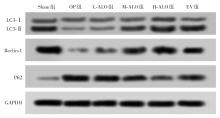
Fig.3
Detection and analysis of the expression levels of LC3, Beclin-1 and P62 by Western blot"

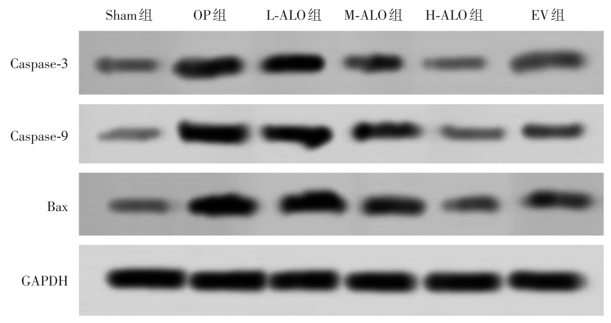
Tab.4
Comparison of apoptosis-related proteins among each group of mice (n = 10)"
| 组别 | Caspase-3 | Caspase-9 | Bax |
|---|---|---|---|
| Sham组 | 0.31 ± 0.05 | 0.19 ± 0.03 | 0.23 ± 0.04 |
| OP组 | 1.23 ± 0.14* | 1.16 ± 0.15* | 1.29 ± 0.17* |
| L-ALO组 | 0.98 ± 0.10# | 0.92 ± 0.09# | 0.92 ± 0.11# |
| M-ALO组 | 0.69 ± 0.08#△ | 0.71 ± 0.05#△ | 0.66 ± 0.07#△ |
| H-ALO组 | 0.34 ± 0.05#▽ | 0.23 ± 0.03#▽ | 0.29 ± 0.04#▽ |
| EV组 | 0.67 ± 0.09# | 0.51 ± 0.08# | 0.61 ± 0.09# |
| F值 | 882.281 | 879.487 | 716.943 |
| P值 | < 0.001 | < 0.001 | < 0.001 |
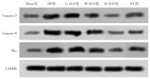
Fig.4
Detection and analysis of the expression levels of apoptosis-related proteins by Western blot"
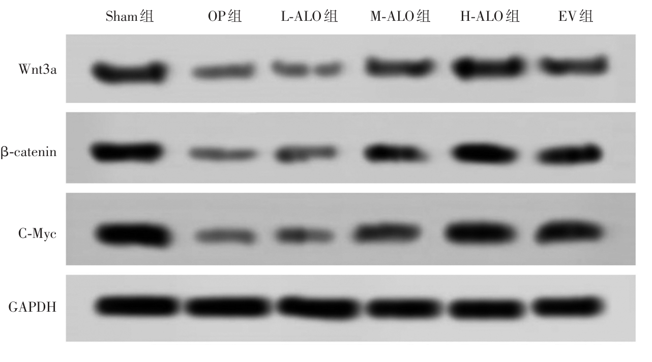
Tab.5
Comparison of proteins related to the Wnt/β-catenin signaling pathway among each group of mice(n = 10)"
| 组别 | Wnt3a | β-catenin | C-Myc |
|---|---|---|---|
| Sham组 | 1.03 ± 0.10 | 1.05 ± 0.11 | 1.12 ± 0.16 |
| OP组 | 0.23 ± 0.04? | 0.29 ± 0.05? | 0.34 ± 0.05? |
| L-ALO组 | 0.45 ± 0.06# | 0.49 ± 0.06# | 0.52 ± 0.07# |
| M-ALO组 | 0.75 ± 0.09#△ | 0.77 ± 0.09#△ | 0.81 ± 0.10#△ |
| H-ALO组 | 0.98 ± 0.10#▽ | 1.01 ± 0.13#▽ | 1.08 ± 0.16#▽ |
| EV组 | 0.78 ± 0.10# | 0.79 ± 0.11# | 0.82 ± 0.11# |
| F值 | 955.455 | 795.038 | 614.914 |
| P值 | < 0.001 | < 0.001 | < 0.001 |
Fig.5
Detection and analysis of the expression levels of Wnt3a, β-catenin and C-Myc proteins by Western blot"
| 1 |
GREGSON C L, ARMSTRONG D J, BOWDEN J, et al. UK clinical guideline for the prevention and treatment of osteoporosis [J]. Arch Osteoporos, 2022, 17(1): 58. doi:10.1007/s11657-022-01061-5
doi: 10.1007/s11657-022-01061-5 |
| 2 |
ONIZUKA N, ONIZUKA T. Disparities in Osteoporosis Prevention and Care: Understanding Gender, Racial, and Ethnic Dynamics [J]. Curr Rev Musculoskelet Med, 2024, 17(9): 365-372. doi:10.1007/s12178-024-09909-8
doi: 10.1007/s12178-024-09909-8 |
| 3 |
FISCHER V, HAFFNER-LUNTZER M. Interaction between bone and immune cells: Implications for postmenopausal osteoporosis [J]. Semin Cell Dev Biol, 2022, 123: 14-21. doi:10.1016/j.semcdb.2021.05.014
doi: 10.1016/j.semcdb.2021.05.014 |
| 4 |
ZHANG Y W, CAO M M, LI Y J, et al. Fecal microbiota transplantation ameliorates bone loss in mice with ovariectomy-induced osteoporosis via modulating gut microbiota and metabolic function [J]. J Orthop Translat, 2022, 37: 46-60. doi:10.1016/j.jot.2022.08.003
doi: 10.1016/j.jot.2022.08.003 |
| 5 |
CHANG Z, ZHANG P, ZHANG M, et al. Aloperine suppresses human pulmonary vascular smooth muscle cell proliferation via inhibiting inflammatory response [J]. Chin J Physiol, 2019, 62(4): 157-165. doi:10.4103/cjp.cjp_27_19
doi: 10.4103/cjp.cjp_27_19 |
| 6 |
HU R, CHEN L, CHEN X, et al. Aloperine improves osteoporosis in ovariectomized mice by inhibiting RANKL-induced NF-κB, ERK and JNK approaches [J]. Int Immunopharmacol, 2021, 97: 107720. doi:10.1016/j.intimp.2021.107720
doi: 10.1016/j.intimp.2021.107720 |
| 7 |
WANG X, TIAN Y, LIANG X, et al. Bergamottin promotes osteoblast differentiation and bone formation via activating the Wnt/β-catenin signaling pathway [J]. Food Funct, 2022, 13(5): 2913-2924. doi:10.1039/d1fo02755g
doi: 10.1039/d1fo02755g |
| 8 | CAI Y, SUN H, SONG X, et al. The Wnt/β-catenin signaling pathway inhibits osteoporosis by regulating the expression of TERT: An in vivo and in vitro study [J]. Aging (Albany NY), 2023, 15(20): 11471-11488. |
| 9 |
LI X, LU Y, WEN P, et al. Matrine restrains the development of colorectal cancer through regulating the AGRN/Wnt/β-catenin pathway [J]. Environ Toxicol, 2023, 38(4): 809-819. doi:10.1002/tox.23730
doi: 10.1002/tox.23730 |
| 10 |
李永志,韩礼军,李智斌,等. 秦岭箭叶淫羊藿对骨质疏松大鼠骨代谢及胫骨骨微结构的影响[J]. 疑难病杂志,2024,23(8):993-998,1001. doi:10.3969/j.issn.1671-6450.2024.08.019
doi: 10.3969/j.issn.1671-6450.2024.08.019 |
| 11 |
罗兰兰,张宇静,任明诗,等. 杜仲汤对去卵巢大鼠骨质疏松症的影响及机制研究[J]. 中药新药与临床药理,2024,35(4):461-468. doi:10.19378/j.issn.1003-9783.2024.04.002
doi: 10.19378/j.issn.1003-9783.2024.04.002 |
| 12 |
OH W T, YANG Y S, XIE J, et al. WNT-modulating gene silencers as a gene therapy for osteoporosis, bone fracture, and critical-sized bone defects [J]. Mol Ther, 2023, 31(2): 435-453. doi:10.1016/j.ymthe.2022.09.018
doi: 10.1016/j.ymthe.2022.09.018 |
| 13 |
WARREN J T, ZOU W, DECKER C E, et al. Correlating RANK ligand/RANK binding kinetics with osteoclast formation and function [J]. J Cell Biochem, 2015, 116(11): 2476-2483. doi:10.1002/jcb.25191
doi: 10.1002/jcb.25191 |
| 14 |
ZADJALI F AL, BROOKS J, O'NEILL T W, et al. Experiences of postmenopausal osteoporosis: A narrative review [J]. Disabil Rehabil, 2024, 46(5): 828-840. doi:10.1080/09638288.2023.2169770
doi: 10.1080/09638288.2023.2169770 |
| 15 |
TRÉMOLLIERES F A, CHABBERT-BUFFET N, PLU-BUREAU G, et al. Management of postmenopausal women: Collège National des Gynécologues et Obstétriciens Français (CNGOF) and Groupe d'Etude sur la Ménopause et le Vieillissement (GEMVi) Clinical Practice Guidelines [J]. Maturitas, 2022, 163: 62-81. doi:10.1016/j.maturitas.2022.05.008
doi: 10.1016/j.maturitas.2022.05.008 |
| 16 |
YUAN F, PENG W, YANG C, et al. Teriparatide versus bisphosphonates for treatment of postmenopausal osteoporosis: A meta-analysis [J]. Int J Surg, 2019, 66: 1-11. doi:10.1016/j.ijsu.2019.03.004
doi: 10.1016/j.ijsu.2019.03.004 |
| 17 |
LIU Y, YU P, PENG X, et al. Hexapeptide-conjugated calcitonin for targeted therapy of osteoporosis [J]. J Control Release, 2019, 304: 39-50. doi:10.1016/j.jconrel.2019.04.042
doi: 10.1016/j.jconrel.2019.04.042 |
| 18 |
REID I R, BILLINGTON E O. Drug therapy for osteoporosis in older adults [J]. Lancet, 2022, 399(10329): 1080-1092. doi:10.1016/s0140-6736(21)02646-5
doi: 10.1016/s0140-6736(21)02646-5 |
| 19 |
PASCHALIS E P, GAMSJAEGER S, HASSLER N, et al. Vitamin D and calcium supplementation for three years in postmenopausal osteoporosis significantly alters bone mineral and organic matrix quality [J]. Bone, 2017, 95: 41-46. doi:10.1016/j.bone.2016.11.002
doi: 10.1016/j.bone.2016.11.002 |
| 20 |
CHEN L R, KO N Y, CHEN K H. Medical Treatment for Osteoporosis: From Molecular to Clinical Opinions [J]. Int J Mol Sci, 2019, 20(9) : 2213. doi:10.3390/ijms20092213
doi: 10.3390/ijms20092213 |
| 21 |
CHOI D, CHOI S, CHANG J, et al. Exposure to oral bisphosphonates and risk of gastrointestinal cancer [J]. Osteoporos Int, 2020, 31(4): 775-782. doi:10.1007/s00198-020-05327-x
doi: 10.1007/s00198-020-05327-x |
| 22 |
CHEN Y J, JIA L H, HAN T H, et al. Osteoporosis treatment: current drugs and future developments [J]. Front Pharmacol, 2024, 15: 1456796. doi:10.3389/fphar.2024.1456796
doi: 10.3389/fphar.2024.1456796 |
| 23 |
TAO X, YIN L, XU L, et al. Dioscin: A diverse acting natural compound with therapeutic potential in metabolic diseases, cancer, inflammation and infections [J]. Pharmacol Res, 2018, 137: 259-269. doi:10.1016/j.phrs.2018.09.022
doi: 10.1016/j.phrs.2018.09.022 |
| 24 |
CAO G, HU S, NING Y, et al. Traditional Chinese medicine in osteoporosis: From pathogenesis to potential activity [J]. Front Pharmacol, 2024, 15: 1370900. doi:10.3389/fphar.2024.1370900
doi: 10.3389/fphar.2024.1370900 |
| 25 |
MUHAMMAD T, SAKHAWAT A, KHAN A A, et al. Aloperine in combination with therapeutic adenoviral vector synergistically suppressed the growth of non-small cell lung cancer [J]. J Cancer Res Clin Oncol, 2020, 146(4): 861-874. doi:10.1007/s00432-020-03157-2
doi: 10.1007/s00432-020-03157-2 |
| 26 |
YU H I, SHEN H C, CHEN S H, et al. Autophagy Modulation in Human Thyroid Cancer Cells following Aloperine Treatment [J]. Int J Mol Sci, 2019, 20(21): 5315. doi:10.3390/ijms20215315
doi: 10.3390/ijms20215315 |
| 27 |
LIU J S, HUO C Y, CAO H H, et al. Aloperine induces apoptosis and G2/M cell cycle arrest in hepatocellular carcinoma cells through the PI3K/Akt signaling pathway [J]. Phytomedicine, 2019, 61: 152843. doi:10.1016/j.phymed.2019.152843
doi: 10.1016/j.phymed.2019.152843 |
| 28 |
TAHIR M, ALI S, ZHANG W, et al. Aloperine: A Potent Modulator of Crucial Biological Mechanisms in Multiple Diseases [J]. Biomedicines, 2022, 10(4): 905. doi:10.3390/biomedicines10040905
doi: 10.3390/biomedicines10040905 |
| 29 |
CHEN X, ZHI X, PAN P, et al. Matrine prevents bone loss in ovariectomized mice by inhibiting RANKL-induced osteoclastogenesis [J]. FASEB J, 2017, 31(11): 4855-4865. doi:10.1096/fj.201700316r
doi: 10.1096/fj.201700316r |
| 30 |
JIANG C, MA Q, WANG S, et al. Oxymatrine Attenuates Osteoclastogenesis via Modulation of ROS-Mediated SREBP2 Signaling and Counteracts Ovariectomy-Induced Osteoporosis [J]. Front Cell Dev Biol, 2021, 9: 684007. doi:10.3389/fcell.2021.684007
doi: 10.3389/fcell.2021.684007 |
| 31 |
BRENT M B. Pharmaceutical treatment of bone loss: From animal models and drug development to future treatment strategies [J]. Pharmacol Ther, 2023, 244: 108383. doi:10.1016/j.pharmthera.2023.108383
doi: 10.1016/j.pharmthera.2023.108383 |
| 32 |
YAMAMOTO H, ZHANG S, MIZUSHIMA N. Autophagy genes in biology and disease [J]. Nat Rev Genet, 2023, 24(6): 382-400. doi:10.1038/s41576-022-00562-w
doi: 10.1038/s41576-022-00562-w |
| 33 |
DERETIC V. Autophagy in inflammation, infection, and immunometabolism [J]. Immunity, 2021, 54(3): 437-453. doi:10.1016/j.immuni.2021.01.018
doi: 10.1016/j.immuni.2021.01.018 |
| 34 |
ZHANG L, GUO Y F, LIU Y Z, et al. Pathway-based genome-wide association analysis identified the importance of regulation-of-autophagy pathway for ultradistal radius BMD [J]. J Bone Miner Res, 2010, 25(7): 1572-1580. doi:10.1002/jbmr.36
doi: 10.1002/jbmr.36 |
| 35 |
TANG N, ZHAO H, ZHANG H, et al. Effect of autophagy gene DRAM on proliferation, cell cycle, apoptosis, and autophagy of osteoblast in osteoporosis rats [J]. J Cell Physiol, 2019, 234(4): 5023-5032. doi:10.1002/jcp.27304
doi: 10.1002/jcp.27304 |
| 36 |
ZHANG L, ZHENG Y L, WANG R, et al. Exercise for osteoporosis: A literature review of pathology and mechanism [J]. Front Immunol, 2022, 13: 1005665. doi:10.3389/fimmu.2022.1005665
doi: 10.3389/fimmu.2022.1005665 |
| 37 |
LIU F, FANG F, YUAN H, et al. Suppression of autophagy by FIP200 deletion leads to osteopenia in mice through the inhibition of osteoblast terminal differentiation [J]. J Bone Miner Res, 2013, 28(11): 2414-2430. doi:10.1002/jbmr.1971
doi: 10.1002/jbmr.1971 |
| 38 |
TANG T, LIANG H, WEI W, et al. Aloperine targets lysosomes to inhibit late autophagy and induces cell death through apoptosis and paraptosis in glioblastoma [J]. Mol Biomed, 2023, 4(1): 42. doi:10.1186/s43556-023-00155-x
doi: 10.1186/s43556-023-00155-x |
| 39 |
OBENG E. Apoptosis (programmed cell death) and its signals-A review [J]. Braz J Biol, 2021, 81(4): 1133-1143. doi:10.1590/1519-6984.228437
doi: 10.1590/1519-6984.228437 |
| 40 |
BERTHELOOT D, LATZ E, FRANKLIN B S. Necroptosis, pyroptosis and apoptosis: An intricate game of cell death [J]. Cell Mol Immunol, 2021, 18(5): 1106-1121. doi:10.1038/s41423-020-00630-3
doi: 10.1038/s41423-020-00630-3 |
| 41 |
RU J Y, WANG Y F. Osteocyte apoptosis: The roles and key molecular mechanisms in resorption-related bone diseases [J]. Cell Death Dis, 2020, 11(10): 846. doi:10.1038/s41419-020-03059-8
doi: 10.1038/s41419-020-03059-8 |
| 42 |
CHANDRA A, RAJAWAT J. Skeletal Aging and Osteoporosis: Mechanisms and Therapeutics [J]. Int J Mol Sci, 2021, 22(7): 3553. doi:10.3390/ijms22073553
doi: 10.3390/ijms22073553 |
| 43 |
WEINSTEIN R S, MANOLAGAS S C. Apoptosis and osteoporosis [J]. Am J Med, 2000, 108(2): 153-164. doi:10.1016/s0002-9343(99)00420-9
doi: 10.1016/s0002-9343(99)00420-9 |
| 44 |
XU Z, WANG P, WANG Z, et al. ER-β accelerates the process of primary osteoporosis by promoting VEGFA-mediated apoptosis of osteoblasts[J]. Genomics, 2023, 115(6): 110743. doi:10.1016/j.ygeno.2023.110743
doi: 10.1016/j.ygeno.2023.110743 |
| 45 |
MORIISHI T, FUKUYAMA R, MIYAZAKI T, et al. Overexpression of BCLXL in Osteoblasts Inhibits Osteoblast Apoptosis and Increases Bone Volume and Strength[J]. J Bone Miner Res, 2016, 31(7): 1366-1380. doi:10.1002/jbmr.2808
doi: 10.1002/jbmr.2808 |
| 46 |
WONG S K, MOHAMAD N V, JAYUSMAN P A, et al. A Review on the Crosstalk between Insulin and Wnt/β-Catenin Signalling for Bone Health[J]. Int J Mol Sci, 2023, 24(15): 12441. doi:10.3390/ijms241512441
doi: 10.3390/ijms241512441 |
| 47 |
LIU J, XIAO Q, XIAO J, et al. Wnt/β-catenin signalling: Function, biological mechanisms, and therapeutic opportunities [J]. Signal Transduct Target Ther, 2022, 7(1): 3. doi:10.1038/s41392-021-00762-6
doi: 10.1038/s41392-021-00762-6 |
| 48 |
VISWESWARAN M, POHL S, ARFUSO F, et al. Multi-lineage differentiation of mesenchymal stem cells-To Wnt, or not Wnt [J]. Int J Biochem Cell Biol, 2015, 68: 139-147. doi:10.1016/j.biocel.2015.09.008
doi: 10.1016/j.biocel.2015.09.008 |
| 49 |
CHENG B F, FENG X, GAO Y X, et al. Neural Cell Adhesion Molecule Regulates Osteoblastic Differentiation Through Wnt/β-Catenin and PI3K-Akt Signaling Pathways in MC3T3-E1 Cells [J]. Front Endocrinol (Lausanne), 2021, 12: 657953. doi:10.3389/fendo.2021.657953
doi: 10.3389/fendo.2021.657953 |
| 50 |
YU W, XIE C R, CHEN F C, et al. LGR5 enhances the osteoblastic differentiation of MC3T3-E1 cells through the Wnt/β-catenin pathway [J]. Exp Ther Med, 2021, 22(2): 889. doi:10.3892/etm.2021.10321
doi: 10.3892/etm.2021.10321 |
| 51 |
ZHAO Y, LIU J, ZHANG Y, et al. Mir-381-3p aggravates ovariectomy-induced osteoporosis by inhibiting osteogenic differentiation through targeting KLF5/Wnt/β-catenin signaling pathway [J]. J Orthop Surg Res, 2024, 19(1): 480. doi:10.1186/s13018-024-04992-6
doi: 10.1186/s13018-024-04992-6 |
| 52 |
LI R, RUAN Q, YIN F, et al. MiR-23b-3p promotes postmenopausal osteoporosis by targeting MRC2 and regulating the Wnt/β-catenin signaling pathway [J]. J Pharmacol Sci, 2021, 145(1): 69-78. doi:10.1016/j.jphs.2020.11.004
doi: 10.1016/j.jphs.2020.11.004 |
| 53 | XIAO X, AO M, XU F, et al. Effect of matrine against breast cancer by downregulating the vascular endothelial growth factor via the Wnt/β-catenin pathway [J]. Oncol Lett, 2018, 15(2): 1691-1697. |
| [1] | Huixia YANG,Ning DING,Runqiu MA,Guizhong LI,Yinju HAO,Shengchao MA,Yideng JIANG,Zhigang. BAI. Role and mechanism of circular RNA mmu_circ_0000818 in dexamethasone⁃induced apoptosis of MC3T3⁃E1 cells [J]. The Journal of Practical Medicine, 2025, 41(4): 478-489. |
| [2] | Zonglin LI,Chunlin FENG,Xin LIU,Xingming SHU,Min. SONG. CCCTC⁃binding factors promote the formation of oxaliplatin related gastric cancer drug-tolerant cells by resisting apoptosis [J]. The Journal of Practical Medicine, 2025, 41(4): 490-499. |
| [3] | Yufen LU,Xiaoming ZHENG,Shaojuan WEI,Liqin CHEN,Tongtong XU,Xiangwei. LÜ. Effects of miR⁃483⁃3p on hypoxia/reoxygenation⁃induced apoptosis and pyroptosis in cardiomyocytes [J]. The Journal of Practical Medicine, 2025, 41(3): 339-346. |
| [4] | Shan LUO,Ying FENG,Dandan FAN,Wenxin ZHENG,Xingrong GUO,Xuzhi. RUAN. ANGPTL8 knockout reduces lipopolysaccharide⁃induced hepatic lipid deposition [J]. The Journal of Practical Medicine, 2024, 40(9): 1197-1203. |
| [5] | Wei HE,Liping LIU,Jingwei ZHUO,Xiaodong ZHANG,Tong YANG,Jubin. FENG. CCR5 blockade reduces tumor growth by inducing apoptosis and impairing immunosuppression of tumor microenvironment [J]. The Journal of Practical Medicine, 2024, 40(9): 1204-1210. |
| [6] | Fangming WANG,Wenxuan SHANG,Jingwen ZHANG,Yingxiao JI,Litao. LI. Research advances on the regulation of microglia polarization by autophagy in ischemic stroke [J]. The Journal of Practical Medicine, 2024, 40(9): 1324-1330. |
| [7] | Lulu CHEN,Meng LUO,Kaiqi SU,Jing GAO,Xiaodong. FENG. Research progress of the endoplasmic reticulum⁃mitochondrial interactions in post⁃stroke cognitive impairment [J]. The Journal of Practical Medicine, 2024, 40(7): 1023-1028. |
| [8] | Wenxin LI,Minjun LU,Li LIN,Yueqin LIU,Xiaolan. ZHU. circRAF1 regulates the proliferation and apoptosis of human ovarian granulosa cells [J]. The Journal of Practical Medicine, 2024, 40(7): 910-917. |
| [9] | Zhen YANG,Shaoru JIANG,Xiaoyan CHEN,Xiaolin CHEN,Weimin DENG,Xinyu. GUO. Effect of Jinghou Zengzhi Granules on ovarian GDF9 secretion and granulosa cells apoptosis in controlled ovarian hyperstimulation rats [J]. The Journal of Practical Medicine, 2024, 40(7): 918-923. |
| [10] | Ying ZHOU,Dajun JIANG,Yong TIAN,Yongxiang GU,Guohui. YANG. Inhibition of TRAF6 ameliorates myocardial inflammatory injury and cardiac dysfunction via regulating cardiomyocyte inflammation in sepsis mice [J]. The Journal of Practical Medicine, 2024, 40(5): 608-614. |
| [11] | Runwei MA,Chunjie MU,Wenting GUI,Yao DENG,Minzhang ZHAO,Min LIU,Yi SONG. LncRNA SENCR targeted miR⁃206 regulates proliferation and apoptosis of human vascular smooth muscle cells of aortic dissection tissues [J]. The Journal of Practical Medicine, 2024, 40(3): 302-308. |
| [12] | Lihong DING,Shijia GENG,Yujie WANG. Effects of wedelobata on apoptosis and secretion of inflammatory cytokines in the alveolar epithelium infected by Streptococcus pneumonia [J]. The Journal of Practical Medicine, 2024, 40(3): 316-320. |
| [13] | Chunfang KONG,Anna LI,Bo KE,Weirong DING,Tingting LIU,Huan FU,Tingting ZHANG,Chenghao JIN,Mei. WU. The inhibitory effect and molecular mechanism of 6-gingerol on human multiple myeloma cells [J]. The Journal of Practical Medicine, 2024, 40(23): 3291-3297. |
| [14] | Xiang JIA,Tianjie XU,Jiaxin FAN,Xiaoling GUO,Kainan LIU,Hui ZHANG,Yongsheng WANG,Qian. WANG. Metformin exerts a protective effect on articular cartilage in osteoarthritis rats by activating the SIRT1/p53 signaling pathway [J]. The Journal of Practical Medicine, 2024, 40(23): 3306-3316. |
| [15] | Bing′er WU,Qing LI,Kerong YANG,Jian ZHANG,Yi YU,Lei LEI,Bo. HU. Impact of spermidine on proliferation and apoptosis in diffuse large B⁃cell lymphoma cell lines [J]. The Journal of Practical Medicine, 2024, 40(22): 3130-3137. |
| Viewed | ||||||
|
Full text |
|
|||||
|
Abstract |
|
|||||