

The Journal of Practical Medicine ›› 2024, Vol. 40 ›› Issue (22): 3138-3145.doi: 10.3969/j.issn.1006-5725.2024.22.004
• Basic Research • Previous Articles Next Articles
Zhe SHI1,2,Xialin ZUO2,3,Linhui PENG2,3,Zhiwei LU1,2,Kongping. LI1,2( )
)
Received:2024-07-12
Online:2024-11-25
Published:2024-11-25
Contact:
Kongping. LI
E-mail:Lkp159123@163.com
CLC Number:
Zhe SHI,Xialin ZUO,Linhui PENG,Zhiwei LU,Kongping. LI. Effect of M1 microglial polarization on secondary damage in the thalamus after cerebral cortical infarction[J]. The Journal of Practical Medicine, 2024, 40(22): 3138-3145.
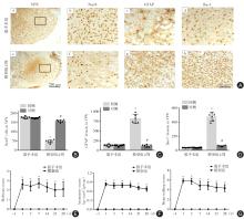
Fig.1
Secondary damage in VPN of thalamus and neurological dysfunction after cerebral cortex infarction in rats"

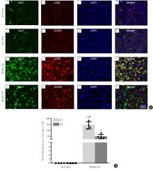
Fig. 2
Polarization of microglia to M1 type in ipsilateral thalamicafter cortical infarction in rats"
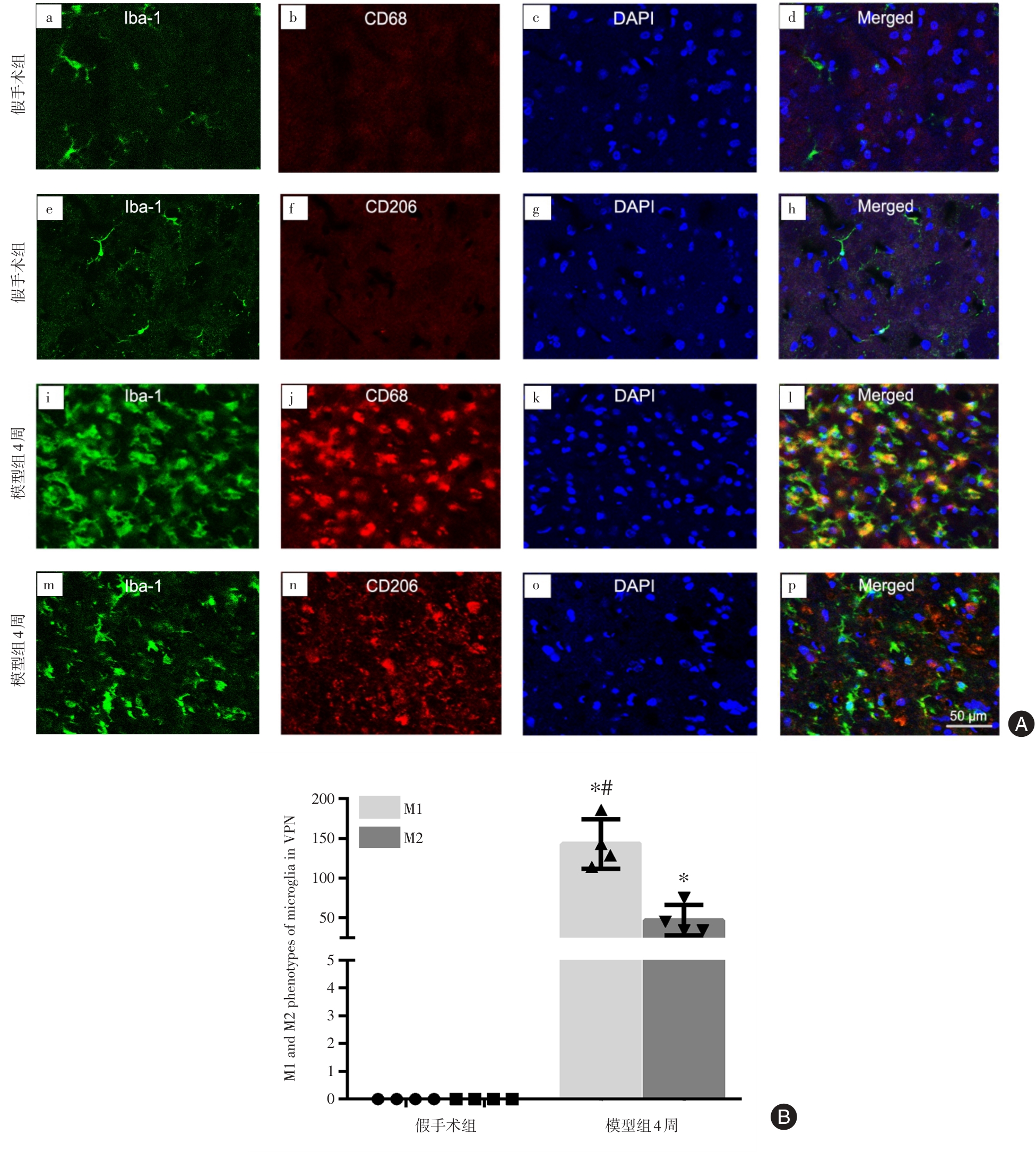

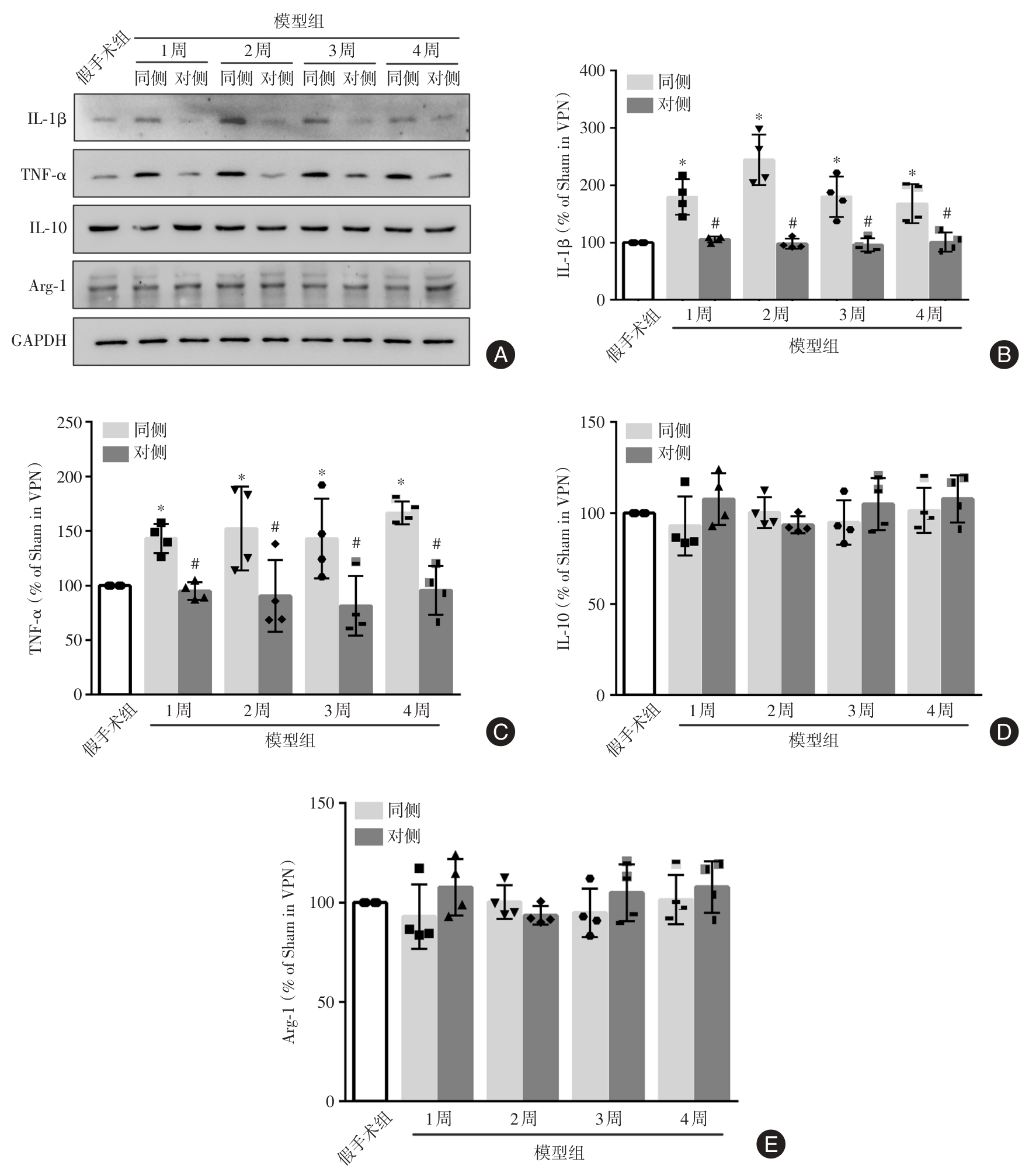
Fig.3
Secondary inflammatory response in ipsilateral thalamus after cortical infarction in rats"
| 1 |
CAO Z, HARVEY S S, BLISS T M, et al. Inflammatory Responses in the Secondary Thalamic Injury After Cortical Ischemic Stroke [J]. Front Neurol, 2020, 11: 236. doi:10.3389/fneur.2020.00236
doi: 10.3389/fneur.2020.00236 |
| 2 |
ZHOU K, TAN Y, ZHANG G, et al. Loss of SARM1 ameliorates secondary thalamic neurodegeneration after cerebral infarction [J]. J Cereb Blood Flow Metab. 2024, 44(2): 224-238. doi:10.1177/0271678x231210694
doi: 10.1177/0271678x231210694 |
| 3 |
李虹莹,沈缘,吴巧凤,等. 小胶质细胞极化信号通路在神经炎症中的研究进展 [J]. 实用医学杂志, 2022, 38 (14): 1838-1846. doi:10.3969/j.issn.1006⁃5725.2022.14.024
doi: 10.3969/j.issn.1006?5725.2022.14.024 |
| 4 |
FAN P L, WANG S S, CHU S F, et al. Time-dependent dual effect of microglia in ischemic stroke [J]. Neurochem Int, 2023, 169:105584. doi:10.1016/j.neuint.2023.105584
doi: 10.1016/j.neuint.2023.105584 |
| 5 |
DARWISH S F, ELBADRY A M M, ELBOKHOMY A S, et al. The dual face of microglia (M1/M2) as a potential target in the protective effect of nutraceuticals against neurodegenerative diseases [J]. Front Aging, 2023, 4:1231706. doi:10.3389/fragi.2023.1231706
doi: 10.3389/fragi.2023.1231706 |
| 6 |
SURUGIU R, CATALIN B, DUMBRAVA D, et al. Intracortical Administration of the Complement C3 Receptor Antagonist Trifluoroacetate Modulates Microglia Reaction after Brain Injury [J]. Neural Plast, 2019, 2019:1071036. doi:10.1155/2019/1071036
doi: 10.1155/2019/1071036 |
| 7 |
LI J, WANG H, DU C, et al. hUC-MSCs ameliorated CUMS-induced depression by modulating complement C3 signaling-mediated microglial polarization during astrocyte-microglia crosstalk [J]. Brain Res Bull, 2020, 163:109-119. doi:10.1016/j.brainresbull.2020.07.004
doi: 10.1016/j.brainresbull.2020.07.004 |
| 8 |
DATTA A, SARMAH D, KALIA K, et al. Advances in Studies on Stroke-Induced Secondary Neurodegeneration (SND) and Its Treatment [J]. Curr Top Med Chem, 2020, 20(13):1154-1168. doi:10.2174/1568026620666200416090820
doi: 10.2174/1568026620666200416090820 |
| 9 |
LI K, PENG L, XING Q, et al. Transplantation of hESCs-Derived Neural Progenitor Cells Alleviates Secondary Damage of Thalamus After Focal Cerebral Infarction in Rats [J]. Stem Cells Transl Med, 2023, 12(8):553-568. doi:10.1093/stcltm/szad037
doi: 10.1093/stcltm/szad037 |
| 10 |
JIANG Z, WEI J, LIANG J, et al. Dl-3-n-Butylphthalide Alleviates Secondary Brain Damage and Improves Working Memory After Stroke in Cynomolgus Monkeys [J]. Stroke,2024, 55(3):725-734. doi:10.1161/strokeaha.123.045037
doi: 10.1161/strokeaha.123.045037 |
| 11 |
KANEMITSU H, NAKAGOMI T, TAMURA A, et al. Differences in the extent of primary ischemic damage between middle cerebral artery coagulation and intraluminal occlusion models [J]. J Cereb Blood Flow Metab, 2002, (10):1196-1204. doi:10.1097/01.wcb.0000037992.07114.95
doi: 10.1097/01.wcb.0000037992.07114.95 |
| 12 |
ZENG L, HU S, ZENG L, et al. Animal Models of Ischemic Stroke with Different Forms of Middle Cerebral Artery Occlusion [J]. Brain Sci,2023, 13(7):1007. doi:10.3390/brainsci13071007
doi: 10.3390/brainsci13071007 |
| 13 | 刘毅, 孙邈, 吉训明, 等. 丘脑供血及丘脑缺血性卒中临床表现[J]. 中国现代神经疾病杂志, 2018,18(12):902-905. |
| 14 |
ONG L K. Beyond the Primary Infarction: Focus on Mechanisms Related to Secondary Neurodegeneration after Stroke[J]. Int J Mol Sci,2022, 23(24):16024. doi:10.3390/ijms232416024
doi: 10.3390/ijms232416024 |
| 15 |
BRUNELLI S, GIANNELLA E, BIZZAGLIA M, et al. Secondary neurodegeneration following Stroke: What can blood biomarkers tell us? [J]. Front Neurol,2023, 14:1198216. doi:10.3389/fneur.2023.1198216
doi: 10.3389/fneur.2023.1198216 |
| 16 |
STUCKEY S M, ONG L K, COLLINS-PRAINO L E, et al. Neuroinflammation as a Key Driver of Secondary Neurodegeneration Following Stroke? [J]. Int J Mol Sci,2021, 22(23):13101. doi:10.3390/ijms222313101
doi: 10.3390/ijms222313101 |
| 17 |
PENG L, LI K, LI D, et al. The p75 neurotrophin receptor attenuates secondary thalamic damage after cortical infarction by promoting angiogenesis [J]. CNS Neurosci Ther,2024, 30(7):e14875. doi:10.1111/cns.14875
doi: 10.1111/cns.14875 |
| 18 |
DUAN M, XU Y, LI Y, et al. Targeting brain-peripheral immune responses for secondary brain injury after ischemic and hemorrhagic stroke [J]. J Neuroinflammation,2024, 21(1):102. doi:10.1186/s12974-024-03101-y
doi: 10.1186/s12974-024-03101-y |
| 19 | KIM G S, HARMON E, GUTIERREZ M, et al. Single-cell analysis identifies Ifi27l2a as a novel gene regulator of microglial inflammation in the context of aging and stroke [J]. Res Sq [Preprint],2023, rs.3.rs-2557290. |
| 20 |
NECULA D, CHO F S, HE A, et al. Secondary thalamic neuroinflammation after focal cortical stroke and traumatic injury mirrors corticothalamic functional connectivity [J]. J Comp Neurol,2022, 530(7):998-1019. doi:10.1002/cne.25259
doi: 10.1002/cne.25259 |
| 21 | 王方明,尚文璇,张靖雯,等. 自噬调控小胶质细胞极化在缺血性脑卒中的研究进展 [J]. 实用医学杂志,2024, 40(9): 1324-1330. |
| 22 |
D'ANMA L, SEARLE G, HARVEY K,et al. Time course of neuroinflammation after human stroke-A pilot study using co-registered PET and MRI [J]. BMC Neurol,2023, 23(1):193. doi:10.1186/s12883-023-03178-7
doi: 10.1186/s12883-023-03178-7 |
| 23 |
ANTTILA J E, ALBERT K, WIRES E S, et al. Post-stroke Intranasal (+)-Naloxone Delivery Reduces Microglial Activation and Improves Behavioral Recovery from Ischemic Injury [J]. eNeuro, 2018, 5(2):ENEURO.0395-17.2018. doi:10.1523/eneuro.0395-17.2018
doi: 10.1523/eneuro.0395-17.2018 |
| 24 |
SHUI X, CHEN J, FU Z, et al. Microglia in Ischemic Stroke: Pathogenesis Insights and Therapeutic Challenges [J]. J Inflamm Res, 2024, 17: 3335-3352. doi:10.2147/jir.s461795
doi: 10.2147/jir.s461795 |
| 25 |
WANG H, LI X, WANG Q, et al. TREM2, microglial and ischemic stroke [J]. J Neuroimmunol, 2023, 381:578108. doi:10.1016/j.jneuroim.2023.578108
doi: 10.1016/j.jneuroim.2023.578108 |
| 26 |
BERNIS M E, SCHLEEHUBER Y, ZWEYER M, et al. Temporal Characterization of Microglia-Associated Pro- and Anti-Inflammatory Genes in a Neonatal Inflammation-Sensitized Hypoxic-Ischemic Brain Injury Model [J]. Oxid Med Cell Longev, 2022, 2022:2479626. doi:10.1155/2022/2479626
doi: 10.1155/2022/2479626 |
| 27 |
CAO Z, HARVEY S S, CHIANG T, et al. Unique Subtype of Microglia in Degenerative Thalamus After Cortical Stroke [J]. Stroke, 2021, 52(2):687-698. doi:10.1161/strokeaha.120.032402
doi: 10.1161/strokeaha.120.032402 |
| 28 |
ZHENG Y, HU Y, YAN F, et al. Dihydroergotamine protects against ischemic stroke by modulating microglial/macrophage polarization and inhibiting inflammation in mice [J]. Neurol Res,2024, 46(4):367-377. doi:10.1080/01616412.2024.2328481
doi: 10.1080/01616412.2024.2328481 |
| [1] | Song LI,Xingyou HE,Dian HE,Bo WANG,Yu ZHAN,Jingjing. SUN. The joint efficacy of NBP and LIPost C in treatment of elderly patients with large atherosclerotic cerebral infarction [J]. The Journal of Practical Medicine, 2024, 40(9): 1286-1292. |
| [2] | Xiangzhi XIAO,Guanxiong CHEN,Zhiwen HU. The relationship between EGCG targeted regulation of Nrf2⁃Keap1 signaling pathway and neuroprotective effect in cerebral infarction [J]. The Journal of Practical Medicine, 2024, 40(3): 309-315. |
| [3] | Guidi ZOU,Xiaokai CHEN,Huihong TAN,Yi LI,Nan LI,Yefan CAO,Hewei. WANG. The clinical efficacy of closed⁃loop rehabilitation therapy by brain⁃computer interface combined with exoskeleton robotic hand for patients with hand dysfunction after cerebral infarction [J]. The Journal of Practical Medicine, 2024, 40(17): 2395-2400. |
| [4] | Juan DU,Xiaoyu ZHOU,Miao ZHANG,Xueling ZHANG,Wenya. LAN. Impact of mechanical thrombectomy using Solitaire stent combined with dual antiplatelet therapy on limb function and vascular reocclusion in patients with acute cerebral infarction [J]. The Journal of Practical Medicine, 2024, 40(14): 1952-1956. |
| [5] | Mengni HU,Xiaolei ZHANG,Zhen RONG,Yao WANG,Ya′nan LI,Jun. MA. Effects of electroacupuncture on MPTP⁃induced FoXO1/NLRP3 pathway mediated neuroinflammation in mice with Parkinson′s disease [J]. The Journal of Practical Medicine, 2024, 40(11): 1494-1499. |
| [6] | Xiaobin GUO,Ping LIU,Wenxia. YU. The association between the waist⁃to⁃height ratio and novel cerebral infarction in the elderly with hypertension [J]. The Journal of Practical Medicine, 2024, 40(11): 1592-1596. |
| [7] | Zijuan FU,Qian LI,Lin LU,Yongqiu. LI. Relationship of serum ANGPTL3 and NFATc1 levels with the severity and prognosis in cerebral infarction patients [J]. The Journal of Practical Medicine, 2024, 40(10): 1407-1411. |
| [8] | Yuanyuan MAO,Jingjing. YUAN. Research advances of long noncoding RNA H19 in central nervous system diseases [J]. The Journal of Practical Medicine, 2023, 39(23): 3021-3026. |
| [9] | DENG Xiren , ZENG Daojun, ZHANG Guanpeng, DUAN Xiaoxia. . The effect of baicalin on cognitive function of cerebral ischemia-reperfusion injury in mice through PGE2 [J]. The Journal of Practical Medicine, 2023, 39(15): 1881-1887. |
| [10] | GUO Xiaobin, LIU Ping, YANG Haohui, ZHAO Jing, KAN Chunna, ZHAI Jingxiu, YU Wenxia, ZHAO Chumin, LIU Zhiwei.. The association between the body height and novel cerebral infarction in the elderly with hypertension [J]. The Journal of Practical Medicine, 2023, 39(13): 1714-1718. |
| [11] |
NIU Wen, QIU Xiaohui, LIU Yichao..
Relationship between perivascular fat density of carotid artery stenosis and cerebral infarction [J]. The Journal of Practical Medicine, 2023, 39(1): 103-108. |
| [12] |
HUANG Jianmin, YUN Yanfang, YANG Guixin, CHEN Haiyan, JIANG Yongming, HUANG Dongxu, LIU Jie, MO Xiaorong, LI Xuebin..
Effects of survivin inhibitor YM155 on microvessel density and angiogenesis⁃related gene expression in rats with cerebral infarction [J]. The Journal of Practical Medicine, 2022, 38(8): 946-951. |
| [13] |
JIN Bowen, LI Dongna, ZHUANG Pengwei, GUO Hong, ZHANG Yanjun.
BThe thalamic BDNF ⁃ TrkB signaling pathway mediates cerebral ischemia ⁃ induced hyperalgesia [J]. The Journal of Practical Medicine, 2022, 38(21): 2663-2669. |
| [14] |
LI Hongy⁃ing , SHEN Yuan, WU Qiaofeng, XIE Lushuang, YU Shuguang..
Role of microglia polarization signaling pathways in neuroinflammation:A literature review
[J]. The Journal of Practical Medicine, 2022, 38(14): 1838-1846.
|
| [15] |
GUO Zhuang, ZHOU Lijun..
Dual role of astrocyte⁃microglia crosstalk in neuroinflammation [J]. The Journal of Practical Medicine, 2021, 37(18): 2432-2436. |
| Viewed | ||||||
|
Full text |
|
|||||
|
Abstract |
|
|||||