

The Journal of Practical Medicine ›› 2024, Vol. 40 ›› Issue (7): 948-954.doi: 10.3969/j.issn.1006-5725.2024.07.012
• Basic Research • Previous Articles Next Articles
Ling XIAO1,Chunlei GAO1,Wei GUO1,Ning WANG1,Xuan ZHANG2,Ming. LIU( )
)
Received:2023-07-22
Online:2024-04-10
Published:2024-04-08
Contact:
Ming. LIU
E-mail:175013446@qq.com
CLC Number:
Ling XIAO,Chunlei GAO,Wei GUO,Ning WANG,Xuan ZHANG,Ming. LIU. Codonopsis polysaccharide protected LPS⁃induced acute lung injury by inhibiting MAPK/NF⁃κB signaling pathway in mice[J]. The Journal of Practical Medicine, 2024, 40(7): 948-954.
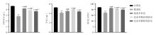
Fig.1
Effect of Codonopsis polysaccharide on lung function indexes in mice"
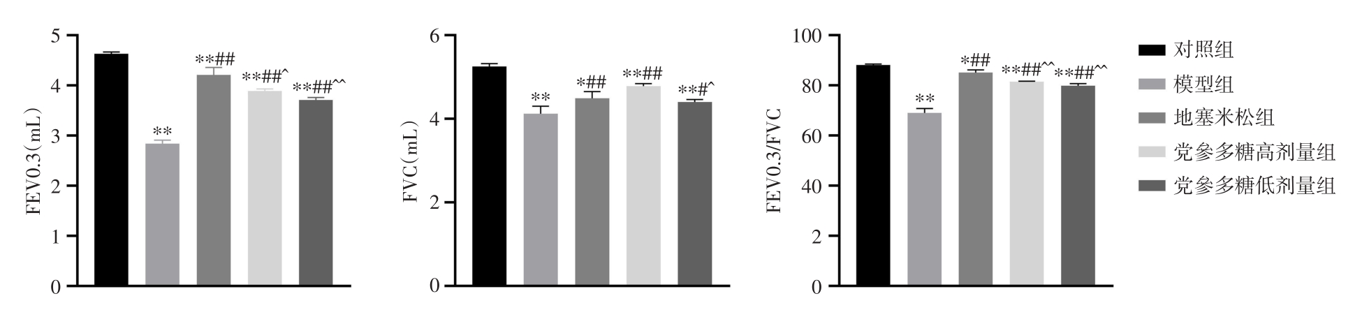
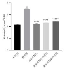
Fig.2
Effect of Codonopsis polysaccharide on wet mass/dry mass in lung tissue of mice in each group"

Fig.3
Effect of Codonopsis polysaccharide on lung histopathology in mice(HE, × 400)"
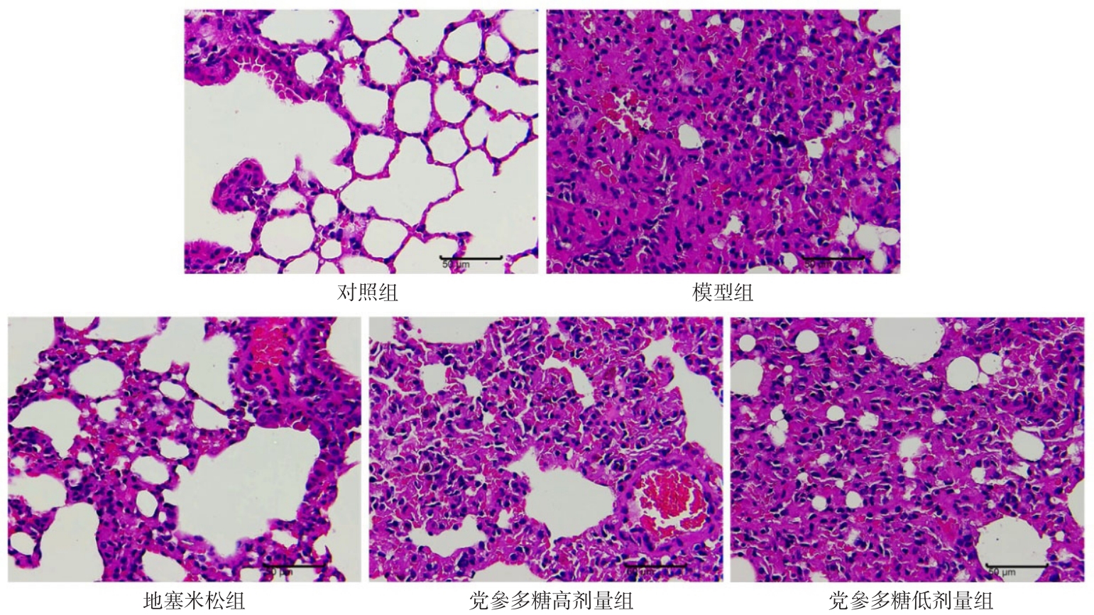
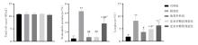
Fig.4
Effect of Codonopsis polysaccharide on number and classification of inflammatory cells in BALF of mice"
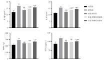
Fig.5
Effect of Codonopsis polysaccharide on the levels of IL-1β, IL-6, MPO and TNF-α in BALF of mice"
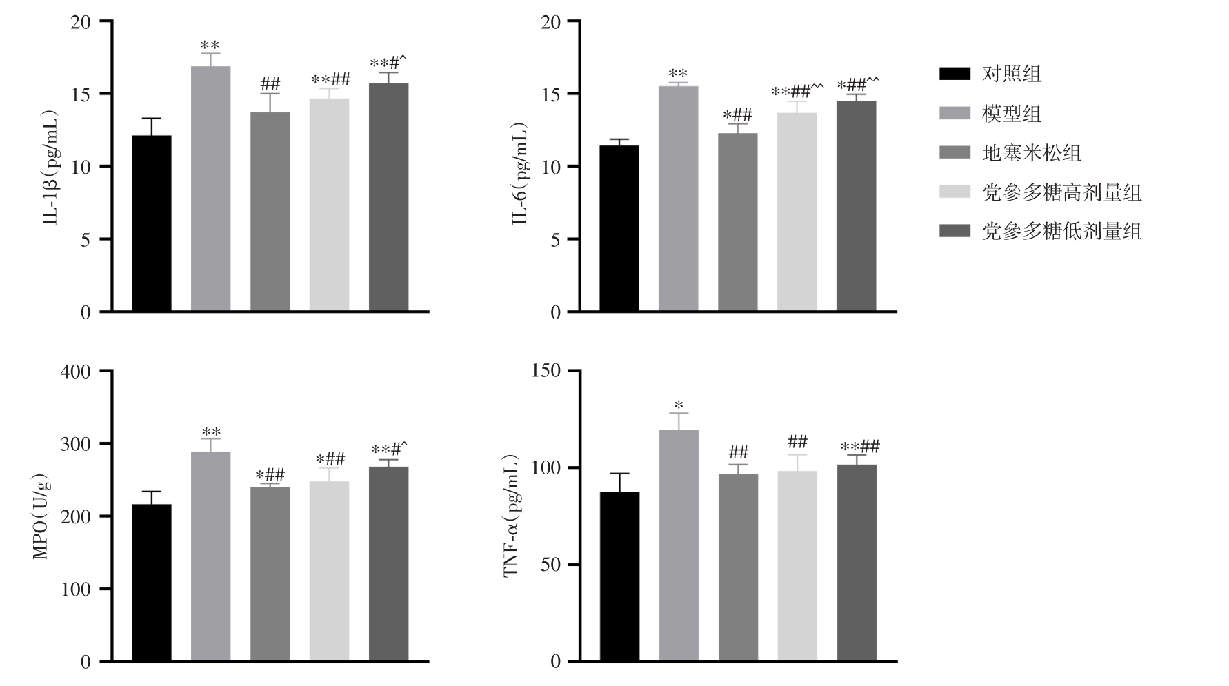
Fig.6
Effect of Codonopsis polysaccharide on the levels of IL-1β, IL-6, MPO and TNF-α in plasma of mice"
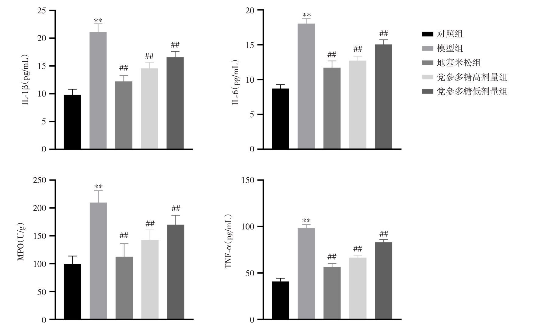
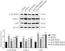
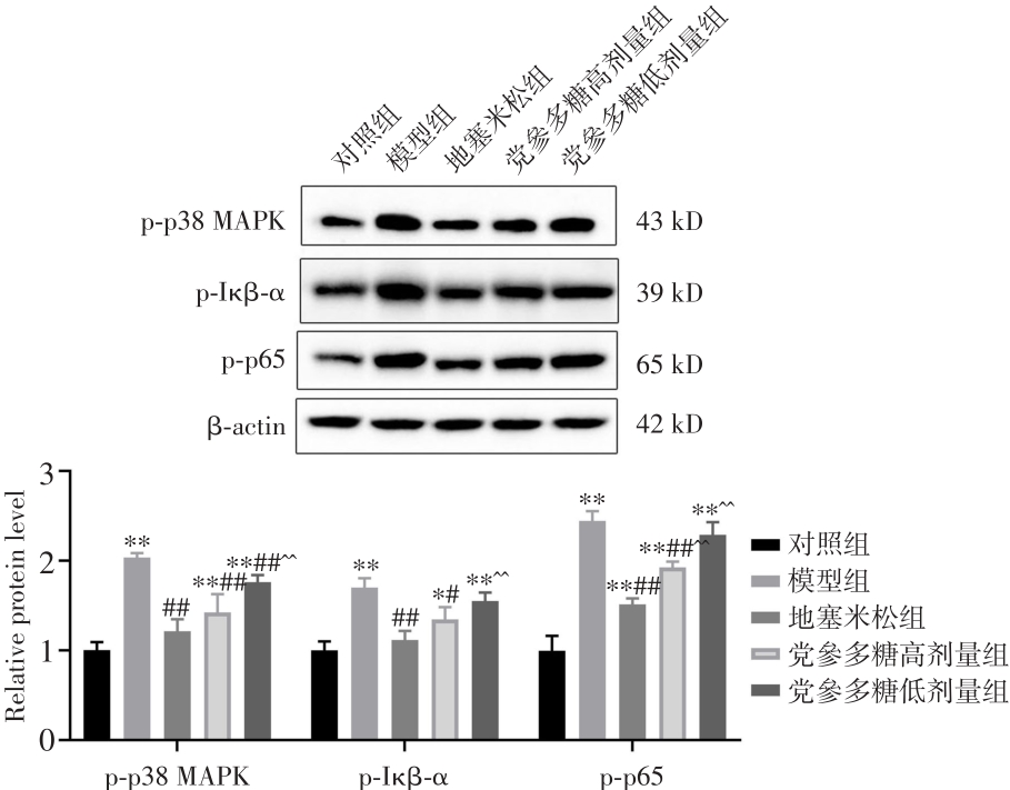
Fig.7
Effect of Codonopsis polysaccharide on expression levels of p-p38 MAPK、p-IκB-α and p-p65 in lung tissue of mice"
| 1 |
SANDEEP B, XIAO Z, ZHAO F, et al. Role of Platelets in Acute Lung Injury after Extracorporeal Circulation in Cardiac Surgery Patients: A systemic review[J]. Curr Probl Cardiol, 2022,47(11):101088. doi:10.1016/j.cpcardiol.2021.101088
doi: 10.1016/j.cpcardiol.2021.101088 |
| 2 |
MOKRÁ D. Acute lung injury - from pathophysiology to treatment[J]. Physiol Res, 2020,69():S353-S366. doi:10.33549/physiolres.934602
doi: 10.33549/physiolres.934602 |
| 3 |
急性肺损伤/急性呼吸窘迫综合征诊断与治疗指南(2006)[J].中华内科杂志,2007,46(5):430-435. doi:10.3760/j.issn:0578-1426.2007.05.037
doi: 10.3760/j.issn:0578-1426.2007.05.037 |
| 4 |
TAN Y Y, ZHOU H Q, LIN Y J, et al. FGF2 is overexpressed in asthma and promotes airway inflammation through the FGFR/MAPK/NF-κB pathway in airway epithelial cells[J]. Mil Med Res,2022,9(1):7. doi:10.1186/s40779-022-00366-3
doi: 10.1186/s40779-022-00366-3 |
| 5 |
YAO M, ZHANG C, NI L, et al. Cepharanthine Ameliorates Chondrocytic Inflammation and Osteoarthritis via Regulating the MAPK/NF-κB-Autophagy Pathway[J]. Front Pharmacol,2022,13854239. doi:10.3389/fphar.2022.854239
doi: 10.3389/fphar.2022.854239 |
| 6 |
CHEN I C, WANG S C, CHEN Y T, et al. Corylin Ameliorates LPS-Induced Acute Lung Injury via Suppressing the MAPKs and IL-6/STAT3 Signaling Pathways[J]. Pharmaceuticals (Basel),2021,14(10):1046. doi:10.3390/ph14101046
doi: 10.3390/ph14101046 |
| 7 |
BARNABEI L, LAPLANTINE E, MBONGO W, et al. NF-κB: At the Borders of Autoimmunity and Inflammation[J]. Front Immunol, 2021,12:716469. doi:10.3389/fimmu.2021.716469
doi: 10.3389/fimmu.2021.716469 |
| 8 |
DAI H, XU D, SU J, et al. Transmembrane protein 106a activates mouse peritoneal macrophages via the MAPK and NF-κB signaling pathways[J]. Sci Rep, 2015,5:12461. doi:10.1038/srep12461
doi: 10.1038/srep12461 |
| 9 |
ZHANG H, CHEN S, ZENG M, et al. Apelin-13 Administration Protects Against LPS-Induced Acute Lung Injury by Inhibiting NF-κB Pathway and NLRP3 Inflammasome Activation[J]. Cell Physiol Biochem, 2018,49(5):1918-1932. doi:10.1159/000493653
doi: 10.1159/000493653 |
| 10 | 李芳, 杨扶德. 党参多糖提取分离、化学组成和药理作用研究进展[J]. 中华中医药学刊, 2023,41(4):42-49. |
| 11 | 杨自豪, 周亨露. 党参多糖调控miR-361-5p/TLR4/NF-κB通路对胃癌细胞AGS增殖、凋亡和炎症因子表达的影响[J]. 免疫学杂志, 2022,38(4):347-353. |
| 12 |
林小玲, 方草, 柯维强. 党参多糖调控NF-κB信号通路对慢性阻塞性肺疾病大鼠T细胞免疫紊乱和气道炎症的影响[J]. 天津中医药, 2021,38(6):788-793. doi:10.11656/j.issn.1672-1519.2021.06.24
doi: 10.11656/j.issn.1672-1519.2021.06.24 |
| 13 |
郜艳雪, 时坤, 李健明, 等. 党参多糖对动物免疫调节作用研究进展[J]. 动物医学进展, 2019,40(9):103-106. doi:10.3969/j.issn.1007-5038.2019.09.021
doi: 10.3969/j.issn.1007-5038.2019.09.021 |
| 14 |
刘雪枫, 乔婧, 高建德, 等. 党参多糖对溃疡性结肠炎大鼠结肠上皮NF-κB信号通路的影响[J]. 中成药, 2021,43(6):1445-1450. doi:10.3969/j.issn.1001-1528.2021.06.010
doi: 10.3969/j.issn.1001-1528.2021.06.010 |
| 15 |
JIANG L, ZHANG L, KANG K, et al. Resveratrol ameliorates LPS-induced acute lung injury via NLRP3 inflammasome modulation[J]. Biomed Pharmacother, 2016,84:130-138. doi:10.1016/j.biopha.2016.09.020
doi: 10.1016/j.biopha.2016.09.020 |
| 16 |
OBADINA E, TORREALBA J, KANNE J. Acute pulmonary injury: high-resolution CT and histopathological spectrum[J]. Br J Radiol, 2013,86(1027):20120614. doi:10.1259/bjr.20120614
doi: 10.1259/bjr.20120614 |
| 17 |
LEI J, WEI Y, SONG P, et al. Cordycepin inhibits LPS-induced acute lung injury by inhibiting inflammation and oxidative stress[J]. Eur J Pharmacol, 2018,818:110-114. doi:10.1016/j.ejphar.2017.10.029
doi: 10.1016/j.ejphar.2017.10.029 |
| 18 |
ZHAO J, YU H, LIU Y, et al. Protective effect of suppressing STAT3 activity in LPS-induced acute lung injury[J]. Am J Physiol Lung Cell Mol Physiol, 2016,311(5):L868-L880. doi:10.1152/ajplung.00281.2016
doi: 10.1152/ajplung.00281.2016 |
| 19 |
LI X, HUANG R, LIU K, et al. Fucoxanthin attenuates LPS-induced acute lung injury via inhibition of the TLR4/MyD88 signaling axis[J]. Aging, 2020,13(2):2655-2667. doi:10.18632/aging.202309
doi: 10.18632/aging.202309 |
| 20 |
王慧, 龚园其, 周仪华, 等. 青藤碱调控Nrf2/Keap1信号通路对脓毒症急性肺损伤的改善作用[J]. 实用医学杂志, 2022,38(15):1896-1900. doi:10.3969/j.issn.1006-5725.2022.15.009
doi: 10.3969/j.issn.1006-5725.2022.15.009 |
| 21 |
ZHAN Y, LING Y, DENG Q, et al. HMGB1-Mediated Neutrophil Extracellular Trap Formation Exacerbates Intestinal Ischemia/Reperfusion-Induced Acute Lung Injury[J]. J Immunol,2022,208(4):968-978. doi:10.4049/jimmunol.2100593
doi: 10.4049/jimmunol.2100593 |
| 22 |
BERETTA E, ROMANÒ F, SANCINI G, et al. Pulmonary Interstitial Matrix and Lung Fluid Balance From Normal to the Acutely Injured Lung[J]. Front Physiol, 2021,12:781874. doi:10.3389/fphys.2021.781874
doi: 10.3389/fphys.2021.781874 |
| 23 |
XIE H, CHAI H, DU X, et al. Overexpressing long non-coding RNA OIP5-AS1 ameliorates sepsis-induced lung injury in a rat model via regulating the miR-128-3p/Sirtuin-1 pathway[J]. Bioengineered, 2021,12(2):9723-9738. doi:10.1080/21655979.2021.1987132
doi: 10.1080/21655979.2021.1987132 |
| 24 |
盛青, 邹威凤, 周强, 等. 巨噬细胞p38丝裂原活化蛋白激酶信号通路促进抗结核菌感染作用[J]. 实用医学杂志, 2019,35(12):1887-1890. doi:10.3969/j.issn.1006-5725.2019.12.006
doi: 10.3969/j.issn.1006-5725.2019.12.006 |
| 25 |
TANG M, TIAN Y, LI D, et al. TNF-α mediated increase of HIF-1α inhibits VASP expression, which reduces alveolar-capillary barrier function during acute lung injury (ALI)[J].PLoS One, 2014,9(7):e102967. doi:10.1371/journal.pone.0102967
doi: 10.1371/journal.pone.0102967 |
| 26 |
LUO Q, YAN X, BOBROVSKAYA L, et al. Anti-neuroinflammatory effects of grossamide from hemp seed via suppression of TLR-4-mediated NF-κB signaling pathways in lipopolysaccharide-stimulated BV2 microglia cells[J]. Mol Cell Biochem, 2017,428:129-137. doi:10.1007/s11010-016-2923-7
doi: 10.1007/s11010-016-2923-7 |
| 27 |
WANG J, LIU Y T, XIAO L, et al. Anti-inflammatory effects of apigenin in lipopolysaccharide-induced inflammatory in acute lung injury by suppressing COX-2 and NF-kB pathway[J]. Inflammation, 2014,37(6):2085-2090. doi:10.1007/s10753-014-9942-x
doi: 10.1007/s10753-014-9942-x |
| 28 | ZHANG Z B, XU Q P. Experimental Study of Ginsenoside Rg1 Combined with Antibiotics in the Treatment of Acute Lung Injury in Mice with Sepsis[J]. Sichuan Da Xue Xue Bao Yi Xue Ban,2020,51(3):371-375. |
| 29 |
ZAMPIERI F, MAZZA B. Mechanical Ventilation in Sepsis: A Reappraisal[J]. Shock (Augusta, Ga.), 2017,47:41-46. doi:10.1097/shk.0000000000000702
doi: 10.1097/shk.0000000000000702 |
| [1] | Yuying HU,Jinming ZHU,Jie LIU,Yawen PAN,Qian ZHANG,Jinqiu FENG. Expression and significance of miR⁃223⁃3p in serum of pregnant women with preterm premature rupture of membranes [J]. The Journal of Practical Medicine, 2024, 40(9): 1275-1279. |
| [2] | Chi ZHANG,Sai HU,Jing WANG,Fengqiang XIA,Xiaoying CHENG,Zeying. GAN. Involvement of RNF99 in potential link between ubiquitination and septic shock via TAK1/NF⁃κB signaling pathway [J]. The Journal of Practical Medicine, 2024, 40(5): 615-620. |
| [3] | Zhouyou WU,Ting LI,Tengwei ZHANG,Qiaoyan FANG,Liu YANG,Qiao LI. Hydroxynonenal alleviates neonatal sepsis⁃induced acute lung injury by inhibiting endothelial cell pyrosis [J]. The Journal of Practical Medicine, 2024, 40(2): 195-201. |
| [4] | Bin ZHANG,Wei HU,Rongzhen TAN,Panpan YANG,Jun HU,Zhong YUAN,Gongtao. JIANG. Effects of Yishen Huayu Xugu prescription combined with Dixumab on IL-6, β-CTX and bone mineral density in elderly patients operated for osteoporotic lumbar vertebral compression fractures [J]. The Journal of Practical Medicine, 2024, 40(19): 2766-2771. |
| [5] | Weifeng ZHANG,Hailong MA,Jinling. ZHANG. Effect of PCSK9 inhibitors on inflammation levels and ventricular remodeling after PCI in ST⁃elevation acute myocardial infarction [J]. The Journal of Practical Medicine, 2024, 40(15): 2142-2147. |
| [6] | Yinbi ZHENG,Yiming SHAO,Zhaoji LI,Shiting LI,Mingdi CHEN,Wenchi ZENG,Hongyu. DONG. Effect of dexmedetomidine on renal function in patients with septic⁃associated acute kidney injury: A cohort study [J]. The Journal of Practical Medicine, 2024, 40(10): 1423-1428. |
| [7] |
WANG Jing , GAO Yuru, CAI Qianwei, ZHUA Weiwei, HUANG Xiao, SUN Dakang, WANG Xiaozhi, WANG Tao..
Remimazolam alleviates LPS ⁃ induced acute lung injury by regulating macrophage polarization [J]. The Journal of Practical Medicine, 2023, 39(9): 1092-1097. |
| [8] |
GAO Yujiu, HU Rong, FANG Chen, LI Panpan, MENG Xiang, GUO Xingrong, FENG Ying..
ANGPTL8 knockout alleviates DEN⁃induced acute liver injury [J]. The Journal of Practical Medicine, 2023, 39(3): 278-284. |
| [9] |
BI Jing, HUANG Bo..
Clinical value of miR⁃92a and miR⁃342 in peripheral blood in the diagnosis and prognosis of acute lung injury [J]. The Journal of Practical Medicine, 2023, 39(3): 360-368. |
| [10] |
HOU Yongzhe, ZHANG Qin, ZHAO Xiaochen, HE Miao, YU Lingling, BAI Hai, WU Tao..
Research progress of extracellular vesicles derived from mesenchymal stem cells in treatment of acute lung injury [J]. The Journal of Practical Medicine, 2023, 39(3): 390-394. |
| [11] |
ZHOU Xiangui, JIANG Yan, HAN Mei, ZHEGN Jie, QIN Song..
miR21⁃5p targets transcriptional activator protein STAT3 to alleviate hyperoxia⁃induced acute lung injury [J]. The Journal of Practical Medicine, 2023, 39(1): 21-27. |
| [12] | JIANG Xiaoling, HE Yihuai, WAN Di⁃anwei, LIU Xia, QIU Longmin. . Research progress of effect of endotoxemia on chronic liver disease [J]. The Journal of Practical Medicine, 2022, 38(5): 638-643. |
| [13] | YUAN Jing, XIA Jinchan, GUO Xiaoqi, JIANG Hua. Progress of traditional chinese medicine in anti⁃acute lung injury effect based on macrophage plasticity [J]. The Journal of Practical Medicine, 2022, 38(5): 644-649. |
| [14] |
JING Xin, SHAO Ping, LI Xueli..
The effect of miR ⁃ 143⁃3p on lipopolysaccharide ⁃induced alveolar epithelial cell injury by regulating the CX3CL1/CX3CR1 signaling pathway [J]. The Journal of Practical Medicine, 2022, 38(21): 2649-2656. |
| [15] |
XU Meng, WANG Ziwen, XIE Xu, ZHANG Linna..
Effects of different doses of dexmedetomidine on inflammatory response,immune function and brain func⁃ tion in patients with sepsis associated encephalopathy [J]. The Journal of Practical Medicine, 2022, 38(20): 2580-2584. |
| Viewed | ||||||
|
Full text |
|
|||||
|
Abstract |
|
|||||