

The Journal of Practical Medicine ›› 2026, Vol. 42 ›› Issue (7): 1177-1182.doi: 10.3969/j.issn.1006-5725.2026.07.009
• Oncology: Diagnosis, Treatment and Prevention • Previous Articles
Lihong CHEN( ),Huichun CHEN,Siyi FENG
Received:2025-10-12
Revised:2025-11-25
Accepted:2025-12-02
Online:2026-04-10
Published:2026-04-13
Contact:
Lihong CHEN
E-mail:992550062@qq.com
CLC Number:
Lihong CHEN,Huichun CHEN,Siyi FENG. Application of modified contrast-enhanced ultrasound Liver Imaging Reporting and Data System in diagnosis of ≤5cm hepatocellular carcinoma[J]. The Journal of Practical Medicine, 2026, 42(7): 1177-1182.
Tab.1
CEUS, AFP/DCP, and LI-RADs findings in 461 lesions"
| 指标 | HCC(n = 379) | 非HCC恶性病灶(n = 49) | 良性病灶(n = 33) | HCC vs.非HCC恶性病灶 | HCCvs.良性病灶 | |||
|---|---|---|---|---|---|---|---|---|
| χ2值 | P值 | χ2值 | P值 | |||||
| 动脉期增强方式 | 1.513 | 0.219 | 7.497 | 0.006 | ||||
| 高增强 | 363 | 45 | 28 | |||||
| 环状增强 | 15 | 4 | 3 | |||||
| 等增强 | 1 | 0 | 2 | |||||
| 开始廓清时间 | 16.166 | <0.001 | 2.060 | 0.151 | ||||
| < 60 s | 115 | 29 | 14 | |||||
| ≥ 60 s | 254 | 0 | 15 | |||||
| 无廓清 | 10 | 20 | 4 | |||||
| AFP/DCP | 8.320 | 0.004 | 67.015 | <0.001 | ||||
| 阳性 | 283 | 27 | 2 | |||||
| 阴性 | 96 | 22 | 31 | |||||
| 调整前LI-RADS | ||||||||
| LR-3 | 1 | 0 | 1 | |||||
| LR-4 | 18 | 1 | 5 | |||||
| LR-5 | 236 | 28 | 12 | |||||
| LR-M | 124 | 30 | 15 | |||||
| 调整后LI-RADS | ||||||||
| LR-3 | 1 | 0 | 1 | |||||
| LR-4 | 9 | 1 | 5 | |||||
| LR-5 | 351 | 33 | 13 | |||||
| LR-M | 18 | 15 | 14 | |||||
Tab.2
LR-5 diagnostic efficiency before and after modified classification for ≤ 5 cm HCC"
| 肿瘤最大径分组 | 灵敏度/% | 特异度/% | PPV/% | NPV/% | 准确度/% | AUC(95%CI) | |
|---|---|---|---|---|---|---|---|
| 总体组(n = 412) | 调整前 | 62.27 | 63.64 | 95.16 | 12.80 | 62.38 | 0.630(0.543 ~ 0.716) |
| 调整后 | 92.61 | 60.61 | 96.43 | 41.67 | 90.05 | 0.766(0.680 ~ 0.852) | |
| Z/χ2值 | -99.869 | 0.064 | 0.605 | 19.828 | 87.000 | -7.106 | |
| P值 | < 0.001 | 0.800 | 0.437 | < 0.001 | < 0.001 | < 0.001 | |
| ≤3 cm组(n = 314) | 调整前 | 61.24 | 56.00 | 94.14 | 11.11 | 60.83 | 0.586(0.483 ~ 0.689) |
| 调整后 | 92.41 | 56.00 | 96.06 | 38.89 | 89.52 | 0.742(0.641 ~ 0.842) | |
| Z/χ2值 | -78.691 | 0.000 | 0.895 | -15.112 | -70.933 | -11.413 | |
| P值 | < 0.001 | 1.000 | 0.344 | < 0.001 | < 0.001 | < 0.001 | |
| 3 ~ 5 cm组(n = 98) | 调整前 | 65.56 | 87.50 | 98.33 | 18.42 | 67.35 | 0.765(0.633 ~ 0.897) |
| 调整后 | 93.33 | 75.00 | 97.67 | 50.00 | 91.84 | 0.842(0.679 ~ 1.000) | |
| Z/χ2值 | -21.263 | -0.410 | -0.076 | -4.727 | -18.092 | -1.143 | |
| P值 | < 0.001 | 0.522 | 0.782 | 0.030 | < 0.001 | 0.253 | |
| 0 ~ 2 cm组(n = 180) | 调整前 | 58.79 | 60.00 | 94.17 | 11.69 | 58.89 | 0.594(0.460 ~ 0.728) |
| 调整后 | 90.30 | 60.00 | 96.13 | 36.00 | 87.78 | 0.752(0.621 ~ 0.882) | |
| Z/χ2值 | -43.182 | 0.000 | -0.533 | -7.676 | -36.828 | 8.687 | |
| P值 | < 0.001 | 1.000 | 0.465 | 0.006 | < 0.001 | < 0.001 | |
| 2 ~ 3 cm组(n = 134) | 调整前 | 64.46 | 50.00 | 93.98 | 10.20 | 63.36 | 0.572(0.404 ~ 0.741) |
| 调整后 | 95.04 | 50.00 | 95.93 | 45.45 | 91.60 | 0.726(0.561 ~ 0.890) | |
| Z/χ2值 | -36.173 | 0.000 | -0.363 | -8.037 | -31.009 | -7.372 | |
| P值 | < 0.001 | 1.000 | 0.547 | 0.005 | < 0.001 | < 0.001 |
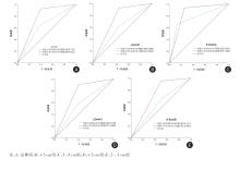
Fig.1
ROC of LR-5 for diagnosing HCC before and after LI-RADS modified classification in groups with different diameters"
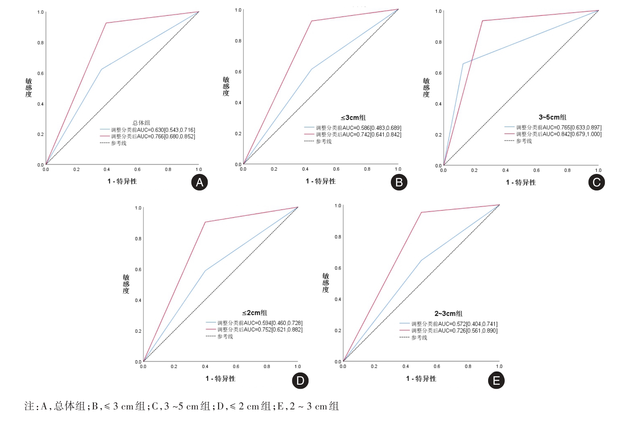
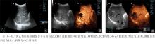
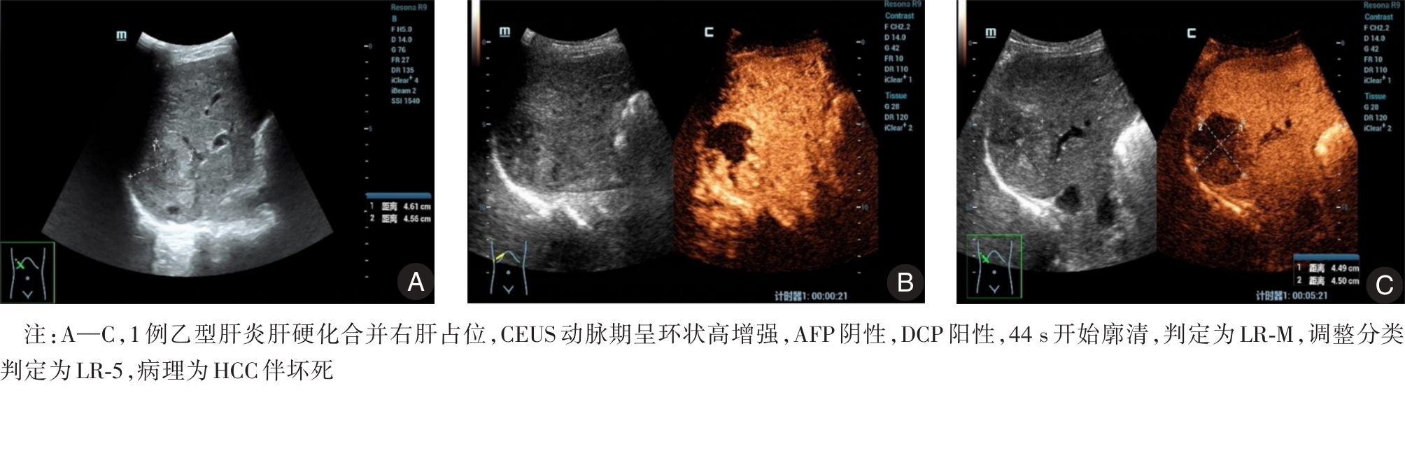
Fig.2
Typical case"
| [1] |
HAN B, ZHENG R, ZENG H, et al. Cancer incidence and mortality in China, 2022[J]. J Natl Cancer Cent,2024,4(1):47-53.doi:10.1016/j.jncc.2024.01.006 .
doi: 10.1016/j.jncc.2024.01.006 |
| [2] |
DONG Y, MAO F, CAO J, et al. Characterization of Focal Liver Lesions Indistinctive on B Mode Ultrasound: Benefits of Contrast-Enhanced Ultrasound[J]. Biomed Res Int,2017,2017:8970156.doi:10.1155/2017/8970156 .
doi: 10.1155/2017/8970156 |
| [3] |
中华人民共和国国家卫生健康委员会医政司. 原发性肝癌诊疗指南(2024年版)[J]. 协和医学杂志,2024,15(3): 532-559.doi:10.12290/xhyxzz.2024-0304 .
doi: 10.12290/xhyxzz.2024-0304 |
| [4] |
BARTOLOTTA T V, TERRANOVA M C, GAGLIARDO C,et al. CEUS LI-RADS: A pictorial review[J]. Insights Imaging,2020,11(1):9.doi:10.1186/s13244-019-0819-2 .
doi: 10.1186/s13244-019-0819-2 |
| [5] |
MAKOYEVA A, KIM T K, JANG H J, et al. Use of CEUS LI-RADS for the Accurate Diagnosis of Nodules in Patients at Risk for Hepatocellular Carcinoma: A Validation Study[J]. Radiol Imaging Cancer,2020,2(2):e190014.doi:10.1148/rycan.2020190014 .
doi: 10.1148/rycan.2020190014 |
| [6] |
WILDNER D, BERNATIK T, GREIS C, et al. CEUS in hepatocellular carcinoma and intrahepatic cholangiocellular carcinoma in 320 patients - early or late washout matters: A subanalysis of the DEGUM multicenter trial[J]. Ultraschall Med,2015,36(2):132-139.doi:10.1055/s-0034-1399147 .
doi: 10.1055/s-0034-1399147 |
| [7] |
LI F, LI Q, LIU Y, et al. Distinguishing intrahepatic cholangiocarcinoma from hepatocellular carcinoma in patients with and without risks: The evaluation of the LR-M criteria of contrast-enhanced ultrasound liver imaging reporting and data system version 2017[J]. Eur Radiol,2020,30(1):461-470. doi:10.1007/s00330-019-06317-2 .
doi: 10.1007/s00330-019-06317-2 |
| [8] |
赫捷, 陈万青, 沈洪兵, 等. 中国人群肝癌筛查指南(2022,北京)[J]. 中华消化外科杂志,2022,21(8):971-996.doi:10.3760/cma.j.cn115610-20220805-00448 .
doi: 10.3760/cma.j.cn115610-20220805-00448 |
| [9] |
朱嫦琳, 陈展泽, 李启欣. 基于决策曲线分析评估血清异常凝血酶原和甲胎蛋白在原发性肝癌中的诊断价值[J]. 实用医学杂志,2021,37(19):2524-2529.doi:10.3969/j.issn.1006-5725.2021.19.018 .
doi: 10.3969/j.issn.1006-5725.2021.19.018 |
| [10] |
KONO Y, LYSHCHIK A, COSGROVE D, et al. Contrast Enhanced Ultrasound (CEUS) Liver Imaging Reporting and Data System (LI-RADS®): the official version by the American College of Radiology (ACR)[J]. Ultraschall Med,2017,38(1):85-86. doi:10.1055/s-0042-124369 .
doi: 10.1055/s-0042-124369 |
| [11] |
ZENG D, XU M, LIANG J Y, et al. Using new criteria to improve the differentiation between HCC and non-HCC malignancies: Clinical practice and discussion in CEUS LI-RADS 2017[J]. Radiol Med, 2022,127(1):1-10. doi:10.1007/s11547-021-01417-w .
doi: 10.1007/s11547-021-01417-w |
| [12] |
WEN R, HUANG F, LIN P, et al. Performance of current versus modified CEUS LI-RADS in the diagnosis of non-hepatocellular carcinoma malignancies[J]. Abdom Radiol (NY), 2023,48(12):3688-3695. doi:10.1007/s00261-023-04043-4 .
doi: 10.1007/s00261-023-04043-4 |
| [13] |
WEN R, PENG Y, LIANG Y, et al. CEUS LI-RADS in Combination With the Serum Biomarker-Based ASAP Model Improves the Diagnostic Performance of HCC in High-Risk Patients[J]. Ultrasound Med Biol,2024,50(11):1739-1744. doi:10.1016/j.ultrasmedbio.2024.08.003 .
doi: 10.1016/j.ultrasmedbio.2024.08.003 |
| [14] |
LI C Q, HUANG H, RUAN S M, et al. An assessment of liver lesions using a combination of CEUS LI-RADS and AFP[J]. Abdom Radiol (NY),2022,47(4):1311-1320.doi:10.1007/s00261-022-03428-1 .
doi: 10.1007/s00261-022-03428-1 |
| [15] |
莫敏, 林静静, 卢鑫, 等. 超声造影参数联合血清CXCL9、IGFBP-3对肝癌介入治疗后复发的预测价值[J]. 实用医学杂志,2025,41(16):2568-2574.doi:10.3969/j.issn.1006-5725.2025.16.019 .
doi: 10.3969/j.issn.1006-5725.2025.16.019 |
| [16] |
HU X Y, SUN Y K, MIAO Y, et al. Preoperative identification of hepatocellular carcinoma from focal liver lesions ≤ 20 mm in high-risk patients using clinical and contrast-enhanced ultrasound features[J]. Eur J Radiol,2025,187:112076.doi:10.1016/j.ejrad.2025.112076 .
doi: 10.1016/j.ejrad.2025.112076 |
| [17] |
李加伍, 凌文武, 陈爽, 等. 肝脏病灶大小及肝细胞癌分化程度对超声造影肝脏影像报告与数据管理系统分类诊断的影响 [J]. 中国医学影像技术,2022,38(9):1356-1360.doi:10.13929/j.issn.1003-3289.2022.09.017 .
doi: 10.13929/j.issn.1003-3289.2022.09.017 |
| [18] |
奚静, 顾美琴, 包作伟. 基于超声造影LI-RADS特征的肝细胞癌微血管侵犯列线图模型的构建及验证[J]. 临床肝胆病杂志,2022,38(11):2520-2525.doi:10.3969/j.issn.1001-5256.2022.11.016 .
doi: 10.3969/j.issn.1001-5256.2022.11.016 |
| [19] |
冒玉香, 王珏, 葛舒. 超声造影LI-RADS鉴别诊断最大径≤3 cm肝脏局灶性病变的临床价值 [J]. 临床超声医学杂志,2023,25(10):824-828.doi:10.3969/j.issn.1008-6978.2023.10.015 .
doi: 10.3969/j.issn.1008-6978.2023.10.015 |
| [20] |
LIN W, HE N, ZENG Q, et al. Efficacy of Multiple Modified Methods of Criteria for LR-M Liver Nodules of Different Sizes: Clinical Practice and Discussion in CEUS LI-RADS Version 2017[J]. J Ultrasound Med,2023,42(12):2739-2748.doi:10.1002/jum.16308 .
doi: 10.1002/jum.16308 |
| Viewed | ||||||
|
Full text |
|
|||||
|
Abstract |
|
|||||