

The Journal of Practical Medicine ›› 2026, Vol. 42 ›› Issue (1): 29-36.doi: 10.3969/j.issn.1006-5725.2026.01.004
• Oncology: Diagnosis, Treatment and Prevention • Previous Articles Next Articles
Meixia YIN1,Jie TANG1( ),Jing ZHAO2,Bin YANG3
Received:2025-09-03
Online:2026-01-10
Published:2026-01-14
Contact:
Jie TANG
E-mail:tjiehone23@163.com
CLC Number:
Meixia YIN,Jie TANG,Jing ZHAO,Bin YANG. Clinical application value of combined detection of uterine artery hemodynamic parameters and elastography score in uterine fibroid degeneration[J]. The Journal of Practical Medicine, 2026, 42(1): 29-36.
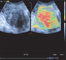
Fig.1
Stress elastography image of a patient with non-degenerated uterine fundus myoma"

Tab.1
Comparison of uterine artery hemodynamic parameters and elastography scores between the two groups of subjects"
| 组别 | RI | PI | Vmin/(cm/s) | EDV/(cm/s) | PSV/(cm/s) | 弹性成像评分/分 |
|---|---|---|---|---|---|---|
| 研究组(n = 158) | 0.65 ± 0.12 | 1.58 ± 0.26 | 15.46 ± 2.84 | 8.79 ± 2.17 | 42.56 ± 4.26 | 4.08 ± 0.62 |
| 健康对照组(n = 150) | 0.94 ± 0.21 | 2.13 ± 0.30 | 18.86 ± 3.43 | 13.63 ± 2.08 | 47.84 ± 5.16 | 1.19 ± 0.28 |
| t值 | 14.974 | 17.219 | 9.495 | 19.964 | 9.813 | 52.251 |
| P值 | 0.000 | 0.000 | 0.000 | 0.000 | 0.000 | 0.000 |
Tab.2
Comparison of clinical data, uterine artery hemodynamic parameters, and elastography scores among subgroup patients in the study group"
| 临床资料 | 非变性组(n = 127 ) | 变性组(n = 31) | t值 | P值 |
|---|---|---|---|---|
| 年龄/岁 | 36.12 ± 10.24 | 35.32 ± 10.17 | 0.390 | 0.697 |
| BMI/(kg/m2) | 22.68 ± 1.42 | 23.16 ± 1.33 | 1.708 | 0.090 |
| 产次/次 | 1.26 ± 0.35 | 1.35 ± 0.28 | 1.330 | 0.185 |
| RI | 0.61 ± 0.13 | 0.65 ± 0.12 | 1.637 | 0.104 |
| PI | 1.45 ± 0.18 | 1.62 ± 0.26 | 3.441 | 0.001 |
| Vmin/(cm/s) | 15.71 ± 2.33 | 14.13 ± 2.15 | 3.434 | 0.001 |
| EDV/(cm/s) | 9.11 ± 2.44 | 8.36 ± 2.12 | 1.572 | 0.118 |
| PSV/(cm/s) | 43.41 ± 4.74 | 41.25 ± 4.15 | 2.328 | 0.021 |
| 弹性成像评分/分 | 4.39 ± 0.50 | 3.98 ± 0.60 | 3.516 | 0.001 |
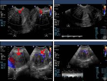
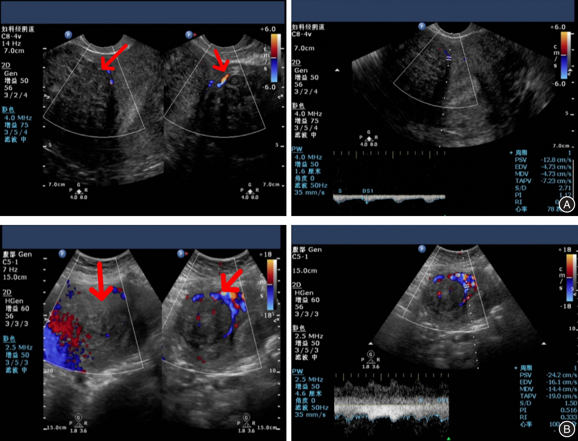
Fig.2
Ultrasonic manifestations of typical cases"
Tab.3
Elastography scores of patients with uterine myoma degeneration"
| 类型 | 例数 | 弹性成像评分/分 |
|---|---|---|
| 玻璃样变性 | 15 | 4.73 ± 0.46 |
| 部分囊性病变 | 10 | 2.20 ± 0.42 |
| 脂肪变性 | 5 | 2.80 ± 0.45 |
| 肉瘤样变性 | 1 | 4 |
Tab.4
Variable assignment"
| 因素 | 变量 | 赋值 |
|---|---|---|
| 子宫肌瘤变性 | Y | 原值代入 |
| PI | X1 | 原值代入 |
| Vmin | X2 | 原值代入 |
| PSV | X3 | 原值代入 |
| 弹性成像评分 | X4 | 玻璃样变性= 1;部分囊性病变= 2;脂肪变性= 3;肉瘤样变性= 4 |
Tab.5
Logistic regression analysis for the diagnosis of uterine myoma degeneration"
| 自变量 | 回归系数 | S.E. | Wald值 | P值 | OR | 95% CI |
|---|---|---|---|---|---|---|
| X1:PI | 4.043 | 1.187 | 11.608 | 0.001 | 57.009 | 5.569 ~ 583.570 |
| X2:Vmin | -0.293 | 0.108 | 7.411 | 0.006 | 0.746 | 0.604 ~ 0.921 |
| X3:PSV | -0.135 | 0.01 | 4.926 | 0.026 | 0.873 | 0.775 ~ 0.984 |
| X4:弹性成像评分 | 1.196 | 0.425 | 7.911 | 0.005 | 3.308 | 1.437 ~ 7.612 |
| 常数 | -2.580 | 4.021 | 0.412 | 0.521 | 0.076 |
Tab.6
Predictive value of uterine artery hemodynamic parameters and stress elastography for degeneration of uterine myoma"
| 检测变量 | AUC | 标准误 | 临界值 | 灵敏度/% | 特异度/% | P值 | 95%CI |
|---|---|---|---|---|---|---|---|
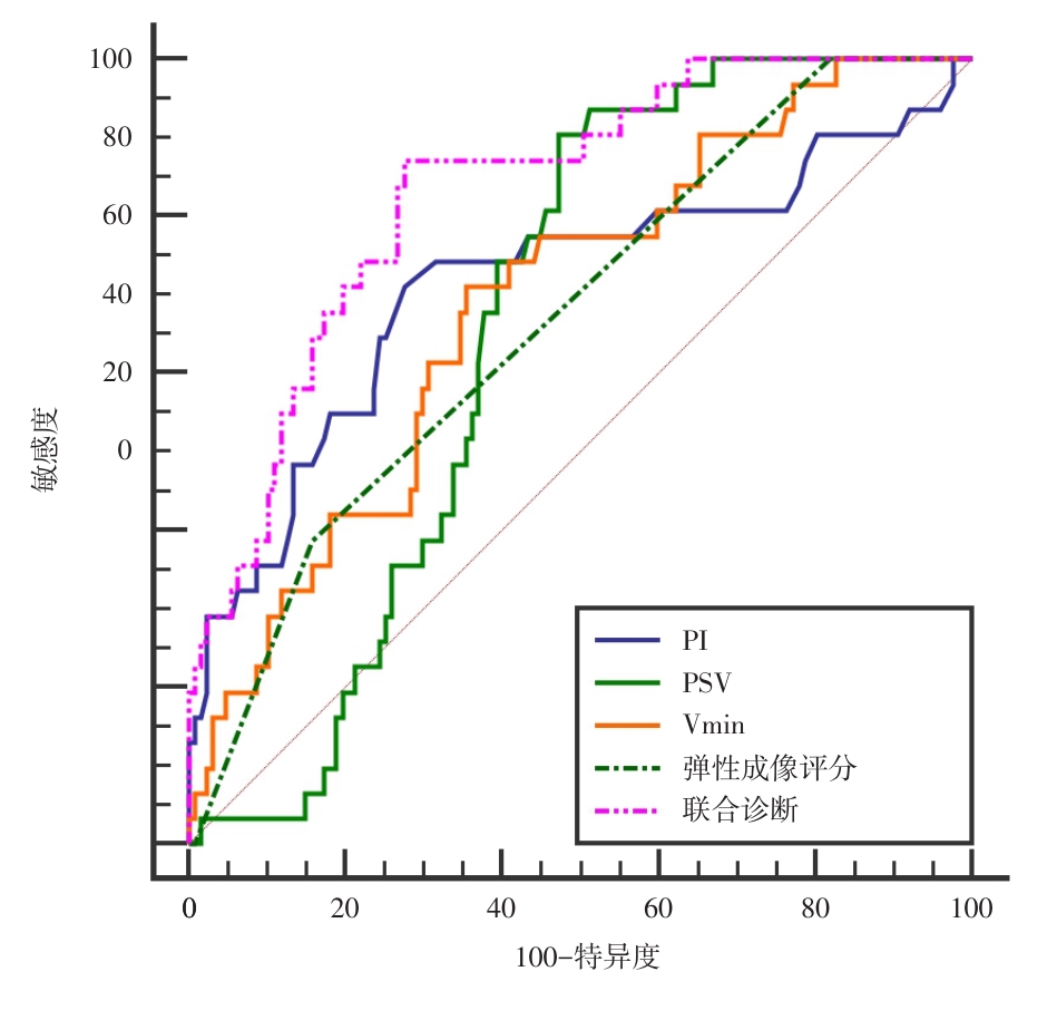
| PI | 0.714 | 0.0603 | 1.53 | 71.00 | 72.40 | 0.000 | 0.637 ~ 0.783 |
| Vmin | 0.690 | 0.0511 | 15.25 cm/s | 70.97 | 64.57 | 0.000 | 0.611 ~ 0.761 |
| PSV | 0.660 | 0.0425 | 43.35 cm/s | 90.32 | 52.76 | 0.000 | 0.581 ~ 0.734 |
| 弹性成像评分 | 0.669 | 0.0419 | 4 | 38.71 | 84.25 | 0.000 | 0.590 ~ 0.742 |
| 联合预测 | 0.832 | 0.0381 | 0.17 | 87.10 | 72.44 | 0.000 | 0.764 ~ 0.887 |
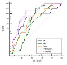
Fig.3
ROC curves for predicting degeneration of uterine myoma using uterine artery hemodynamic parametersand stress elastography"
| [1] |
汪海金,王克功,龚廷,等. 腹腔镜引导下双侧子宫骶韧带区域神经阻滞在妇科腔镜子宫肌瘤剔除手术中的应用[J]. 实用医学杂志,2023,39(9):1127-1131.doi:10.3969/j.issn.1006-5725.2023.09.011 .
doi: 10.3969/j.issn.1006-5725.2023.09.011 |
| [2] |
韦玮,方梓羽,马艳群,等. 经皮神经电刺激联合加速康复外科多模式镇痛在腹腔镜全子宫切除术后的镇痛效果[J]. 实用医学杂志,2022,38(10):1251-1254.doi:10.3969/j.issn.1006-5725.2022.10.015 .
doi: 10.3969/j.issn.1006-5725.2022.10.015 |
| [3] | 周蓉. 子宫肌瘤对生育力的影响及其围手术期保护[J]. 实用妇产科杂志, 2024, 40(2):84-87. |
| [4] |
SOTOMAYOR C G, PARRA C, MIRANDA M, et al. Hyaline and Cystic Degeneration of Uterine Leiomyomas: CT and MR Imaging with Histopathological Sample Analyses[J]. Diagnostics (Basel),2023,13(20):3230.doi: 10.3390/diagnostics13203230 .
doi: 10.3390/diagnostics13203230 |
| [5] |
DUNPHY L, GEE M, FORD J. Red degeneration of fibroid presenting with abdominal pain in pregnancy[J]. BMJ Case Rep, 2025,18(8):e263747. doi: 10.1136/bcr-2024-263747 .
doi: 10.1136/bcr-2024-263747 |
| [6] |
JIANG S, XU Z, SHAO X, et al. Diagnostic value and clinical significance of serum miR-134-5p combined with uterine artery color Doppler ultrasound parameters in endometriosis[J]. BMC Womens Health, 2024,24(1):544.doi: 10.1186/s12905-024-03372-w .
doi: 10.1186/s12905-024-03372-w |
| [7] |
WU J, LI Y, ZHANG X, et al. Assessment of blood flow around the knee joint in patients with knee osteoarthritis by color Doppler ultrasound[J]. Eur J Radiol, 2023, 166:111005.doi: 10.1016/j.ejrad.2023.111005 .
doi: 10.1016/j.ejrad.2023.111005 |
| [8] |
HARAKE J EL, SAYSENG V, GRONDIN J, et al. Preliminary Feasibility of Stress Myocardial Elastography for the Detection of Coronary Artery Disease[J]. Ultrasound Med Biol, 2023,49(2):549-559.doi: 10.1016/j.ultrasmedbio.2022.10.007 .
doi: 10.1016/j.ultrasmedbio.2022.10.007 |
| [9] |
JIA W, LUO T, DONG Y, et al. Breast Elasticity Imaging Techniques: Comparison of Strain Elastography and Shear-Wave Elastography in the Same Population[J]. Ultrasound Med Biol, 2021,47(1):104-113. doi: 10.1016/j.ultrasmedbio.2020.09.022 .
doi: 10.1016/j.ultrasmedbio.2020.09.022 |
| [10] |
MILLER B S, SINGH S S, FLAXMAN T E. The effect of uterine fibroid region and depth on endometrial stress and strain: a finite element approach[J]. Comput Methods Biomech Biomed Engin, 2024,6:1-10. doi: 10.1080/10255842.2024.2431653 .
doi: 10.1080/10255842.2024.2431653 |
| [11] | 孔北华,马丁,段涛. 妇产科学[M]. 10版.北京:人民卫生出版社,2024:316-320. |
| [12] |
田士峰,刘爱连,牛淼,等. 扩散峰度成像定量参数直方图分析鉴别子宫癌肉瘤与变性子宫肌瘤的价值[J]. 中国临床医学影像杂志,2020,31(4):281-284. doi:10.12117/jccmi. 2020. 04.012 .
doi: 10.12117/jccmi. 2020. 04.012 |
| [13] |
蒋晓敏,纪瑞云,周宇佳.影响子宫肌瘤剔除术后妊娠及其结局的因素[J].实用医学杂志,2020,36(24):3385-3389.doi:10.3969/j.issn.1006-5725.2020.24.016 .
doi: 10.3969/j.issn.1006-5725.2020.24.016 |
| [14] |
VANNUCCINI S, PETRAGLIA F, CARMONA F, et al. The modern management of uterine fibroids-related abnormal uterine bleeding[J]. Fertil Steril, 2024,122(1):20-30.doi:10.1016/j.fertnstert.2024.04.041 .
doi: 10.1016/j.fertnstert.2024.04.041 |
| [15] |
DOLMANS M M, PETRAGLIA F, CATHERINO W H, et al. Pathogenesis of uterine fibroids: Current understanding and future directions[J]. Fertil Steril, 2024,122(1):6-11.doi: 10.1016/j.fertnstert.2024.02.048 .
doi: 10.1016/j.fertnstert.2024.02.048 |
| [16] |
杨红玉,孙莉,杨波,等. 18~25岁子宫肌瘤临床及病理特征分析[J]. 临床误诊误治,2020,33(8):80-84. doi:10.3969/j.issn.1002-3429 .
doi: 10.3969/j.issn.1002-3429 |
| [17] |
张小敬,方群,刘赵一,等. 彩色多普勒成像联合子宫动脉血流参数诊断子宫肌瘤效果[J]. 中国计划生育学杂志, 2024, 32(3):643-646. doi:10.3969/j.issn.1004-8189.2024.03.033 .
doi: 10.3969/j.issn.1004-8189.2024.03.033 |
| [18] |
PIEROH P, LI ZL, KAWATA S,et al. The arterial blood supply of the symphysis pubis-Spatial orientated and highly variable[J]. Ann Anat, 2021,234:151649. doi: 10.1016/j.aanat.2020.151649 .
doi: 10.1016/j.aanat.2020.151649 |
| [19] |
张逸群,张春莲,薛璐. 不同磁共振T2WI信号对子宫肌瘤患者术前超声造影血流灌注的评估价值[J]. 中国计划生育学杂志,2020,28(8):1274-1277. doi:10.3969/j.issn.1004-8189.2020.08.031 .
doi: 10.3969/j.issn.1004-8189.2020.08.031 |
| [20] |
PENG J, WANG J, SHU Q, et al. Systematic review and meta-analysis of current evidence in uterine artery embolization vs myomectomy for symptomatic uterine fibroids[J].Sci Rep, 2024,14(1):19252.doi:10.1038/s41598-024-69754-0 .
doi: 10.1038/s41598-024-69754-0 |
| [21] |
SOTOMAYOR C G, PARRA C, MIRANDA M, et al. Hyaline and Cystic Degeneration of Uterine Leiomyomas: CT and MR Imaging with Histopathological Sample Analyses[J]. Diagnostics (Basel), 2023,13(20):3230.doi: 10.3390/diagnostics13203230 .
doi: 10.3390/diagnostics13203230 |
| [22] |
TAKEUCHI M, MATSUZAKI K, BANDO Y, et al. Evaluation of red degeneration of uterine leiomyoma with susceptibility-weighted MR imaging[J]. Mag Resonance Med Sci,2019,18(2):158-162.doi: 10.2463/mrms.mp.2018-0074 .
doi: 10.2463/mrms.mp.2018-0074 |
| [23] |
WEN S, PENG B, WEI X,et al. Convolutional Neural Network-Based Speckle Tracking for Ultrasound Strain Elastography: An Unsupervised Learning Approach[J]. IEEE Trans Ultrason Ferroelectr Freq Control, 2023,70(5):354-367. doi:10.1109/TUFFC.2023.3243539 .
doi: 10.1109/TUFFC.2023.3243539 |
| [24] |
THOMSEN C R, JENSEN M S S, BOR P, et al. Recommendations for strain elastography of the uterine cervix[J].Arch Gynecol Obstet, 2024,310(4):2023-2033. doi: 10.1007/s00404-024-07693-x .
doi: 10.1007/s00404-024-07693-x |
| [25] |
张菁菁,徐海飞,李统怀,等. 声辐射力脉冲成像联合应力式弹性成像诊断子宫肌瘤变性的应用价值[J]. 临床超声医学杂志,2020,22(11):843-846. doi: 10.3969/j.issn.1008-6978.2020.11.013 .
doi: 10.3969/j.issn.1008-6978.2020.11.013 |
| [26] |
YAMASHITA Y, YAMAZAKI H, SHIMOKAWA T, et al. Shear-wave versus strain elastography in endoscopic ultrasound for the diagnosis of chronic pancreatitis[J]. Pancreatol, 2023,23(1):35-41.doi:10.1016/j.pan.2022.11.009 .
doi: 10.1016/j.pan.2022.11.009 |
| Viewed | ||||||
|
Full text |
|
|||||
|
Abstract |
|
|||||