

The Journal of Practical Medicine ›› 2025, Vol. 41 ›› Issue (16): 2581-2589.doi: 10.3969/j.issn.1006-5725.2025.16.021
• Medical Examination and Clinical Diagnosis • Previous Articles
Zelin XU1,Zhenhao ZHENG1,Yaqian DENG1,Guanming ZENG2,Tingting DU1,Peishan ZHU1,Wen LIU1,Jun. LI1,3( )
)
Received:2025-05-26
Online:2025-08-25
Published:2025-08-28
Contact:
Jun. LI
E-mail:1287424798@qq.com
CLC Number:
Zelin XU,Zhenhao ZHENG,Yaqian DENG,Guanming ZENG,Tingting DU,Peishan ZHU,Wen LIU,Jun. LI. Predictive value of dual‑modality ultrasound combined with S‑Detect for cervical lymph node metastasis in papillary thyroid carcinoma[J]. The Journal of Practical Medicine, 2025, 41(16): 2581-2589.
Tab.1
Comparison of conventional ultrasound, ultrasound elastography, and S-Detect characteristic parameters between meta static and non-metastatic PTC patients"
| 指标 | 未转移组 (n=84) | 转移组 (n=51) | Z值 | P值 |
|---|---|---|---|---|
| 年龄 | 6.619 | 0.006 | ||
| ≥ 55 岁 | 58(69) | 23 (45) | ||
| < 55 岁 | 26(31) | 28 (55) | ||
| 性别 | 0.474 | 0.385 | ||
| 女 | 48(57.1) | 33(64.7) | ||
| 男 | 36(42.9) | 18(35.3) | ||
| 体质量[M(P25,P75)]/kg | 68.0(62.75,73.25) | 67.0(61.5,72.0) | 89.945 | 0.700 |
| 身高[M(P25,P75)]/cm | 166.0(160.0,170.0) | 166.0(159.0,171.5) | 2 074.000 | 0.632 |
| ECI[M(P25,P75)] | 1.25(0.84,1.93) | 1.62(1.13,2.53) | 1 666.000 | 0.023 |
| ES-ECI | 23.691 | 0.001 | ||
| 1 | 50(59.5) | 11(21.6) | ||
| 2 | 28(33.3) | 26(51.0) | ||
| 3 | 4(4.8) | 4(7.8) | ||
| 4 | 2(2.4) | 10(19.6) | ||
| 弹性硬度 | 8.824 | 0.003 | ||
| 软 | 78(92.9) | 37(72.5) | ||
| 硬 | 6(7.1) | 1(27.5) | ||
| 包膜侵犯 | 1(1.2) | 9(17.6) | 12.530 | 0.007 |
| 最大径 | 9.039 | 0.002 | ||
| < 1 cm | 58(69.0) | 21(41.2) | 0.002 | |
| ≥ 1 cm | 26(31.0) | 30(58.8) | ||
| 回声 | 3.812 | 0.987 | ||
| 等回声/高回声 | 6(7.1) | 0(0.0) | ||
| 低回声 | 78(92.9) | 51(100.0) | ||
| 纵横比 | 1.351 | 0.177 | ||
| < 1 | 22(26.2) | 19(37.3) | ||
| ≥ 1 | 62(73.8) | 32(62.7) | ||
| 边界 | 3.961 | 0.031 | ||
| 清晰 | 60(71.4) | 27(52.9) | ||
| 不清晰 | 24(28.6) | 24(47.1) | ||
| 边缘 | 0.837 | 0.365 | ||
| 光滑 | 78(92.9) | 45(88.2) | ||
| 粗糙 | 6(7.1) | 6(11.8) | ||
| 钙化 | 1.179 | 0.199 | ||
| 无/大钙化 | 66(78.6) | 35(68.6) | ||
| 微钙化 | 18(21.4) | 16(31.4) | ||
| 成分 | 1.191 | 0.299 | ||
| 囊性/囊实性 | 5(6.0) | 1(2.0) | ||
| 实性 | 79(94.0) | 50(98.0) |
Tab.2
Comparison of S-Detect characteristic parameters between metastatic and non-metastatic PTC patients 例(%)"
| 指标 | 横切面 | 纵切面 | ||||||
|---|---|---|---|---|---|---|---|---|
未转移组 (n=84) | 转移组 (n=51) | Z值 | P值 | 未转移组 (n=84) | 转移组 (n=51) | Z值 | P值 | |
| 识别结果 | 0.300 | 0.585 | 0.300 | 0.585 | ||||
| 可能良性 | 6(7.1) | 5(9.8) | 5 (6.0) | 3(5.9) | ||||
| 可能恶性 | 78(92.9) | 46(90.2) | 79(94.0) | 48(94.1) | ||||
| 成分 | 0.686 | 0.412 | 1.956 | 0.179 | ||||
| 部分囊性 | 5(6.0) | 5(9.8) | 9 (10.7) | 2 (3.9) | ||||
| 实性 | 79(94.0) | 46(90.2) | 75(89.3) | 49 (96.1) | ||||
| 回声强度 | 0.182 | 0.670 | 0.266 | 0.608 | ||||
| 等回声/高回声 | 5(6.0) | 4(7.8) | 5(6.0) | 2 (3.9) | ||||
| 轻度低回声 | 79(94.0) | 47(92.2) | 79(94.0) | 49(96.1) | ||||
| 方向 | 0.174 | 0.552 | 0.003 | 0.959 | ||||
| 平行 | 45(53.6) | 30(58.8) | 58(69.0) | 35(68.6) | ||||
| 不平行 | 39(46.4) | 21(41.2) | 26(31.0) | 16(31.4) | ||||
| 形状 | 7.162 | 0.005 | 1.008 | 0.231 | ||||
| 圆形到椭圆形 | 71(84.5) | 32(62.7) | 64(76.2) | 34(66.7) | ||||
| 不规则 | 13(15.5) | 19(37.3) | 20(23.8) | 17(33.3) | ||||
| 钙化 | 4.828 | 0.019 | 1.964 | 0.116 | ||||
| 无/大钙化 | 49(58.3) | 19(37.3) | 48(57.1) | 22(43.1) | ||||
| 微钙化 | 35(41.7) | 32(62.7) | 36(42.9) | 29(56.9) | ||||
| 边缘 | 1.950 | 0.108 | 0.008 | 0.741 | ||||
| 光滑 | 12(14.3) | 13(25.5) | 13(15.5) | 9(17.6) | ||||
| 针状/浅分叶状/不明确 | 72(85.7) | 38(74.5) | 71(84.5) | 42(82.4) | ||||
Tab.3
Multivariate logistic regression analysis of risk parameters for CLNM in PTC"
| 变量 | B | SE | Z值 | Wald χ2 | P值 | OR | 95%CI |
|---|---|---|---|---|---|---|---|
| 形状-SD横切面 | 1.817 | 0.581 | 3.127 | 9.779 | 0.002 | 6.151 | 1.970 ~ 19.203 |
| 最大径 | 1.103 | 0.522 | 2.114 | 4.470 | 0.035 | 3.013 | 1.084 ~ 8.374 |
| 包膜外侵犯 | 4.469 | 1.330 | 3.360 | 11.288 | 0.001 | 87.293 | 6.439 ~ 1 183.368 |
| ES-ECI | 1.331 | 0.542 | 2.455 | 6.026 | 0.014 | 3.785 | 1.308 ~ 10.958 |
| ECI | -0.228 | 0.305 | -0.746 | 0.556 | 0.456 | 0.796 | 0.438 ~ 1.449 |
| 钙化-SD横切面 | 0.912 | 0.536 | 1.703 | 2.900 | 0.089 | 2.490 | 0.871 ~ 7.116 |
| 边界-常规超声 | 1.224 | 0.528 | 2.317 | 5.370 | 0.020 | 3.402 | 1.208 ~ 9.585 |
| 年龄 | 1.596 | 0.534 | 2.988 | 8.926 | 0.003 | 4.935 | 1.732 ~ 14.065 |
| 弹性硬度 | 0.310 | 1.309 | 0.237 | 0.056 | 0.813 | 1.363 | 0.105 ~ 17.727 |
Tab.4
comparison of performance of different diagnostic models in predicting CLNM in PTC patients"
| 模型 | AUC(95%CI) | SEN | SPE | PPV | NPV | ACC | P值 | Z值 |
|---|---|---|---|---|---|---|---|---|
| 常规超声模型 | 0.813(0.742 ~ 0.884) | 0.706 | 0.774 | 0.655 | 0.812 | 0.748 | 0.008 | 2.645 |
| S-Detect模型 | 0.609(0.531 ~ 0.686) | 0.373 | 0.845 | 0.594 | 0.689 | 0.667 | < 0.001 | 6.728 |
| 弹性超声模型 | 0.721(0.639 ~ 0.803) | 0.784 | 0.595 | 0.541 | 0.820 | 0.667 | < 0.001 | 4.256 |
| 联合预测模型 | 0.890(0.835 ~ 0.945) | 0.824 | 0.833 | 0.750 | 0.886 | 0.830 | / | / |
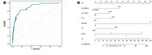
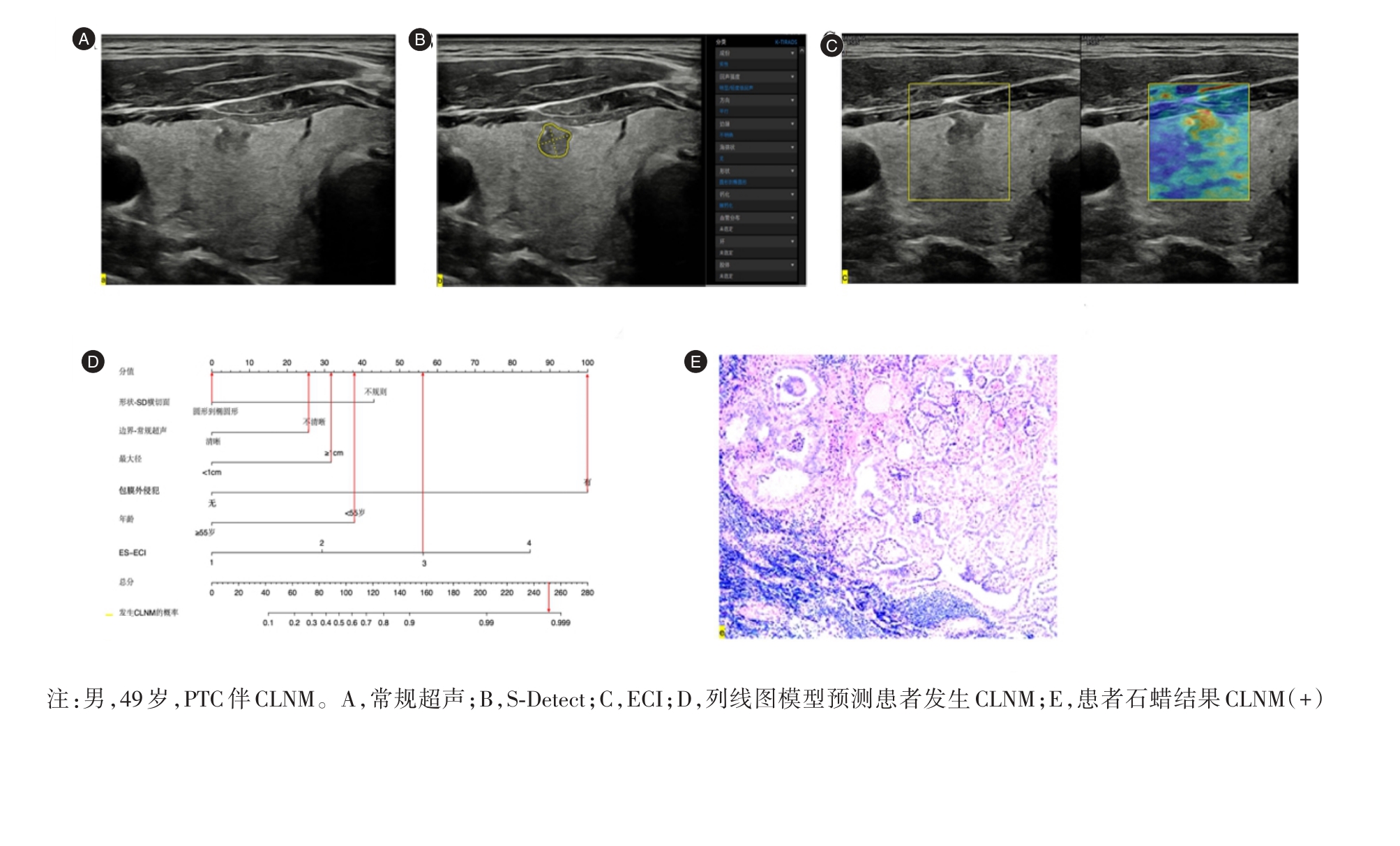
Fig.1
ROC curves and nomogram for CLNM prediction in PTC patients"
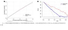
Fig.2
Calibration curves and decision curves of the nomogram prediction model for CLNM in PTC patients"
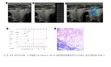
Fig.3
Clinical application cases"
| [1] |
MIRANDA-FILHO A, LORTET-TIEULENT J, BRAY F, et al. Thyroid cancer incidence trends by histology in 25 countries: A population-based study[J]. Lancet Diabetes Endocrinol, 2021, 9(4): 225-234. doi:10.1016/s2213-8587(21)00027-9
doi: 10.1016/s2213-8587(21)00027-9 |
| [2] |
CAO M, LI H, SUN D, et al. Cancer burden of major cancers in China: A need for sustainable actions[J]. Cancer Commun, 2020, 40(5): 205-210. doi:10.1002/cac2.12025
doi: 10.1002/cac2.12025 |
| [3] |
SIEGEL R L, MILLER K D, WAGLE N S, et al. Cancer statistics, 2023[J]. CA Cancer J Clin, 2023, 73(1): 17-48. doi:10.3322/caac.21763
doi: 10.3322/caac.21763 |
| [4] |
ALABOUSI M, ALABOUSI A, ADHAM S, et al. Diagnostic Test Accuracy of Ultrasonography vs Computed Tomography for Papillary Thyroid Cancer Cervical Lymph Node Metastasis: A Systematic Review and Meta-analysis[J]. JAMA Otolaryngol Head Neck Surg, 2022, 148(2): 107-116. doi:10.1001/jamaoto.2021.3387
doi: 10.1001/jamaoto.2021.3387 |
| [5] |
MONPEYSSEN H, TRAMALLONI J, POIRÉE S, et al. Elastography of the thyroid[J]. Diagn Interv Imaging, 2013, 94(5): 535-544. doi:10.1016/j.diii.2013.01.023
doi: 10.1016/j.diii.2013.01.023 |
| [6] |
HU X, LIU Y, QIAN L. Diagnostic potential of real-time elastography (RTE) and shear wave elastography (SWE) to differentiate benign and malignant thyroid nodules: A systematic review and meta-analysis[J]. Medicine (Baltimore), 2017, 96(43): e8282. doi:10.1097/md.0000000000008282
doi: 10.1097/md.0000000000008282 |
| [7] |
CHOI W J, PARK J S, KOO H R, et al. Ultrasound elastography using carotid artery pulsation in the differential diagnosis of sonographically indeterminate thyroid nodules[J]. AJR Am J Roentgenol, 2015, 204(2): 396-401. doi:10.2214/ajr.14.12871
doi: 10.2214/ajr.14.12871 |
| [8] |
CONG P, WANG X M, ZHANG Y F. Comparison of artificial intelligence, elastic imaging, and the thyroid imaging reporting and data system in the differential diagnosis of suspicious nodules[J]. Quant Imaging Med Surg, 2024, 14(1): 711-721. doi:10.21037/qims-23-788
doi: 10.21037/qims-23-788 |
| [9] | 付泽辉, 陈卉, 李梦园, 等. S-Detect技术联合C-TIRADS对超声表现为滤泡性肿瘤样甲状腺结节的诊断价值[J]. 中国超声医学杂志, 2023, 39(12): 1321-1323. |
| [10] |
SANT V R, RADHACHANDRAN A, IVEZIC V, et al. From Bench-to-Bedside: How Artificial Intelligence is Changing Thyroid Nodule Diagnostics, a Systematic Review[J]. J Clin Endocrinol Metab, 2024, 109(8): dgae277. doi:10.1210/clinem/dgae277
doi: 10.1210/clinem/dgae277 |
| [11] | 张海琳, 朱梅, 杨阳, 等. 超声引导FNA及FNA-Tg识别甲状腺乳头状癌淋巴结转移的临床价值[J]. 昆明医科大学学报, 2022, 43(12): 105-110. |
| [12] |
RILEY R D, ENSOR J, SNELL K I E, et al. Calculating the sample size required for developing a clinical prediction model[J]. BMJ, 2020, 368: m441. doi:10.1136/bmj.m441
doi: 10.1136/bmj.m441 |
| [13] |
KWAK J Y, HAN K H, YOON J H, et al. Thyroid Imaging Reporting and Data System for US Features of Nodules: A Step in Establishing Better Stratification of Cancer Risk[J]. Radiology, 2011, 260(3): 892-899. doi:10.1148/radiol.11110206
doi: 10.1148/radiol.11110206 |
| [14] |
ASTERIA C, GIOVANARDI A, PIZZOCARO A, et al. US-elastography in the differential diagnosis of benign and malignant thyroid nodules[J]. Thyroid, 2008, 18(5): 523-531. doi:10.1089/thy.2007.0323
doi: 10.1089/thy.2007.0323 |
| [15] | 毛培, 郝真真, 吕会敏. 超声弹性成像联合S-Detect技术用于诊断TI-RADS4类甲状腺结节良恶性价值分析[J]. 医药论坛杂志, 2024, 45(3): 324-327. |
| [16] | 赵雯婷, 杨晓, 徐杰, 等. S-Detect联合SWE及ACR TI-RADS对甲状腺结节的诊断价值[J]. 中国医学影像学杂志, 2023, 31(9): 910-914. |
| [17] |
XU Y, WU D, WU W, et al. Diagnostic value of cytology, thyroglobulin, and combination of them in fine-needle aspiration of metastatic lymph nodes in patients with differentiated thyroid cancer: A systematic review and network meta-analysis[J]. Medicine (Baltimore), 2019, 98(45): e17859. doi:10.1097/md.0000000000017859
doi: 10.1097/md.0000000000017859 |
| [18] | 汪彩英, 张才智, 叶娟, 等. TI-RADS、UE、CEUS及FNAC在预测甲状腺乳头状癌颈部淋巴结转移中的意义[J]. 湖北科技学院学报(医学版), 2022, 36(4): 318-323. |
| [19] |
ZHANG F X, XU P, ZHANG L J, et al. RARγ promotes the invasion and metastasis of thyroid carcinoma by activating the JAK1-STAT3-CD24/MMPs axis[J]. Int Immunopharmacol, 2023, 125: 111129. doi:10.1016/j.intimp.2023.111129
doi: 10.1016/j.intimp.2023.111129 |
| [20] |
LIU L, WU B, CAI H, et al. Tiam1 promotes thyroid carcinoma metastasis by modulating EMT via wnt/β-catenin signaling[J]. Exp Cell Res, 2018, 362(2): 532-540. doi:10.1016/j.yexcr.2017.12.019
doi: 10.1016/j.yexcr.2017.12.019 |
| [21] | ZHOU J, YIN L, WEI X, et al. 2020 Chinese guidelines for ultrasound malignancy risk stratification of thyroid nodules: The C-TIRADS[J]. Endocrine, 2020, 70(2): 256-279. |
| [22] | HAUGEN B R, ALEXANDER E K, BIBLE K C, et al. 2015 american thyroid association management guidelines for adult patients with thyroid nodules and differentiated thyroid cancer: The american thyroid association guidelines task force on thyroid nodules and differentiated thyroid cancer[J]. Thyroid, 2016, 26(1): 1-133. |
| [23] |
TESSLER F N, MIDDLETON W D, GRANT E G, et al. ACR Thyroid Imaging, Reporting and Data System (TI-RADS): White Paper of the ACR TI-RADS Committee[J]. J Am Coll Radiol, 2017, 14(5): 587-595. doi:10.1016/j.jacr.2017.01.046
doi: 10.1016/j.jacr.2017.01.046 |
| [24] |
LI F, PAN D, HE Y, et al. Using ultrasound features and radiomics analysis to predict lymph node metastasis in patients with thyroid cancer[J]. BMC Surg, 2020, 20(1): 315. doi:10.1186/s12893-020-00974-7
doi: 10.1186/s12893-020-00974-7 |
| [25] | 陈璟泰, 侯令密, 唐云辉, 等. S-Detect对甲状腺结节良恶性鉴别诊断价值的Meta分析[J]. 中国全科医学, 2021, 24(30): 3814-3820. |
| [26] | 田菊, 张蕾, 勇强, 等. 常规超声联合S-Detect技术和超声造影评价甲状腺结节良恶性的价值[J]. 中国超声医学杂志, 2021, 37(9): 968-971. |
| [27] | 方明娣, 彭梅, 毕玉. 人工智能S-Detect技术结合钙化特征对甲状腺结节的诊断价值[J]. 中华医学超声杂志(电子版), 2021, 18(2): 177-181. |
| [28] |
HUANG X P, YE T T, ZHANG L, et al. Sonographic features of papillary thyroid microcarcinoma predicting high-volume central neck lymph node metastasis[J]. Surg Oncol, 2018, 27(2): 172-176. doi:10.1016/j.suronc.2018.03.004
doi: 10.1016/j.suronc.2018.03.004 |
| [29] |
JEON M J, CHUNG M S, KWON H, et al. Features of papillary thyroid microcarcinoma associated with lateral cervical lymph node metastasis[J]. Clin Endocrinol (Oxf), 2017, 86(6): 845-851. doi:10.1111/cen.13322
doi: 10.1111/cen.13322 |
| [30] |
JIANG W, WEI H Y, ZHANG H Y, et al. Value of contrast-enhanced ultrasound combined with elastography in evaluating cervical lymph node metastasis in papillary thyroid carcinoma[J]. World J Clin Cases, 2019, 7(1): 49-57. doi:10.12998/wjcc.v7.i1.49
doi: 10.12998/wjcc.v7.i1.49 |
| [31] |
HONG Y R, YAN C X, MO G Q, et al. Conventional US, elastography and contrast enhanced US features of papillary thyroid microcarcinoma predict central compartment lymph node metastases[J]. Sci Rep, 2015, 5: 7748. doi:10.1038/srep07748
doi: 10.1038/srep07748 |
| [32] |
XU J M, XU X H, XU H X, et al. Prediction of cervical lymph node metastasis in patients with papillary thyroid cancer using combined conventional ultrasound, strain elastography, and acoustic radiation force impulse (ARFI) elastography[J]. Eur Radiol, 2016, 26(8): 2611-2622. doi:10.1007/s00330-015-4088-2
doi: 10.1007/s00330-015-4088-2 |
| [33] |
WANG H, ZHAO L, XIN X, et al. Diagnostic value of elastosonography for thyroid microcarcinoma[J]. Ultrasonics, 2014, 54(7): 1945-1949. doi:10.1016/j.ultras.2014.04.027
doi: 10.1016/j.ultras.2014.04.027 |
| [34] |
MOON H J, KIM E K, YOON J H, et al. Clinical Implication of Elastography as a Prognostic Factor of Papillary Thyroid Microcarcinoma[J]. Ann Surg Oncol, 2012, 19(7): 2279-2287. doi:10.1245/s10434-011-2212-3
doi: 10.1245/s10434-011-2212-3 |
| [35] | 孙芳, 石岩, 杨智, 等. 剪切波联合二维超声对甲状腺乳头状癌中央区淋巴结转移的预测价值[J]. 实用医学杂志, 2021, 37(14): 1866-1871. |
| [36] | 李高峰, 石岩, 徐翠, 等. 超声弹性对比指数联合常规超声预测甲状腺乳头状癌淋巴结转移的研究[J]. 实用医学杂志, 2020, 36(5): 661-666. |
| [37] |
TANG Z, GAO L, WANG X, et al. Metastases to the thyroid gland: Ultrasonographic findings and diagnostic value of fine-needle aspiration cytology[J]. Front Oncol, 2022, 12: 939965. doi:10.3389/fonc.2022.939965
doi: 10.3389/fonc.2022.939965 |
| [38] |
ZHU Z, SU C, CHEN G, et al. Significance of FNAC, BRAF mutation, and intraoperative frozen section in surgical decision‐Making of thyroid nodules[J]. Diagn Cytopathol, 2023, 51(7): 441-448. doi:10.1002/dc.25135
doi: 10.1002/dc.25135 |
| [1] | Changhui WU,Zhiping HUANG,Huiping DAI,Huifang QIU,Chun HE,Fang. TANG. Energy efficiency of contrast⁃enhanced ultrasound combined with TERT promoter mutation to construct a nomogram model for the prediction of concomitant cervical lymph node metastasis in PTMC [J]. The Journal of Practical Medicine, 2025, 41(5): 756-765. |
| [2] | Yaqian DENG,Wenxiao LI,Zelin XU,Jinmei MA,Tingting DU,Wen LIU,Jun LI. Predictive value of growth orientation quantification combined with S⁃Detect technique for axillary lymph node metastasis in breast cancer [J]. The Journal of Practical Medicine, 2025, 41(1): 100-107. |
| [3] |
LIU Tingting, LUO Deqin, DENG Cheng, MENG Xubiao..
Expression of receptor tyrosine kinase⁃like orphan receptor 2 in papillary thyroid carcinoma and its clini⁃ cal significance [J]. The Journal of Practical Medicine, 2023, 39(8): 985-990. |
| [4] | WU Tianqi , ZHANG Jing. . Application progress of carbon nanoparticles in thyroid papillary carcinoma surgery [J]. The Journal of Practical Medicine, 2023, 39(8): 1063-1066. |
| [5] | Jianghao PAN,Jianming SUN,Jiaming SONG,Guangyin FU,Yong LEI,Xiaojie. ZHANG. Evaluation value of monitoring changes of ultrasonic elastic imaging parameters before and after CDT combined with ART in patients with acute deep venous thrombosis of lower extremities [J]. The Journal of Practical Medicine, 2023, 39(18): 2362-2367. |
| [6] | SUN Kang, WANG Xiaoming, WANG Jianguo.. Clinical characteristics of papillary thyroid carcinoma with concomitant Hashimoto′ s thyroiditis [J]. The Journal of Practical Medicine, 2023, 39(13): 1641-1646. |
| [7] |
QIN Xiaojing, FAN Huili, LIN Xu, WANG Dongmei, XUE Gang, WU Jingfang. .
Expression and mechanism of microRNA⁃7⁃5p in papillary thyroid carcinoma [J]. The Journal of Practical Medicine, 2022, 38(5): 565-570. |
| [8] |
FAN Huili, ZHAO Ruhua, ZHANG He, LIN Xu, XUE Gang, WU Jingfang..
Expression and biological role of E2F7 in papillary thyroid carcinoma [J]. The Journal of Practical Medicine, 2022, 38(17): 2138-2144. |
| [9] |
SUN Fang, SHI Yan, YANG Zhi, TANG Liwei, CUI Guanghe, GAO Yanbing, DONG Jingyun.
The predictive value of shear wave elastography combined with two ⁃ dimensional ultrasound in central lymph node metastasis of papillary thyroid carcinoma [J]. The Journal of Practical Medicine, 2021, 37(14): 1866-1871. |
| [10] |
LIU Chengkai, ZHANG Lei, WANG Xiaodong. .
Risk factors of cervical lymph nodule metastasis on papillary thyroid carcinoma based on C⁃TIRADS [J]. The Journal of Practical Medicine, 2021, 37(12): 1587-1591. |
| [11] |
LIU Meilian, SU Faming, LI Xiaoling, PENG Xiaoxia, CHEN Xiaoming. .
R736.1 Research on the predictive value of VEGF and Ang⁃2 in thyroid papillary carcinoma and its concomitant cervical lymph node metastasis [J]. The Journal of Practical Medicine, 2021, 37(11): 1441-1444. |
| Viewed | ||||||
|
Full text |
|
|||||
|
Abstract |
|
|||||