

The Journal of Practical Medicine ›› 2025, Vol. 41 ›› Issue (21): 3442-3448.doi: 10.3969/j.issn.1006-5725.2025.21.021
• Medical Examination and Clinical Diagnosis • Previous Articles
Guoliang WEN1,Hang FANG1,Wei. ZHANG2,3( )
)
Received:2025-07-30
Online:2025-11-10
Published:2025-11-13
Contact:
Wei. ZHANG
E-mail:holly2yang@126.com
CLC Number:
Guoliang WEN,Hang FANG,Wei. ZHANG. Value of conventional radiological features and ct radiomics features in differentiating parotid adenolymphoma from malignant tumors[J]. The Journal of Practical Medicine, 2025, 41(21): 3442-3448.
Tab.1
Comparison of conventional radiological features and Rad-score between AL and MPGT"
| 因素 | AL(n = 128) | MPGT(n = 39) | t//U/ χ2值 | P值 |
|---|---|---|---|---|
| 轴位最大直径[M(P25, P75)]/cm | 2.52(2.07,2.97) | 2.68(2.18,3.39) | -1.653 | 0.098 |
| 形状 | 19.828 | < 0.001 | ||
| 规则 | 105(82.0) | 18(46.2) | ||
| 不规则 | 23(18.0) | 21(53.8) | ||
| 边界 | 15.601 | < 0.001 | ||
| 清楚 | 122(95.3) | 28(71.8) | ||
| 不清楚 | 6(4.7) | 11(28.2) | ||
| 囊变 | 4.177 | 0.041 | ||
| 有 | 95(74.2) | 35(89.7) | ||
| 无 | 33(25.8) | 4(10.3) | ||
| 强化程度 | 9.907 | 0.002 | ||
| 明显 | 69(53.9) | 32(80.8) | ||
| 不明显 | 59(46.1) | 7(19.2) | ||
| -10.894 | < 0.001 | |||
| Rad-score (x ± s) | -1.78 ± 0.71 | -0.36 ± 0.73 |
Tab.2
Comparison of conventional radiological features and Rad-score between training set and validation set"
| 因素 | 训练集(n = 116) | 验证集(n = 51) | t//U/χ2值 | P值 |
|---|---|---|---|---|
| 轴位最大直径[M(P25, P75)]/cm | 2.68(2.11,3.25) | 2.49(2.01,2.84) | -1.713 | 0.087 |
| 形状 | 1.847 | 0.174 | ||
| 规则 | 89(76.7) | 34(66.7) | ||
| 不规则 | 27(23.3) | 17(33.3) | ||
| 边界 | 1.010 | 0.315 | ||
| 清楚 | 106(91.4) | 44(71.8) | ||
| 不清楚 | 10(8.6) | 7(28.2) | ||
| 囊变 | 5.319 | 0.021 | ||
| 有 | 96(82.8) | 34(66.7) | ||
| 无 | 20(17.2) | 17(33.3) | ||
| 强化程度 | 1.176 | 0.278 | ||
| 明显 | 67(53.9) | 34(66.7) | ||
| 不明显 | 49(46.1) | 17(33.3) | ||
| -0.247 | 0.809 | |||
| Rad-score(x ± s) | -1.46 ± 0.96 | -1.42 ± 0.89 |
Tab.3
Univariate and multivariate logistic regression analysis of conventional radiological features and Rad-score between AL and MPGT"
| 因素 | 单因素OR(95%CI) | P值 | 多因素OR(95%CI) | P值 |
|---|---|---|---|---|
| 轴位最大直径 | 1.865(1.246 ~ 2.790) | 0.002 | 1.103(0.562 ~ 2.165) | 0.776 |
| 形状 | 5.326(2.455 ~ 11.556) | < 0.001 | 2.621(0.737 ~ 9.320) | 0.137 |
| 边界 | 7.988(2.723 ~ 23.433) | < 0.001 | 1.075(0.135 ~ 8.589) | 0.945 |
| 囊变 | 3.039(1.004 ~ 9.202) | 0.049 | 1.067(0.228 ~ 4.992) | 0.934 |
| 强化程度 | 3.909(1.607 ~ 9.505) | 0.003 | 4.912(1.197 ~ 20.161) | 0.027 |
| Rad-score | 14.188(5.990 ~ 33.606) | < 0.001 | 13.188(4.938 ~ 35.219) | < 0.001 |
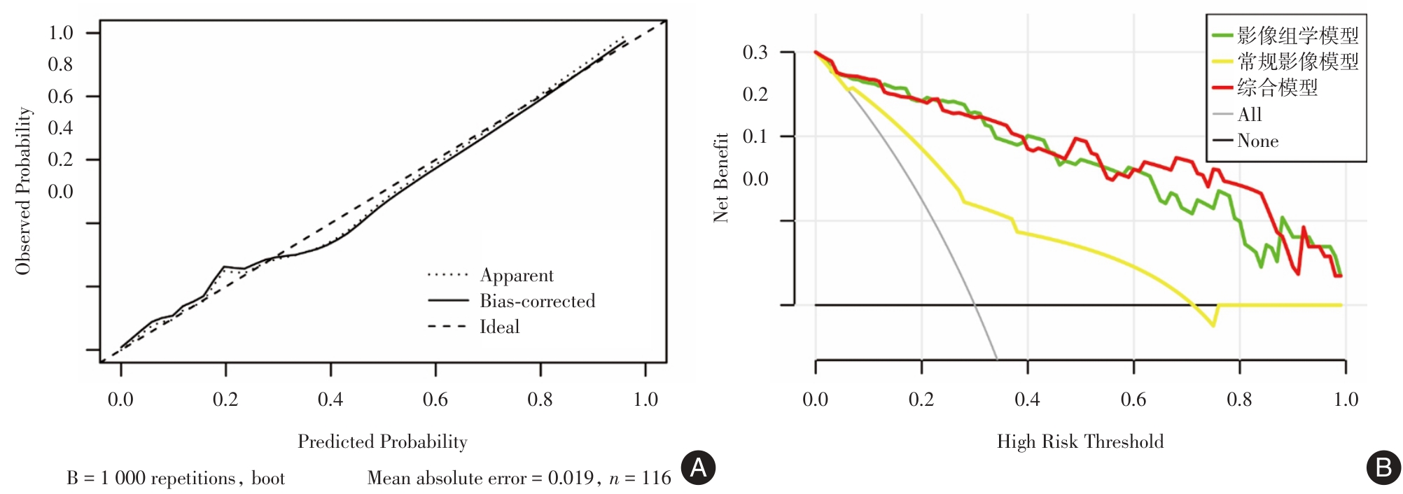
Tab.4
The performance of the radiomics model, conventional radiological model and comprehensive model"
| 诊断方法 | AUC(95%CI) | 特异度 | 灵敏度 | |
|---|---|---|---|---|
| 影像组学模型 | 训练集 | 0.938(0.887 ~ 0.988) | 0.856 | 0.923 |
| 验证集 | 0.858(0.742 ~ 0.975) | 0.711 | 0.923 | |
| 常规影像模型 | 训练集 | 0.785(0.697 ~ 0.873) | 0.856 | 0.538 |
| 验证集 | 0.692(0.540 ~ 0.845) | 0.737 | 0.538 | |
| 综合模型 | 训练集 | 0.944(0.899 ~ 0.990) | 0.844 | 0.923 |
| 验证集 | 0.879(0.777 ~ 0.980) | 0.737 | 0.923 |
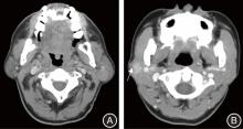
Fig.1
The lesion in the axial slice of CT"
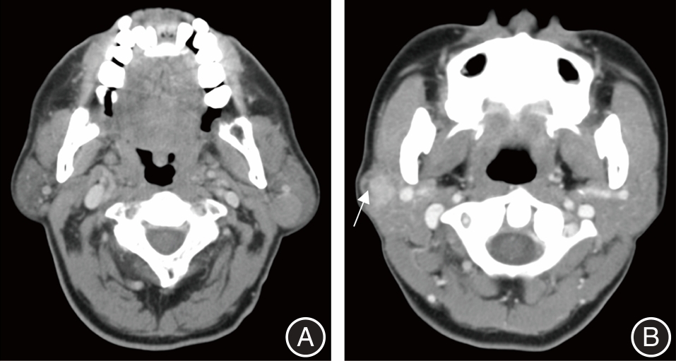
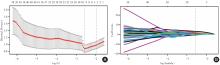
Fig.2
LASSO radiomics feature screening"
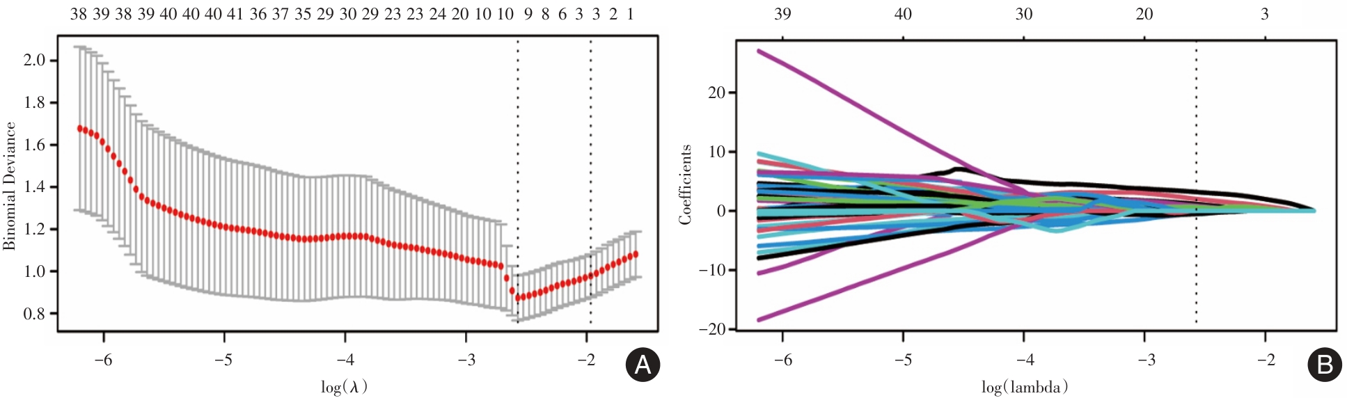
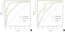
Fig.3
The ROC curves"
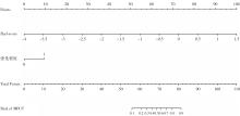
Fig.4
Nomogram"
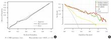
Fig.5
The diagnostic efficacy of the comprehensive model"
| [1] |
LIU S, YU B, ZHENG X, et al. Construction and Application of a Nomogram for Predicting Benign and Malignant Parotid Tumors[J]. J Comput Assist Tomogr,2024,48(1):143-149. doi:10.1097/rct.0000000000001522
doi: 10.1097/rct.0000000000001522 |
| [2] |
QUER M, HERNANDEZ-PRERA J C, SILVER C E, et al. Current Trends and Controversies in the Management of Warthin Tumor of the Parotid Gland[J]. Diagnostics (Basel),2021,11(8):1467. doi:10.3390/diagnostics11081467
doi: 10.3390/diagnostics11081467 |
| [3] | 陈飞,王利伟,布静秋. 腮腺沃辛瘤555例临床分析[J]. 北京口腔医学,2022,30(4):284-286. |
| [4] |
DI SANTO D, DERETTI A, VANDER POORTEN V. Current surgical management of malignant parotid tumors[J]. Curr Opin Otolaryngol Head Neck Surg,2025,33(2):79-84. doi:10.1097/moo.0000000000001039
doi: 10.1097/moo.0000000000001039 |
| [5] |
GEIGER J L, ISMAILA N, BEADLE B, et al. Management of Salivary Gland Malignancy: ASCO Guideline[J]. J Clin Oncol,2021,39(17):1909-1941. doi:10.1200/jco.21.00449
doi: 10.1200/jco.21.00449 |
| [6] |
ESPINOZA S, FELTER A, MALINVAUD D, et al. Warthin's tumor of parotid gland: Surgery or follow-up? Diagnostic value of a decisional algorithm with functional MRI[J]. Diagn Interv Imaging,2016,97(1):37-43. doi:10.1016/j.diii.2014.11.024
doi: 10.1016/j.diii.2014.11.024 |
| [7] |
CENGIZ A B, TANSUKER H D, GUL R, et al. Comparison of preoperative diagnostic accuracy of fine needle aspiration and core needle biopsy in parotid gland neoplasms[J]. Eur Arch Otorhinolaryngol,2021,278(10):4067-4074. doi:10.1007/s00405-021-07022-x
doi: 10.1007/s00405-021-07022-x |
| [8] |
LAMBIN P, RIOS-VELAZQUEZ E, LEIJENAAR R, et al. Radiomics: Extracting more information from medical images using advanced feature analysis[J]. Eur J Cancer,2012,48(4):441-446. doi:10.1016/j.ejca.2011.11.036
doi: 10.1016/j.ejca.2011.11.036 |
| [9] | 何尚鹏,黄炜贤,江燕辉,等. 超声特征与超声影像组学对Bethesda Ⅲ类甲状腺结节良恶性再评价的价值[J]. 实用医学杂志,2025,41(12):1892-1898. |
| [10] |
YE J, CHEN Y, PAN J, et al. US-based Radiomics Analysis of Different Machine Learning Models for Differentiating Benign and Malignant BI-RADS 4A Breast Lesions[J]. Acad Radiol,2025,32(1):67-78. doi:10.1016/j.acra.2024.08.024
doi: 10.1016/j.acra.2024.08.024 |
| [11] |
黄忠江,姜增誉,李健丁,等. 基于增强CT影像组学联合机器学习鉴别均质性肾透明细胞癌与肾乏脂肪血管平滑肌脂肪瘤[J]. 实用医学杂志,2021,37(17):2266-2270. doi:10.3969/j.issn.1006-5725.2021.17.020
doi: 10.3969/j.issn.1006-5725.2021.17.020 |
| [12] |
CHEN F, GE Y, LI S, et al. Enhanced CT-based texture analysis and radiomics score for differentiation of pleomorphic adenoma, basal cell adenoma, and Warthin tumor of the parotid gland[J]. Dentomaxillofac Radiol,2023,52(2):20220009. doi:10.1259/dmfr.20220009
doi: 10.1259/dmfr.20220009 |
| [13] |
HU Z, GUO J, FENG J, et al. Value of T2-weighted-based radiomics model in distinguishing Warthin tumor from pleomorphic adenoma of the parotid[J]. Eur Radiol,2023,33(6):4453-4463. doi:10.1007/s00330-022-09295-0
doi: 10.1007/s00330-022-09295-0 |
| [14] |
FISHER R, RONEN O. Cytologic diagnosis of parotid gland Warthin tumor: Systematic review and meta-analysis[J]. Head Neck,2022,44(10):2277-2287. doi:10.1002/hed.27099
doi: 10.1002/hed.27099 |
| [15] | 王张嵩,谢舒乐,张汉卿,等. 2456例唾液腺肿瘤临床病理分析[J]. 口腔疾病防治,2020,28(5):298-302. |
| [16] |
LEE D H, JUNG E K, LEE J K, et al. Comparative analysis of benign and malignant parotid gland tumors: A retrospective study of 992 patients[J]. Am J Otolaryngol,2023,44(2):103690. doi:10.1016/j.amjoto.2022.103690
doi: 10.1016/j.amjoto.2022.103690 |
| [17] | 张亮,张莉,刘华,等. 312例腮腺区肿瘤临床回顾性分析[J]. 临床口腔医学杂志,2022,38(9):534-537. |
| [18] |
YU H, CALDWELL C, MAH K, et al. Automated radiation targeting in head-and-neck cancer using region-based texture analysis of PET and CT images[J]. Int J Radiat Oncol Biol Phys,2009,75(2):618-625. doi:10.1016/j.ijrobp.2009.04.043
doi: 10.1016/j.ijrobp.2009.04.043 |
| [19] |
LIU Y, ZHENG J, LU X, et al. Radiomics-based comparison of MRI and CT for differentiating pleomorphic adenomas and Warthin tumors of the parotid gland: A retrospective study[J]. Oral Surg Oral Med Oral Pathol Oral Radiol,2021,131(5):591-599. doi:10.1016/j.oooo.2021.01.014
doi: 10.1016/j.oooo.2021.01.014 |
| [20] |
ZHENG Y, ZHOU D, LIU H, et al. CT-based radiomics analysis of different machine learning models for differentiating benign and malignant parotid tumors[J]. Eur Radiol,2022,32(10):6953-6964. doi:10.1007/s00330-022-08830-3
doi: 10.1007/s00330-022-08830-3 |
| [21] |
LI Q, JIANG T, ZHANG C, et al. A nomogram based on clinical information, conventional ultrasound and radiomics improves prediction of malignant parotid gland lesions[J]. Cancer Lett,2022,527:107-114. doi:10.1016/j.canlet.2021.12.015
doi: 10.1016/j.canlet.2021.12.015 |
| [22] |
MUNTEAN D D, DUDEA S M, BĂCIUȚ M, et al. The Role of an MRI-Based Radiomic Signature in Predicting Malignancy of Parotid Gland Tumors[J]. Cancers (Basel),2023,15(13):3319. doi:10.3390/cancers15133319
doi: 10.3390/cancers15133319 |
| [23] |
ZHAO F, HUANG X, HE J, et al. A nomogram for distinguishing benign and malignant parotid gland tumors using clinical data and preoperative blood markers: Development and validation[J]. J Cancer Res Clin Oncol,2023,149(13):11719-11733. doi:10.1007/s00432-023-05032-2
doi: 10.1007/s00432-023-05032-2 |
| [24] | 陈函,陈贤明,王茂鑫,等. 外周血中性粒细胞/淋巴细胞比值在腮腺恶性肿瘤诊断中的价值[J]. 中国耳鼻咽喉头颈外科,2021,28(7):419-422. |
| [25] | 秦硕,李冉,李春梅,等. 术前外周血炎症指标在腮腺恶性肿瘤中的诊断及预后价值分析[J]. 口腔颌面外科杂志,2022,32(6):362-367. |
| [1] | Danhui LAI,Yanhui JIANG,Siting YE,Shulian ZHUANG,Shuang YANG,Wen XUE,Jianxing ZHANG. Analysis of prediction of carotid in-stent restenosis based on ultrasonographic carotid plaque radiomics [J]. The Journal of Practical Medicine, 2025, 41(5): 742-750. |
| [2] | Yangchun DU,Hongyu ZHENG,Haining CHEN,Wenwen GUO,Jinxiu YAO,Tongliu LAN,Yanju XIAO. Ultrasound⁃based deep learning radiomics nomogram to differentiate type Ⅰ and type Ⅱ epithelial ovarian cancer [J]. The Journal of Practical Medicine, 2025, 41(18): 2920-2927. |
| [3] | Zhaoyang WANG,Nan. ZHANG. Constructing a Nomogram prediction model for early recurrence of hepatocellular carcinoma radical hepatectomy based on CT imaging omics and traditional Chinese medicine tongue image features [J]. The Journal of Practical Medicine, 2025, 41(16): 2590-2596. |
| [4] | Huiliang CAI,Qianying ZHANG,Ying HUANG,Weisheng PENG,Chengli WANG,Cuiting YANG,Na DENG,Sizhu ZHANG,Nina XU,Xiaobing HAN. Assessments of ki⁃67 expression in hepatocellular carcinoma using enhanced MRI intratumoral and peritumoral radiomics and clinical imaging features [J]. The Journal of Practical Medicine, 2025, 41(15): 2311-2319. |
| [5] | Shangpeng HE,Weixian HUANG,Yanhui JIANG,Xiongqiang PENG,Lingcui MENG,Jianxing ZHANG. Value of ultrasound radiomics in re⁃evaluating the benign or malignant of Bethesda Ⅲ nodules [J]. The Journal of Practical Medicine, 2025, 41(12): 1892-1898. |
| [6] | Minghong SHI,Zhanwei LIU,Yang ZHU,Lili XIA,Gang CHENG,Youguo LU. Influence of oxycodone-acetaminophen combined with local radiotherapy on serum cytokines in elderly patients with malignant tumor bone metastasis complicated with cancer pain [J]. The Journal of Practical Medicine, 2025, 41(11): 1718-1723. |
| [7] | ZHOU Huien, CHEN Wanming, WANG Mengdie, LUO Sijia, CHEN Jialin, HE Kun, GUO Xinmin. . The Value of Nomogram Model Based on Clinical and Ultrasound Radiomics for Predicting Preterm Birth [J]. The Journal of Practical Medicine, 2023, 39(14): 1835-1841. |
| [8] |
LIAO Shuting, YU Xiangrong. .
Application of spectral CT and artificial intelligence in the diagnosis of thyroid cancer [J]. The Journal of Practical Medicine, 2022, 38(2): 129-1133. |
| [9] |
TIAN Tian, JING Hui, FU Jia..
Nomogram prediction model of delirium risk in patients with malignant tumors [J]. The Journal of Practical Medicine, 2021, 37(20): 2641-2646. |
| [10] |
HUANG Zhongjiang, JIANG Zengyu, LI Jianding, ZHANG Zhixing, CHEN Wenqing.
Application of enhanced CT radiomics in differentiation of renal angiomyolipoma without visible fat from homogeneous clear cell renal cell carcinoma [J]. The Journal of Practical Medicine, 2021, 37(17): 2266-2270. |
| [11] |
LI Ling, HU Datao, XIA Chunhua, LI Hongxia..
Prediction of lymph node metastasis of head and neck malignant tumor based on CT radiomics nomogram [J]. The Journal of Practical Medicine, 2021, 37(14): 1872-1878. |
| Viewed | ||||||
|
Full text |
|
|||||
|
Abstract |
|
|||||