实用医学杂志 ›› 2025, Vol. 41 ›› Issue (2): 232-237.doi: 10.3969/j.issn.1006-5725.2025.02.012

• 临床研究 • 上一篇    

α-肌动蛋白1在皮肤鳞状细胞癌中的表达及与临床-病理特征、预后的相关性

郭金兰1,张晓宁1,江佳慧2,袁涛2()   

  1. 1.新乡市中心医院皮肤科 (河南 新乡 453000 )
    2.皖南医学院第一附属医院(弋矶山医院)皮肤科 (安徽 芜湖 241000 )
  • 收稿日期:2024-08-27 出版日期:2025-01-25 发布日期:2025-01-26
  • 通讯作者: 袁涛 E-mail:yuantao@yjsyy.com
  • 基金资助:
    安徽省自然科学基金青年项目(2308085QH269);安徽省高校自然科学研究重点项目(2023AH051781)

Expression of ACTN1 in cutaneous squamous cell carcinoma and its correlation with clinicopathological features and prognosis

Jinlan GUO1,Xiaoning ZHANG1,Jiahui JIANG2,Tao. YUAN2()   

  1. *.Department of Dermatology,Xinxiang Central Hospital,Xinxiang 453000,He′nan,China
  • Received:2024-08-27 Online:2025-01-25 Published:2025-01-26
  • Contact: Tao. YUAN E-mail:yuantao@yjsyy.com

摘要:

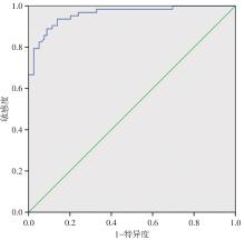
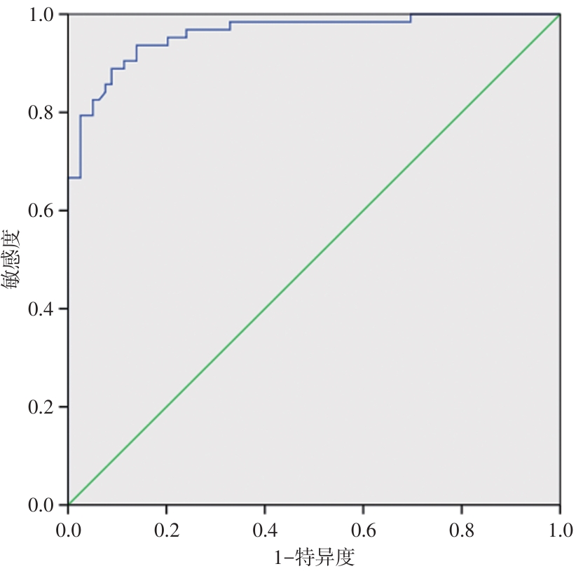
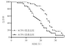
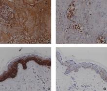
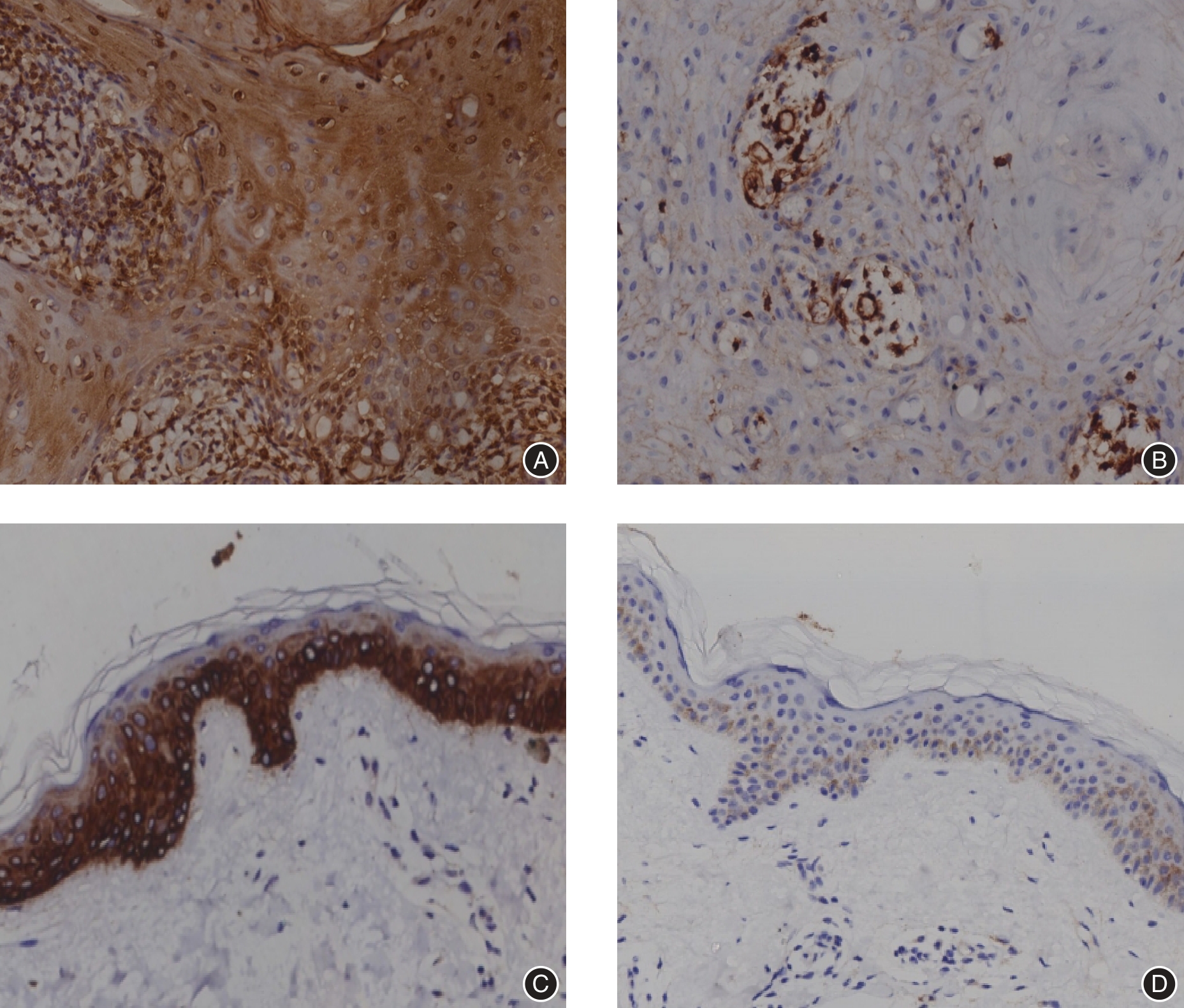
目的 探讨α-肌动蛋白1(ACTN1)在皮肤鳞状细胞癌(CSCC)中的表达及与临床-病理特征、预后的相关性。 方法 纳入150例CSCC患者(CSCC组)和100例正常体检者(对照组)作为研究对象。血清中ACTN1表达水平采用ELISA法检测。对CSCC患者术后进行随访并分为预后不良组(n = 69)和预后良好组(n = 81),预后不良组中69例CSCC患者根据ACTN1中位表达水平分为ACTN1低表达组(n = 35)和ACTN1高表达组(n = 34)。logistic回归分析CSCC患者发生预后不良的危险因素,血清中ACTN1表达水平预测CSCC患者发生预后不良的临床效能采用ROC曲线分析,K-M曲线分析血清中ACTN1表达水平与CSCC患者发生预后不良中位时间的关系。纳入40例CSCC患者作为独立验证样本,采用免疫组化法比较ACTN1在CSCC组织及癌旁组织中的表达差异。 结果 对照组和CSCC组血清中ACTN1表达水平分别为(4.56 ± 1.02)和(12.12 ± 2.26)ng/mL,与对照组相比,CSCC组血清中ACTN1表达水平显著升高,差异有统计学意义(t = 31.37,P < 0.001)。预后不良组肿瘤直径≥ 5 cm、肿瘤细胞分化程度为低分化、肿瘤浸润深度为脂肪下层、有淋巴结转移占比及血清ACTN1表达水平显著高于预后良好组,差异均有统计学意义(P < 0.05)。logistic回归分析显示,淋巴结转移(OR = 3.253)、ACTN1(OR = 2.894)是CSCC患者术后出现预后不良的独立危险因素。血清中ACTN1表达水平预测CSCC患者术后发生预后不良的曲线下面积为0.911,当截断值为13.19 ng/mL,诊断敏感度和特异度分别为89.78%和92.12%。ACTN1低表达组和高表达组发生预后不良的中位时间分别为25个月和18.5个月,相对于ACTN1低表达组,ACTN1高表达组发生预后不良的中位时间显著缩短(HR= 6.627,P < 0.001)。40例CSCC组织中ACTN1高表达32例,ACTN1低表达8例;CSCC癌旁组织中ACTN1高表达11例,ACTN1低表达29例,与癌旁组织相比,ACTN1高表达率在CSCC组织中显著升高(χ2 = 22.175,P < 0.001)。 结论 CSCC患者血清中ACTN1表达水平显著升高,可作为CSCC患者术后评估预后的一项生物学标志物。

关键词: 皮肤鳞状细胞癌, α-肌动蛋白1, 淋巴结转移, 预后

Abstract:

Objective To investigate the expression of ACTN1 in cutaneous squamous cell carcinoma (CSCC) and its association with clinicopathological features and prognosis. Methods A total of 150 patients with cutaneous squamous cell carcinoma (CSCC) and 100 healthy controls were enrolled in this study. The serum expression levels of ACTN1 were measured using ELISA. All CSCC patients underwent post?surgical follow?up and were categorized into a poor prognosis group (n = 69) and a good prognosis group (n = 81). Additionally, the 69 patients in the poor prognosis group were further classified into an ACTN1 lower expression subgroup (n = 35) and an ACTN1 higher expression subgroup (n = 34) based on the median expression level of ACTN1. Logistic regression analysis was employed to identify risk factors associated with poor prognosis in CSCC patients. ROC curve analysis was conducted to evaluate the clinical utility of serum ACTN1 expression levels in predicting poor prognosis in CSCC patients. Kaplan?Meier (K?M) survival analysis was utilized to assess the relationship between serum ACTN1 expression levels and the median time to poor prognosis in the 69 patients with poor prognosis. Furthermore, 40 additional CSCC patients were recruited to compare the expression levels of ACTN1 in CSCC tissues and adjacent tissues using immunohistochemistry. Results The serum expression levels of ACTN1 in the Control group and the CSCC group were (12.12 ± 2.26) ng/mL and (4.56 ± 1.02) ng/mL, respectively. Compared to the Control group, the serum expression level of ACTN1 in the CSCC group was significantly decreased, and the difference was statistically significant (t = 31.37, P < 0.001)。 In the poor prognosis group, the proportion of tumors with a diameter ≥ 5 cm, low degree of tumor cell differentiation, subadipocyte invasion depth, lymph node metastasis incidence, and serum ACTN1 expression levels were all significantly higher compared to the good prognosis group, with statistical significance (P < 0.05). Logistic regression analysis revealed that lymph node metastasis (OR = 3.253) and elevated ACTN1 expression (OR = 2.894) were independent risk factors for poor prognosis following CSCC surgery. The area under the curve (AUC) for serum ACTN1 expression in predicting poor prognosis post?surgery in CSCC patients was 0.911. At a cut?off value of 13.19 ng/mL for ACTN1, the diagnostic sensitivity and specificity were 89.78% and 92.12%, respectively. The median time to poor prognosis was 25 months in the ACTN1 low?expression group and 18.5 months in the ACTN1 high?expression group, respectively. The median time to poor prognosis was significantly shorter in the ACTN1 high?expression group compared to the low?expression group (HR = 6.627, P < 0.001). Among 40 CSCC tissue samples, 32 cases exhibited higher ACTN1 expression, while 8 cases showed lower expression. In contrast, among 40 paracancerous tissue samples, 11 cases had higher ACTN1 expression and 29 cases had lower expression. The higher expression rate of ACTN1 in CSCC tissues was significantly elevated compared to paracancerous tissues (χ2 = 22.175, P < 0.001). Conclusion The serum expression level of ACTN1 in CSCC patients was significantly elevated, suggesting its potential as a biomarker for assessing post?surgical prognosis in CSCC patients.

Key words: cutaneous squamous cell carcinoma, ACTN1, lymph node metastasis, prognosis

中图分类号: