实用医学杂志 ›› 2025, Vol. 41 ›› Issue (2): 153-161.doi: 10.3969/j.issn.1006-5725.2025.02.001

• 基础研究 •    

瑞马唑仑调节ROS/RAGE/NF-κB信号通路对LPS诱导的小胶质细胞炎症的影响

吴兴卫,王建营,郭成晓,刘紫仪,孙超,于飞()   

  1. 滨州医学院附属医院麻醉科 (山东 滨州 256699 )
  • 收稿日期:2024-10-12 出版日期:2025-01-25 发布日期:2025-01-26
  • 通讯作者: 于飞 E-mail:yufei001@bzmc.edu.cn
  • 基金资助:
    国家自然科学基金项目(81900269)

The effect of remimazolam on modulating the ROS/RAGE/NF-κB signaling pathway in LPS-induced microglial inflammation

Xingwei WU,Jianying WANG,Chengxiao GUO,Ziyi LIU,Chao SUN,Fei. YU()   

  1. Anesthesia Department of Binzhou Medical University Hospital,Binzhou 256699,Shandong,China
  • Received:2024-10-12 Online:2025-01-25 Published:2025-01-26
  • Contact: Fei. YU E-mail:yufei001@bzmc.edu.cn

摘要:

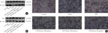
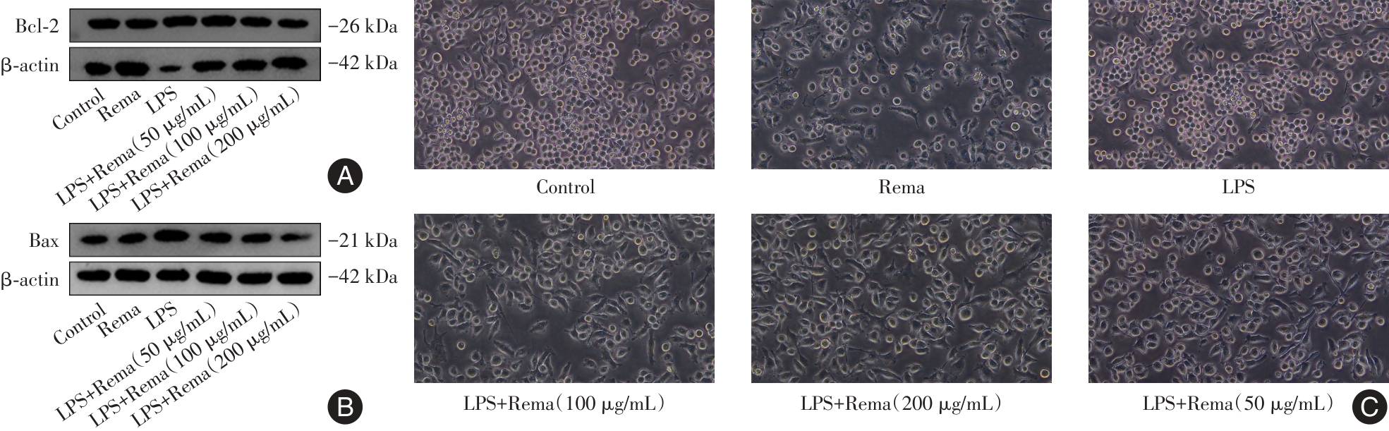
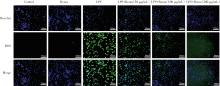
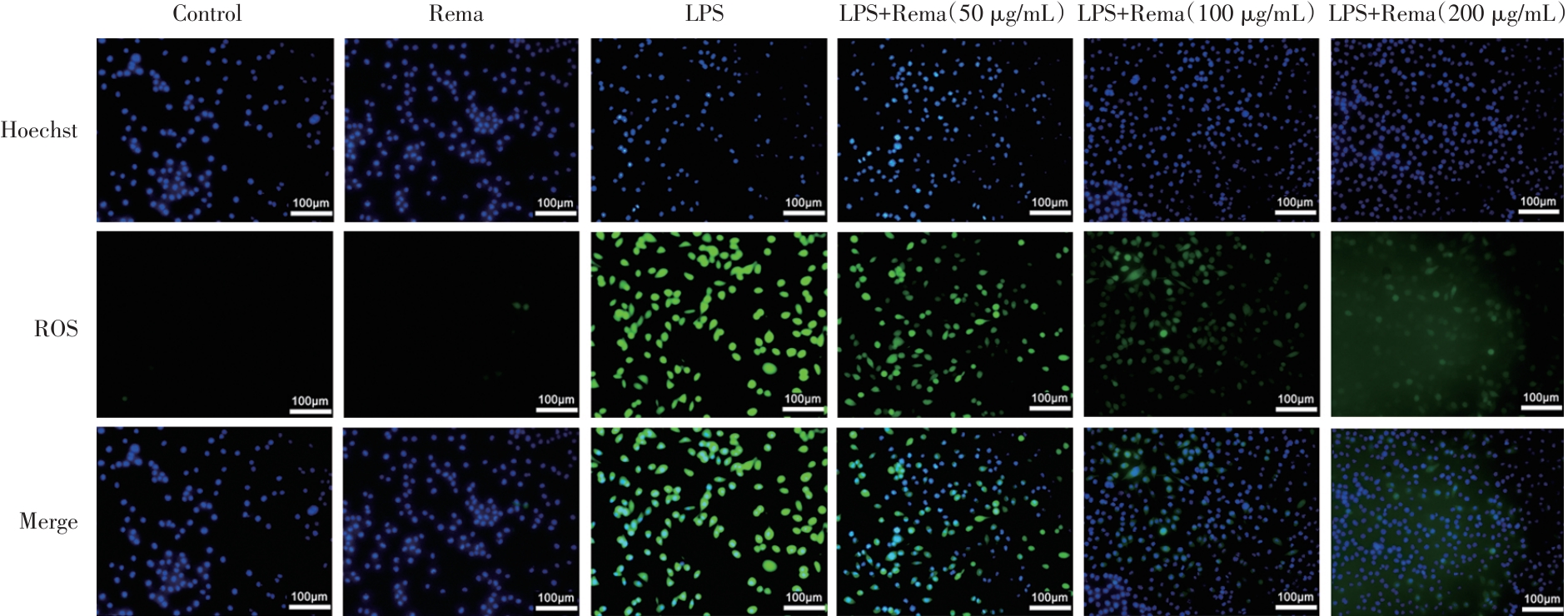
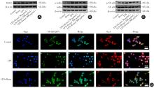
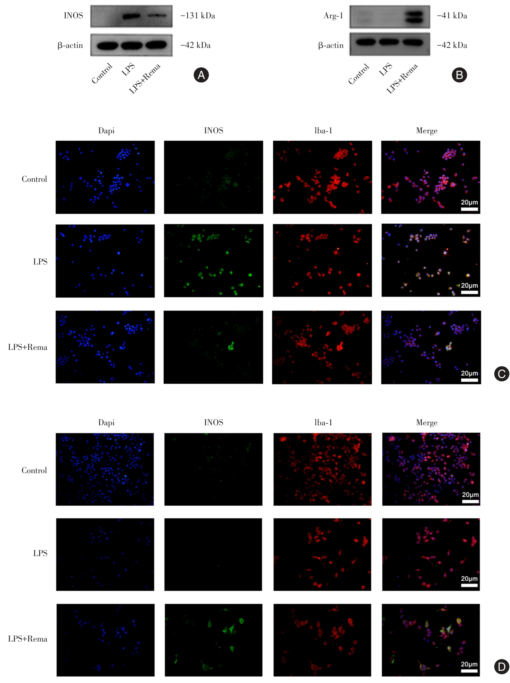
目的 探讨瑞马唑仑对小胶质细胞的炎症保护作用及潜在分子机制。 方法 选取小鼠小胶质细胞系(BV2)作为研究对象,设对照组(完全培养基)、Rema组(200 μg/mL瑞马唑仑)、模型组(1 μg/mL LPS)和不同浓度给药组(1 μg/mL LPS + 50、100、200 μg/mL Rema)。Rema组单纯用200 μg/mL的瑞马唑仑处理细胞26 h,模型组用LPS处理细胞24 h,不同浓度给药组先用不同浓度的瑞马唑仑预处理细胞2 h,再加LPS处理24 h。用光学显微镜观察评估LPS和瑞马唑仑对BV2细胞形态学的影响;CCK-8法检测细胞毒性;实时荧光定量PCR、ELISA法检测炎症因子的表达与分泌;荧光探针法检测细胞ROS活性;试剂盒检测细胞中的丙二醛(MDA)含量及超氧化物歧化酶(SOD)和谷胱甘肽过氧化物酶(GSH)活性;Western blot法检测细胞中的Bax、Bcl-2、IL-1β、RAGE、NF-κB、P- NF-κB、IκBα、p-IκBα、INOS和Arg-1的蛋白表达。免疫荧光法观察NF-κB核转位以及细胞M1/M2极化情况。 结果 与对照组相比,模型组BV2细胞活性降低,炎症因子(TNF-α、IL-6、IL-1β)基因表达与分泌增加,SOD和GSH活性下降,细胞内MDA及ROS含量增加,RAGE蛋白水平上升,IκBα和NF-κB蛋白磷酸化水平增加,NF-κB发生核转位,M1极化标志物INOS表达增加,M2极化标志物Arg-1表达减少差异有统计学意义(P < 0.05)。与模型组相比,LPS+Rema组细胞活性增加,炎症因子基因表达与分泌减少,SOD和GSH活性升高,细胞内MDA及ROS含量减少,RAGE蛋白水平下降,IκBα和NF-κB蛋白磷酸化水平减少,抑制NF-κB核转位发生,M1极化标志物INOS表达减少,M2极化标志物Arg-1表达增加差异有统计学意义(P < 0.05)。 结论 瑞马唑仑通过调节NF-κB通路以及ROS的产生使小胶质细胞从M1表型转移到M2表型,从而减轻LPS诱导的炎症反应。

关键词: 神经炎症, 瑞马唑仑, NF-κB, 氧化应激, 小胶质细胞极化

Abstract:

Objective To investigate the anti?inflammatory protective effects of remimazolam on microglial cells and elucidates the potential molecular mechanisms underlying these effects. Methods The mouse microglial cell line (BV2) was selected as the research object. The following groups were set up: the control group (complete medium), the Rema group (200 μg/mL remimazolam), the model group (1 μg/mL lipopolysaccharide, LPS), and different?concentration administration groups (1 μg/mL LPS + 50, 100, 200 μg/mL remimazolam). In the Rema group, cells were treated with 200 μg/mL remimazolam alone for 26 h. In the model group, cells were treated with LPS for 24 h. In the different?concentration administration groups, cells were pre?treated with different concentrations of remimazolam for 2 h, and then treated with LPS for 24 h. The effects of LPS and remimazolam on the morphology of BV2 cells were observed and evaluated using an optical microscope. Cell viability was determined using the CCK?8 assay, while the expression and secretion of inflammatory cytokines were quantified by quantitative real?time PCR and ELISA. Reactive oxygen species (ROS) levels were measured using a fluorescent probe. Additionally, malondialdehyde (MDA) content, superoxide dismutase (SOD) activity, and glutathione peroxidase (GSH) activity were evaluated using respective assay kits. Western blot analysis was conducted to examine the protein expression levels of Bax, Bcl?2, IL?1β, RAGE, NF?κB, p?NF?κB, IκBα, p?IκBα, iNOS, and Arg?1. Immunofluorescence staining was employed to visualize NF?κB nuclear translocation and M1/M2 polarization in the cells. Results Compared to the control group, LPS?treated BV2 cells demonstrated significantly reduced cell viability, elevated expression and secretion of inflammatory cytokines (TNF?α, IL?6, IL?1β), decreased activities of SOD and GSH, and increased intracellular levels of MDA and ROS. Additionally, RAGE protein levels were upregulated, along with enhanced phosphorylation of IκBα and NF?κB, leading to observable NF?κB nuclear translocation. The expression of the M1 marker iNOS was upregulated, while that of the M2 marker Arg?1 was downregulated. In contrast, in the LPS+Rema group, cell viability was restored, expression and secretion of inflammatory cytokines were attenuated, SOD and GSH activities were improved, and levels of MDA and ROS were reduced compared to the LPS group. Furthermore, RAGE protein expression and phosphorylation levels of IκBα and NF?κB were diminished, inhibiting NF?κB nuclear translocation. The expression of the M1 marker iNOS was downregulated, while that of the M2 marker Arg?1 was upregulated. Conclusion Remimazolam mitigates LPS?induced inflammation by facilitating the transition of microglial cells from the M1 to the M2 phenotype via modulation of the NF?κB pathway and reduction of ROS production.

Key words: neuroinflammation, remimazolam, NF-κB pathway, oxidative stress, microglial polarization

中图分类号: