实用医学杂志 ›› 2024, Vol. 40 ›› Issue (11): 1560-1567.doi: 10.3969/j.issn.1006-5725.2024.11.016

• 临床研究 • 上一篇    下一篇

联合术前MRI和术后病理评估淋巴结转移在直肠癌患者预后预测中的价值

曾志明,朱攀,马德财,邸小慧,李桂婷,邹文彬,潘希敏()   

  1. 中山大学附属第六医院放射诊断科 (广州 510655);广东省结直肠盆底疾病研究重点实验室 (广州 510655);广州市黄埔区中六生物医学创新研究院 (广州 510655 )
  • 收稿日期:2023-12-25 出版日期:2024-06-10 发布日期:2024-06-13
  • 通讯作者: 潘希敏 E-mail:panxm9@mail.sysu.edu.cn
  • 基金资助:
    国家临床重点专科项目;广东省自然科学基金项目(2023A1515011292);广东省消化系统疾病临床研究中心项目(2020B1111170004)

Prognostic value of combined preoperative MRI and postoperative pathological assessment of lymph node metastasis in rectal cancer patients

Zhiming ZENG,Pan ZHU,Decai MA,Xiaohui DI,Guiting LI,Wenbin ZHOU,Ximin. PAN()   

  1. Department of Radiology,the Sixth Affiliated Hospital,Sun Yat?Sen University,Guangzhou 510655,China; Guangdong Provincial Key Laboratory of Colorectal and Pelvic Floor Diseases,the Sixth Affiliated Hospital,Sun Yat?sen University,Guangzhou 510655,China; Biomedical Innovation Center,the Sixth Affiliated Hospital,Sun Yat?sen University,Guangzhou 510655,China
  • Received:2023-12-25 Online:2024-06-10 Published:2024-06-13
  • Contact: Ximin. PAN E-mail:panxm9@mail.sysu.edu.cn

摘要:

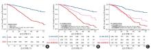
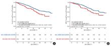
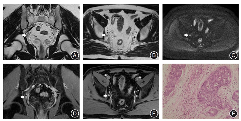
目的 探究联合术前磁共振成像(MRI)和术后病理评估淋巴结转移预测直肠癌总生存期的价值。 方法 回顾性收集2016年1月至2021年12月2610例在中山大学附属第六医院组织病理学证实为直肠腺癌患者的临床、病理和影像学资料。所有患者均行MRI检查,并根据淋巴结术前MRI( MRI N)和术后病理( P N)评估的转移情况将患者分为3组:MRI N+但 P N-( MRI N+组)、 P N+但 MRI N-( P N+组)和 MRI N+且 P N+( MRI-P N+组)。使用逆概率加权(IPW)校正混杂因素。采用Kaplan-Meier曲线估计总生存率,并用log-rank检验进行组间比较。使用单因素Cox回归模型分析肿瘤特征与总生存期的相关性,采用双向逐步Cox回归模型确定总生存期的独立危险因素。 结果 MRI-P N+组比 P N+组、 MRI N+组具有更高的肿瘤分期、更频繁的神经周围侵犯、更多的远处转移和更高的死亡风险(均P < 0.05)。Kaplan-Meier曲线显示 MRI N+组、 P N+组和 MRI-P N+组的3年生存率分别为90.5%、79.1%和76.4%;5年生存率分别为85.7%、71.5%和59.2%。双向逐步Cox回归分析显示年龄、肿瘤位置、癌胚抗原、糖类抗原19-9、淋巴结送检个数、病理肿瘤分期、脉管内癌栓、神经周围侵犯、远处转移、新辅助治疗和辅助治疗、MRI-病理淋巴结转移是影响直肠癌患者总生存期的独立危险因素(均P < 0.05)。 结论 联合术前MRI和术后病理评估淋巴结转移有助于更准确预测直肠癌患者的总生存期。

关键词: 直肠癌, 磁共振成像, 淋巴结, 预后

Abstract:

Objective To investigate the value of combining preoperative magnetic resonance imaging(MRI) and postoperative pathological assessment of lymph node metastasis in predicting overall survival in rectal cancer patients. Methods This retrospective study collected clinical, pathological and image information of 2610 patients histopathologically confirmed with rectal adenocarcinoma at the Sixth Affiliated Hospital of Sun Yat-sen University between January 2016 and December 2021. All patients underwent MRI scans and were divided into three groups according to lymph node status assessed by preoperative MRI( MRI N) and postoperative pathology( P N): MRI N+ but P N-( MRI N+ group), P N+ but MRI N-( P N+ group), MRI N+ and P N+( MRI-P N+ group). Inverse probability weighting(IPW) was used to adjust for confounding factors. Kaplan-Meier curves were used to estimate overall survival and log-rank tests were used to compare the difference. Univariate Cox regression models were used to analyze the correlation between tumor characteristics and overall survival, and bidirectional stepwise Cox regression models were used to identify independent risk factors for overall survival. Results The MRI-P N+ group showed higher tumor staging, more frequent perineural invasion, more distant metastases, and a higher risk of death compared to the P N+ group and MRI N+ group(all P < 0.05). Kaplan-Meier curves showed that the 3-year survival rates for the MRI N+ group, P N+ group, and MRI-P N+ group were 90.5%, 79.1%, and 76.4%, respectively; the 5-year survival rates were 85.7%, 71.5%, and 59.2%, respectively. Stepwise Cox regression showed that age, tumor location, carcinoembryonic antigen, carbohydrate antigen 19-9, lymph nodes number, pathological tumor stage, lymphovascular invasion, perineural invasion, distant metastasis, neoadjuvant therapy and adjuvant therapy, and MRI-pathology lymph node status were independent risk factors for overall survival in rectal cancer(all P < 0.05). Conclusion Evaluating the lymph node status by combining preoperative MRI and postoperative pathology helps predict overall survival in rectal cancer patients more accurately.

Key words: rectal cancer, MRI, lymph node, prognosis

中图分类号: