

实用医学杂志 ›› 2026, Vol. 42 ›› Issue (13): 2404-2418.doi: 10.3969/j.issn.1006-5725.2026.13.017
• 论著·机制与实践 • 上一篇
向雪飞1,高鹤友1,2,陈芷芸1,胡清川1,何流1,王宇1,2( )
)
收稿日期:2026-02-10
出版日期:2026-07-10
发布日期:2026-07-14
通讯作者:
王宇
E-mail:wangyuscu@foxmail.com
基金资助:
Xuefei XIANG1,Heyou GAO1,2,Zhiyun CHEN1,Qingchuang HU1,Liu HE1,Yu WANG1,2()
Received:2026-02-10
Online:2026-07-10
Published:2026-07-14
Contact:
Yu WANG
E-mail:wangyuscu@foxmail.com
摘要:
目的 探讨EPO/pDA/BCP复合支架的促成骨效能及分子机制,重点验证其是否通过激活PI3K/AKT通路并上调MTSS1与CYTL1,从而促进骨髓间充质干细胞(BMSCs)的成骨分化及体内临界骨缺损修复。 方法 采用双相磷酸钙支架并接枝多巴胺和促红细胞生成素构建EPO/pDA/BCP复合材料;体外实验分4组:空白对照组、BCP组、EPO/pDA/BCP组及加入PI3K/AKT通路抑制剂的EPO/pDA/BCP + LY294002组,通过CCK-8法、碱性磷酸酶活性测定、RT-qPCR和免疫印迹法评估细胞增殖及成骨分化能力。通过RT-qPCR和免疫印迹检测PI3K/AKT通路及下游基因MTSS1、CYTL1的表达水平;体内实验通过建立SD大鼠股骨髁临界骨缺损模型,随机分为空白组、BCP组和EPO/pDA/BCP组进行干预。术后4周和12周进行Micro-CT、苏木精-伊红染色、马松染色及免疫组化分析骨再生情况。 结果 CCK-8实验证实EPO/pDA/BCP复合材料具有良好的生物相容性。碱性磷酸酶活性测定、RT-qPCR及Western blot结果显示,与BCP组和对照组相比,EPO/pDA/BCP组能够显著促进BMSCs的成骨分化(P 0.05)。而加入抑制剂LY294002后,该促成骨效应明显减弱,且PI3K/AKT信号通路相关分子(p-PI3K、p-AKT、CYTL1和MTSS1)表达下调(P 0.05)。Micro-CT扫描显示,EPO/pDA/BCP组骨缺损区域的新生骨体积分数和骨密度等均显著高于空白组与BCP组(P 0.05);组织学染色结果提示,相较于其他两组,EPO/pDA/BCP组骨组织再生更为完整和成熟;免疫组化染色进一步显示,EPO/pDA/BCP组骨缺损区内成骨标志物骨形态发生蛋白-2与骨钙素的阳性染色强度均显著高于BCP组和空白对照组。 结论 EPO/pDA/BCP生物陶瓷在体内外均具有良好的促成骨性能,该作用可能与激活PI3K/AKT信号通路蛋白表达有关。
中图分类号:
向雪飞,高鹤友,陈芷芸,胡清川,何流,王宇. PI3K/AKT通路在促红细胞生成素调控钙磷陶瓷表面成骨中的作用[J]. 实用医学杂志, 2026, 42(13): 2404-2418.
Xuefei XIANG,Heyou GAO,Zhiyun CHEN,Qingchuang HU,Liu HE,Yu WANG. Study on the role of the PI3K/AKT pathway in the regulation of osteogenesis on the surface of calcium phosphate ceramics by erythropoietin[J]. The Journal of Practical Medicine, 2026, 42(13): 2404-2418.
表1
各基因引物序列"
| 基因 | 引物序列(5′-3′) |
|---|---|
| Runx2 | 正义链:AAG GAG CAG ACC GTC AAA GG 反义链:CCC AGA GTC CAC CTC TCT GA |
| Osterix | 正义链:GCA TCT GAA AGC CCA CTT GC 反义链:AGT GGT CGC TTC GGG TAA AG |
| OPN | 正义链:GAT GAC GAC GAC GAT GAC GA 反义链:GCT GGC AGT GAA GGA CTC AT |
| CYTL1 | 正义链:CAC CGT CCT CCA GAG TGA AC 反义链:CGG ACC CAG TTT GGA TGG AA |
| MTSS1 | 正义链;GCG GTT TTC TCT GAC CCA AG 反义链:CCC CAA GTC CCA ATC CGA AA |
| GAPDH | 正义链:GAC ATG CCG CCT GGA GAA AC 反义链:AGC CCA GGA TGC CCT TTA GT |

图1
EPO/pDA/BCP复合支架的大体观察结果注:A—B,BCP陶瓷大体观;C—D,EPO/pDA/BCP陶瓷大体观。A、C为用于体内动物实验的直径3.5 mm,厚度4 mm的小型圆柱体支架;B、D是用于体外实验的直径12 mm,厚度2 mm的圆盘状支架"
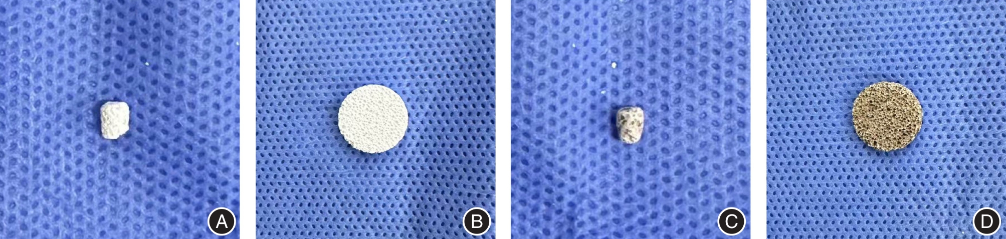
图2
大鼠BMSCs的鉴定结果注:A,原代大鼠BMSCs的形态学观察图;B,第3代BMSCs的形态学观察图(比例尺:500 μm);C—D,不同倍镜下显示的成骨诱导21 d后茜素红S染色显示钙结节形成红色(比例尺分别为500 μm和200 μm);E—F,不同倍镜下显示的成脂诱导21 d后油红O染色显示脂滴形成(比例尺分别为200 μm和50 μm);G,流式细胞仪检测BMSCs的表面标志物表达。结果显示细胞表面标记CD29和CD44阳性,CD34和CD45阴性"
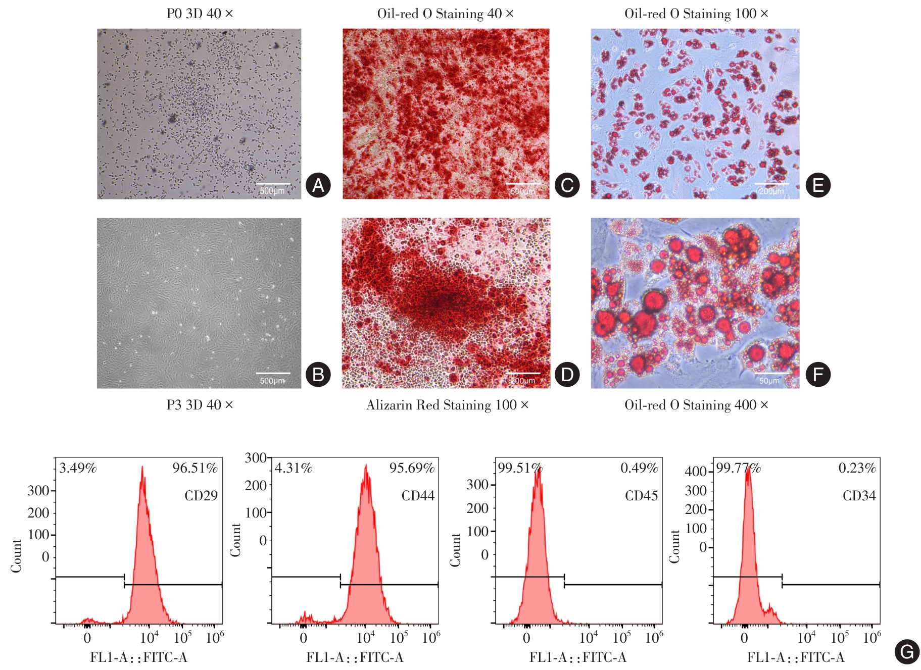
表2
各组BMSCs不同时间段的CCK-8检测结果(n = 3) (x ± s)"
| 组别 | 第1天 | 第3天 | 第7天 |
|---|---|---|---|
| 空白组 | 0.28 ± 0.03 | 0.47 ± 0.06 | 1.02 ± 0.07 |
| BCP组 | 0.32 ± 0.01 | 0.47 ± 0.06 | 0.99 ± 0.06 |
| EPO/pDA/BCP组 | 0.27 ± 0.01 | 0.41 ± 0.01 | 1.04 ± 0.04 |
图 3
基于生信挖掘的成骨阶段特征及PI3K/AKT响应网络分析结果注:A,GSE80614定义的标准成骨参考框架;B,GSE80614严格成骨signatures的功能富集分析;C,GSE46315与GSE80614参考signatures的对接分析;D,机制相关候选基因跨数据集热图;E,BMSCs成骨核心基因表达特征"
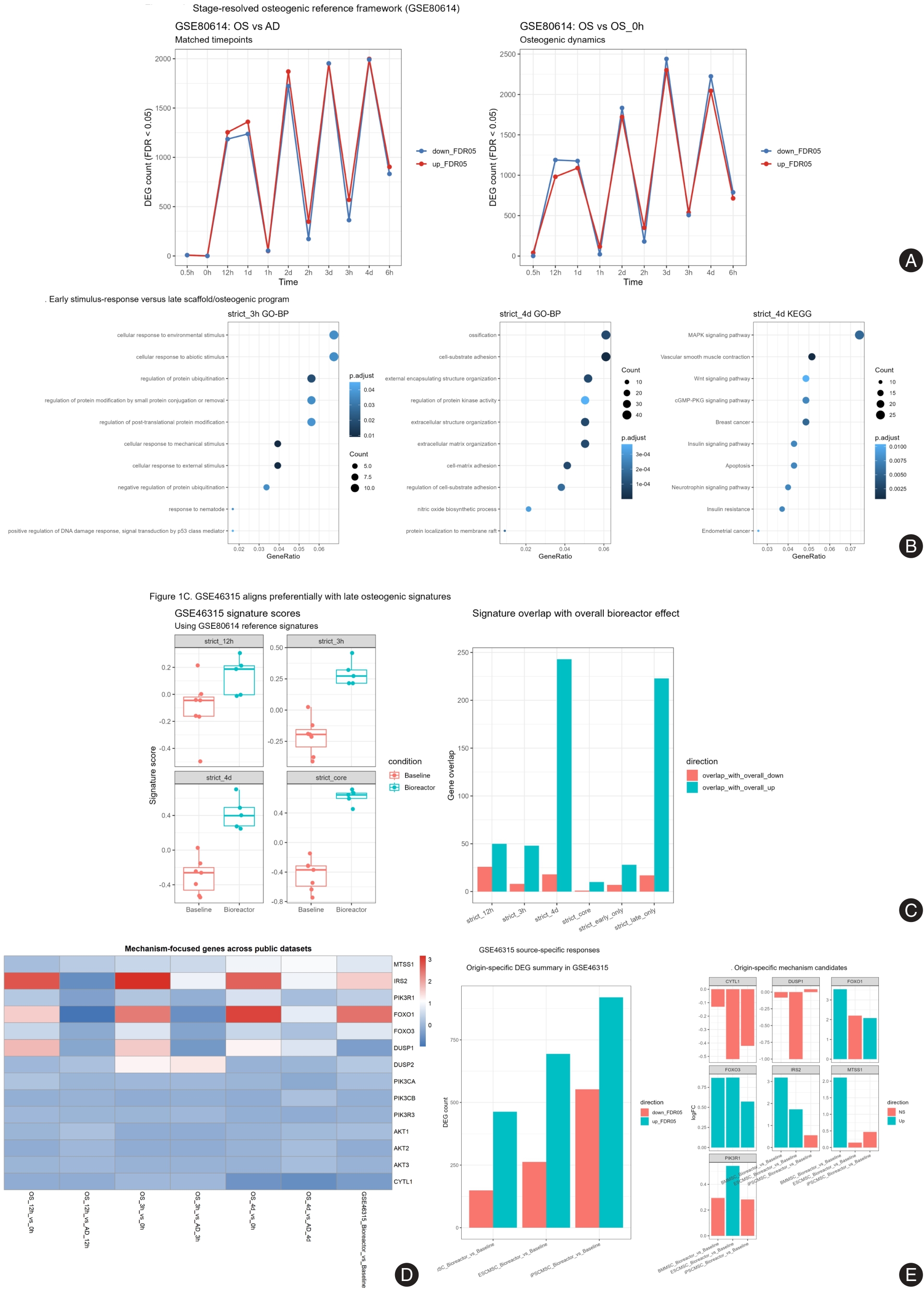
表3
成骨诱导后各组BMSCs的碱性磷酸活性检测结果(n = 3) (x ± s)"
| 组别 | 第3天 | 第7天 |
|---|---|---|
| 空白组 | 0.02 ± 0.00 | 0.05 ± 0.00 |
| BCP组 | 0.04 ± 0.01* | 0.07 ± 0.00* |
| EPO/pDA/BCP组 | 0.06 ± 0.01*# | 0.08 ± 0.00*# |
| EPO/pDA/BCP+LY组 | 0.04 ± 0.00△ | 0.05 ± 0.01△ |
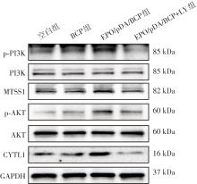
图4
各组BMSCs中PI3K/AKT通路相关蛋白表达的Western blot条带图"
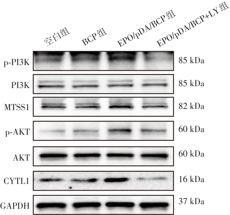
表4
各组BMSCs中PI3K/AKT通路相关蛋白表达的Western blot定量分析结果(n = 3) (x ± s)"
| 组别 | p-PI3K/PI3K | p-AKT/AKT | CYTL1 | MTSS1 |
|---|---|---|---|---|
| 空白组 | 0.29 ± 0.13 | 0.24 ± 0.07 | 0.46 ± 0.03 | 0.41 ± 0.08 |
| BCP组 | 0.47 ± 0.07 | 0.48 ± 0.06 | 0.48 ± 0.04 | 0.51 ± 0.10 |
| EPO/pDA/BCP组 | 0.85 ± 0.09*# | 0.80 ± 0.03*# | 0.61 ± 0.05*# | 0.71 ± 0.05*# |
| EPO/pDA/BCP+LY组 | 0.49 ± 0.13△ | 0.39 ± 0.12△ | 0.39 ± 0.05△ | 0.40 ± 0.06△ |
表5
各组BMSCs中MTSS1和CYTL1的RT-qPCR定量分析结果(n = 3) (x ± s)"
| 组别 | CYTL1 | MTSS1 |
|---|---|---|
| 空白组 | 1.01 ± 0.17 | 1.00 ± 0.15 |
| BCP组 | 2.50 ± 0.55 | 2.56 ± 0.41 |
| EPO/pDA/BCP组 | 4.10 ± 0.75*# | 3.34 ± 0.12*# |
| EPO/pDA/BCP+LY组 | 2.11 ± 0.12△ | 2.21 ± 0.11△ |
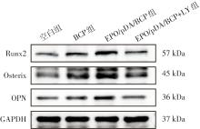
图5
各组BMSCs中成骨相关蛋白表达的Western blot条带图"
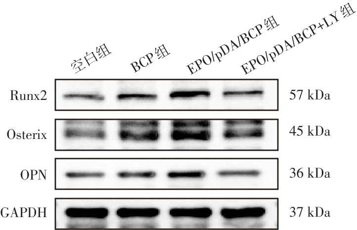
表6
各组BMSCs成骨相关蛋白表达的Western blot定量分析结果(n = 3) (x ± s)"
| 组别 | Runx2 | Osterix | OPN |
|---|---|---|---|
| 空白组 | 0.27 ± 0.07 | 0.38 ± 0.01 | 0.41 ± 0.05 |
| BCP组 | 0.49 ± 0.08* | 0.58 ± 0.06* | 0.51 ± 0.02 |
| EPO/pDA/BCP组 | 0.83 ± 0.12*# | 0.80 ± 0.11*# | 0.75 ± 0.08*# |
| EPO/pDA/BCP+LY组 | 0.45 ± 0.02△ | 0.54 ± 0.07△ | 0.49 ± 0.13△ |
表7
各组BMSCs成骨相关基因的RT-qPCR定量分析结果(n = 3) (x ± s)"
| 组别 | Runx2 | Osterix | OPN |
|---|---|---|---|
| 空白组 | 1.01 ± 0.18 | 1.00 ± 0.09 | 1.01 ± 0.15 |
| BCP组 | 3.36 ± 0.26* | 3.05 ± 0.11* | 1.35 ± 0.11* |
| EPO/pDA/BCP组 | 4.87 ± 1.02*# | 4.15 ± 0.33*# | 1.84 ± 0.16*# |
| EPO/pDA/BCP+LY组 | 2.34 ± 0.38△ | 1.34 ± 0.19△ | 1.05 ± 0.03△ |
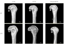
图6
术后4周和12周各组SD大鼠股骨部位缺损显微CT重建图像"
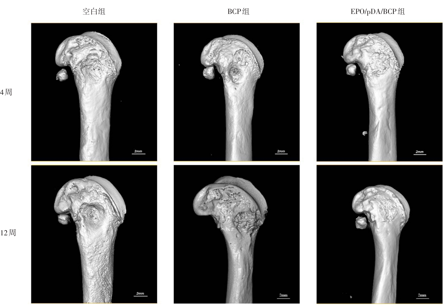
表8
术后4周和12周骨体积分数、骨小梁厚度、骨密度、骨小梁数量的半定量分析结果(n = 6) (x ± s)"
| 组别 | 骨体积分数/% | 骨小梁厚度/mm | 骨密度/(mg/cm3) | 骨小梁数量/(1/mm) |
|---|---|---|---|---|
| 空白组(4周) | 25.15 ± 8.02 | 0.30 ± 0.06 | 162.0 ± 60.67 | 0.42 ± 0.03 |
| BCP组(4周) | 45.90 ± 1.06* | 0.49 ± 0.03* | 312.72 ± 25.24* | 0.59 ± 0.04* |
| EPO/pDA/BCP组(4周) | 56.09 ± 3.55*# | 0.69 ± 0.07*# | 423.73 ± 11.54*# | 0.75 ± 0.09*# |
| 空白组(12周) | 30.20 ± 3.31 | 0.40 ± 0.06 | 238.85 ± 29.61 | 0.54 ± 0.03 |
| BCP组(12周) | 53.31 ± 1.57* | 0.55 ± 0.02* | 389.68 ± 28.29* | 0.80 ± 0.06* |
| EPO/pDA/BCP组(12周) | 72.83 ± 1.70*# | 0.80 ± 0.07*# | 631.11 ± 51.07*# | 0.96 ± 0.06*# |
图7
术后4及12周各组大鼠股骨缺损部位组织形态学分析结果注:A,支架植入术4周后股骨缺损部位组织形态学分析结果(比例尺为200 μm);B,支架植入手术12周后股骨缺损部位组织形态学分析结果(比例尺为200 μm)"
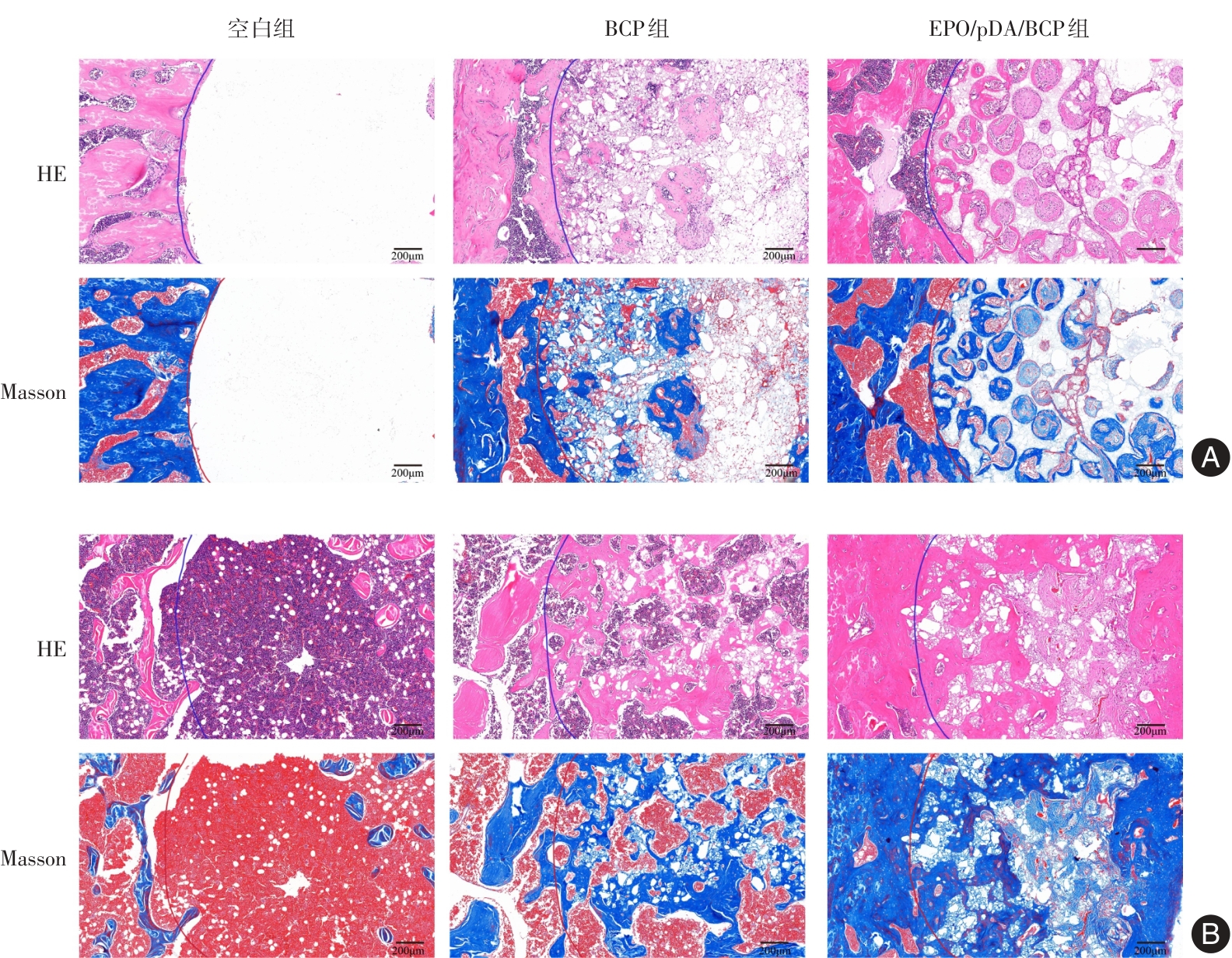
图8
术后4、12周各组大鼠股骨缺损部位免疫组化染色分析结果注:免疫组化染色(棕色为阳性信号,苏木精复染核为蓝色)。A,OCN在不同组骨缺损区域中的表达情况;B,BMP-2在不同组骨缺损区域中的表达情况。比例尺:200 μm"
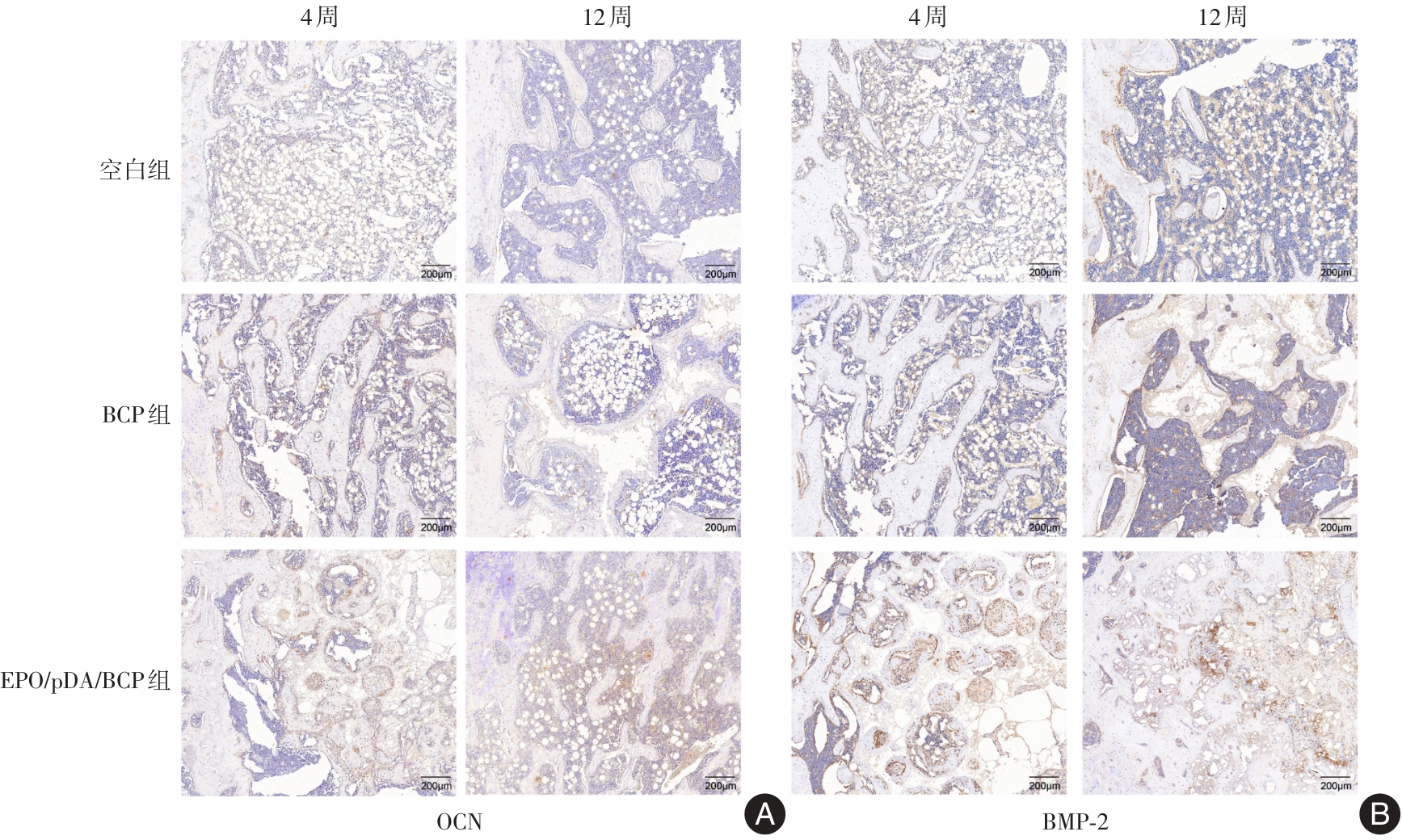
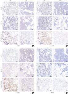
图9
术后4、12周各组大鼠股骨缺损部位免疫组化染色分析结果注:免疫组化染色(棕色为阳性信号,苏木精复染核为蓝色)。A,p-PI3K在不同组骨缺损区域中的表达情况;B,p-AKT在不同组骨缺损区域中的表达情况;C,CYTL1在不同组骨缺损区域中的表达情况;D,MTSS1在不同组骨缺损区域中的表达情况。比例尺:200 μm"

| [1] |
LIU J, MENG Z, SONG J, et al. Yoda1-Loaded Microfibrous Scaffolds Accelerate Osteogenesis through Piezo1-F-Actin Pathway-Mediated YAP Nuclear Localization and Functionalization[J]. ACS Appl Mater Interfaces, 2025, 17(21): 30559-30572. doi: 10.1021/acsami.5c03093 .
doi: 10.1021/acsami.5c03093 |
| [2] |
QIU S, CAO L, XIANG D, et al. Enhanced osteogenic differentiation in 3D hydrogel scaffold via macrophage mitochondrial transfer[J]. J Nanobiotechnology, 2024, 22(1): 540. doi: 10.1186/s12951-024-02757-1 .
doi: 10.1186/s12951-024-02757-1 |
| [3] |
XUE N, DING X, HUANG R, et al. Bone tissue engineering in the treatment of bone defects[J]. Pharmaceuticals, 2022, 15(7): 879. doi: 10.3390/ph15070879 .
doi: 10.3390/ph15070879 |
| [4] |
宋李治, 陈慧坤, 张奕纯, 等. 二甲双胍在骨缺损修复中的研究进展和临床应用[J]. 实用医学杂志, 2022, 38(12): 1443-1448. doi: 10.3969/j.issn.1006-5725.2022.12.001 .
doi: 10.3969/j.issn.1006-5725.2022.12.001 |
| [5] |
栾非凡, 陈嘉怡, 郑臣校, 等. 伤科九味健骨片通过调控基质细胞衍生因子-1α/C-X-C趋化因子受体4型轴对骨髓间充质干细胞成骨分化及牵张成骨的影响[J]. 实用医学杂志, 2026, 42(2): 285-294. doi: 10.3969/j.issn.1006-5725.2026.02.014 .
doi: 10.3969/j.issn.1006-5725.2026.02.014 |
| [6] |
DELYANEE M, TABATABAEE S, SAMANIPOUR R, et al. Effects of gamma radiation as terminal sterilization on mechanical properties and organic composition of bone and skin allograft[J]. J Mater Res, 2025, 40(9): 1374-1389. doi: 10.1557/s43578-025-01584-w .
doi: 10.1557/s43578-025-01584-w |
| [7] |
吴妍廷, 李宇, 廖金凤. 氧化镁纳米粒调控成骨与血管生成相关基因表达促进骨缺损愈合[J]. 中国组织工程研究, 2026, 30(8): 1885-1895. doi: 10.12307/2026.030 .
doi: 10.12307/2026.030 |
| [8] |
HOU X, ZHANG L, ZHOU Z, et al. Calcium phosphate-based biomaterials for bone repair[J]. J Funct Biomater, 2022, 13(4): 187. doi: 10.3390/jfb13040187 .
doi: 10.3390/jfb13040187 |
| [9] |
王宁宁, 徐志民, 郑叶, 等. 聚多巴胺涂层3D打印双相磷酸钙组织工程支架的制备及其评价[J]. 吉林大学学报(医学版), 2019, 45(4): 955-959. doi: 10.13481/j.1671-587x.20190438 .
doi: 10.13481/j.1671-587x.20190438 |
| [10] |
YANG Z, XIE L, ZHANG B, et al. Preparation of BMP-2/PDA-BCP bioceramic scaffold by DLP 3D printing and its ability for inducing continuous bone formation[J]. Front Bioeng Biotechnol, 2022, 10: 854693. doi: 10.3389/fbioe.2022.854693 .
doi: 10.3389/fbioe.2022.854693 |
| [11] |
杨玉芳, 杨芷姗, 段棉棉, 等. 促红细胞生成素在骨组织工程中的应用及前景[J]. 中国组织工程研究, 2024, 28(9): 1443-1449. doi: 10.12307/2024.014 .
doi: 10.12307/2024.014 |
| [12] |
TSIFTSOGLOU A S. Erythropoietin (EPO) as a key regulator of erythropoiesis, bone remodeling and endothelial transdifferentiation of multipotent mesenchymal stem cells (MSCs): Implications in regenerative medicine[J]. Cells, 2021, 10(8): 2140. doi: 10.3390/cells10082140 .
doi: 10.3390/cells10082140 |
| [13] |
ZHANG X, LIU J, LI Y, et al. Erythropoietin Functionalized Hierarchical Scaffolds Enable Immunomodulated and Vascularized Bone Repair in Periodontal Defect[J]. ACS Appl Mater Interfaces, 2026, 18(9): 14256-14273. doi: 10.1021/acsami. 5c24484 .
doi: 10.1021/acsami. 5c24484 |
| [14] |
WU X, ZHU Y, QI Y, et al. Erythropoietin, as a biological macromolecule in modification of tissue engineered constructs: A review[J]. Int J Biol Macromol, 2021, 193: 2332-2342. doi: 10.1016/j.ijbiomac.2021.11.065 .
doi: 10.1016/j.ijbiomac.2021.11.065 |
| [15] |
SENATOV F, AMANBEK G, ORLOVA P, et al. Biomimetic UHMWPE/HA scaffolds with rhBMP-2 and erythropoietin for reconstructive surgery[J]. Mater Sci Eng C, 2020, 111: 110750. doi: 10.1016/j.msec.2020.110750 .
doi: 10.1016/j.msec.2020.110750 |
| [16] |
SENATOV F, ZIMINA A, CHUBRIK A, et al. Effect of recombinant BMP-2 and erythropoietin on osteogenic properties of biomimetic PLA/PCL/HA and PHB/HA scaffolds in critical-size cranial defects model[J]. Mater Sci Eng C, 2022, 135: 112680. doi: 10.1016/j.msec.2022.112680 .
doi: 10.1016/j.msec.2022.112680 |
| [17] |
WANG Y, WANG P, WU Q, et al. Loading of erythropoietin on biphasic calcium phosphate bioceramics promotes osteogenesis and angiogenesis by regulating EphB4/EphrinB2 molecules[J]. J Mater Sci Mater Med, 2022, 33(2): 19. doi: 10.1007/s10856-022-06644-9 .
doi: 10.1007/s10856-022-06644-9 |
| [18] |
YU Y, MA L, ZHANG H, et al. EPO could be regulated by HIF-1 and promote osteogenesis and accelerate bone repair[J]. Artif Cells Nanomed Biotechnol, 2020, 48(1): 206-217. doi: 10.1080/21691401.2019.1699827 .
doi: 10.1080/21691401.2019.1699827 |
| [19] |
QIN Q, LIU Y, YANG Z, et al. Hypoxia-inducible factors signaling in osteogenesis and skeletal repair[J]. Int J Mol Sci, 2022, 23(19): 11201. doi: 10.3390/ijms231911201 .
doi: 10.3390/ijms231911201 |
| [20] |
卢燕勤, 易芳, 鞠巍, 等. Ca-P涂层镁合金支架负载缓释微球修复股骨缺损[J]. 中国组织工程研究, 2019, 23(2): 232-238. doi: 10.3969/j.issn.2095-4344.0689 .
doi: 10.3969/j.issn.2095-4344.0689 |
| [21] |
MA S, LIU X, XIAO B, et al. An Artificial Intelligence‐Selected Multifunctional Oligopeptide Coated Biphasic Calcium Phosphate (BCP) Scaffold Promotes Bone Defect Repair[J]. Adv Healthc Mater, 2026, 15(16): e05458. doi: 10.1002/adhm.202505458 .
doi: 10.1002/adhm.202505458 |
| [22] |
XIANG M, ZHU F, HAO Z, et al. Vat-Photopolymerized Biphasic Calcium Phosphate Bioceramics with Cortical Bone-Like Properties via Microwave Sintering[J]. Ceram Int, 2026. doi: 10.1016/j.ceramint.2026.03.379 .
doi: 10.1016/j.ceramint.2026.03.379 |
| [23] |
KIM S E, PARK K. Recent advances of biphasic calcium phosphate bioceramics for bone tissue regeneration[J]. Adv Exp Med Biol, 2020, 1250: 177-188. doi: 10.1007/978-981-15-3262-7_12 .
doi: 10.1007/978-981-15-3262-7_12 |
| [24] |
YASUOKA Y, IZUMI Y, SANDS J M, et al. Progress in the Detection of Erythropoietin in Blood, Urine, and Tissue[J]. Molecules, 2023, 28(11): 4446. doi: 10.3390/molecules28114446 .
doi: 10.3390/molecules28114446 |
| [25] |
RÖLFING J H D, BAATRUP A, STIEHLER M, et al. The osteogenic effect of erythropoietin on human mesenchymal stromal cells is dose-dependent and involves non-hematopoietic receptors and multiple intracellular signaling pathways[J]. Stem Cell Rev Rep, 2014, 10(1): 69-78. doi: 10.1007/s12015-013-9476-x .
doi: 10.1007/s12015-013-9476-x |
| [26] |
LIU S, WANG Z, LI Y, et al. Erythropoietin‐stimulated macrophage‐derived extracellular vesicles in chitosan hydrogel rescue BMSCs fate by targeting EGFR to alleviate inflammatory bone loss in periodontitis[J]. Adv Sci, 2025, 12(23): 2500554. doi: 10.1002/advs.202500554 .
doi: 10.1002/advs.202500554 |
| [27] |
KIM J, JUNG Y, SUN H, et al. Erythropoietin mediated bone formation is regulated by mTOR signaling[J]. J Cell Biochem, 2012, 113(1): 220-228. doi: 10.1002/jcb.23347 .
doi: 10.1002/jcb.23347 |
| [28] |
VASILEVA R, CHAPRAZOV T. Evaluation of recombinant human erythropoietin as a promoter of bone healing in cats with femoral fractures[J]. Open Vet J, 2024, 14(4): 1012. doi: 10.5455/OVJ.2024.v14.i4.8 .
doi: 10.5455/OVJ.2024.v14.i4.8 |
| [29] |
HOLSTEIN J H, ORTH M, SCHEUER C, et al. Erythropoietin stimulates bone formation, cell proliferation, and angiogenesis in a femoral segmental defect model in mice[J]. Bone, 2011, 49(5): 1037-1045. doi: 10.1016/j.bone.2011.08.004 .
doi: 10.1016/j.bone.2011.08.004 |
| [30] |
CHAPRAZOV T, VASILEVA R, ATLIEV K, et al. Diagnostic imaging studies on local and systemic erythropoietin application for promoting bone regeneration in rat calvarial defects[J]. Vet Sci, 2022, 9(10): 578. doi: 10.3390/vetsci9100578 .
doi: 10.3390/vetsci9100578 |
| [31] |
LI X, TANG J, GUO W, et al. Polydopamine Nanocomposite Hydrogel for Drug Slow-Release in Bone Defect Repair: A Review of Research Advances[J]. Gels, 2025, 11(3): 190. doi: 10.3390/gels11030190 .
doi: 10.3390/gels11030190 |
| [32] |
BEDHIAFI T, IDOUDI S, ALHAMS A A, et al. Applications of polydopaminic nanomaterials in mucosal drug delivery[J]. J Control Release, 2023, 353: 842-849. doi: 10.1016/j.jconrel.2022. 12.037 .
doi: 10.1016/j.jconrel.2022. 12.037 |
| [33] |
GHORBANI F, KIM M, GHALANDARI B, et al. Architecture of β-lactoglobulin coating modulates bioinspired alginate dialdehyde-gelatine/polydopamine scaffolds for subchondral bone regeneration[J]. Acta Biomater, 2024, 181: 188-201. doi: 10.1016/j.actbio.2024.04.028 .
doi: 10.1016/j.actbio.2024.04.028 |
| [34] |
HUANG S, LIANG N, HU Y, et al. Polydopamine-assisted surface modification for bone biosubstitutes[J]. Biomed Res Int, 2016, 2016(1): 2389895. doi: 10.1155/2016/2389895 .
doi: 10.1155/2016/2389895 |
| [35] |
LI D, LI Q, XU T, et al. Pro-vasculogenic fibers by PDA-mediated surface functionalization using cell-free fat extract (CEFFE)[J]. Biomacromolecules, 2024, 25(3): 1550-1562. doi: 10.1021/acs.biomac.3c01124 .
doi: 10.1021/acs.biomac.3c01124 |
| [36] |
SUN X, JIAO X, WANG Z, et al. Polydopamine-coated 3D-printed β-tricalcium phosphate scaffolds to promote the adhesion and osteogenesis of BMSCs for bone-defect repair: mRNA transcriptomic sequencing analysis[J]. J Mater Chem B, 2023, 11(8): 1725-1738. doi: 10.1039/d2tb02280j .
doi: 10.1039/d2tb02280j |
| [37] |
WANG P, YIN H M, LI X, et al. Simultaneously constructing nanotopographical and chemical cues in 3D-printed polylactic acid scaffolds to promote bone regeneration[J]. Mater Sci Eng C, 2021, 118: 111457. doi: 10.1016/j.msec.2020.111457 .
doi: 10.1016/j.msec.2020.111457 |
| [38] |
SWETHA S, RAO G N, MAHALAKSHMI V, et al. Alkaline phosphatase and acid phosphatase in health and disease-A systematic review[J]. J Oral Maxillofac Pathol, 2025, 29(2): 324-334. doi: 10.4103/jomfp.jomfp_304_24 .
doi: 10.4103/jomfp.jomfp_304_24 |
| [39] |
VIMALRAJ S. Alkaline phosphatase: Structure, expression and its function in bone mineralization[J]. Gene, 2020, 754: 144855. doi: 10.1016/j.gene.2020.144855 .
doi: 10.1016/j.gene.2020.144855 |
| [40] |
WANG Y, PAN J, ZHANG Y, et al. Wnt and Notch signaling pathways in calcium phosphate‐enhanced osteogenic differentiation: A pilot study[J]. J Biomed Mater Res B Appl Biomater, 2019, 107(1): 149-160. doi: 10.1002/jbm.b.34105 .
doi: 10.1002/jbm.b.34105 |
| [41] |
胡俊, 谈荣珍, 袁忠, 等. 锁阳多糖通过激活PI3K/Akt/GSK3β/β-catenin信号通路促进MC3T3-E1细胞成骨分化[J]. 实用医学杂志, 2022, 38(22): 2774-2779. doi: 10.3969/j.issn.1006-5725.2022.22.003 .
doi: 10.3969/j.issn.1006-5725.2022.22.003 |
| [42] |
LI J, HUANG Z, LI B, et al. Mobilization of transplanted bone marrow mesenchymal stem cells by erythropoietin facilitates the reconstruction of segmental bone defect[J]. Stem Cells Int, 2019, 2019(1): 5750967. doi: 10.1155/2019/5750967 .
doi: 10.1155/2019/5750967 |
| [43] |
刘岚, 魏松, 张丞, 等. 小檗碱通过PI3K/AKT信号通路调控人牙周膜干细胞的增殖和成骨分化[J]. 中国细胞生物学学报, 2024, 46(3): 494-501. doi: 10.11844/cjcb.2024.03.0012 .
doi: 10.11844/cjcb.2024.03.0012 |
| [44] |
陶恩东, 夏伟杰, 王福林, 等.骨髓间充质干细胞来源的外泌体通过PI3K/AKT信号通路逆转高糖诱导的骨髓间充质干细胞成骨功能障碍[J]. 温州医科大学学报, 2024, 54(8): 641-649. doi: 10.3969/j.issn.2095-9400.2024.08.005 .
doi: 10.3969/j.issn.2095-9400.2024.08.005 |
| [45] |
LI B, ZHANG S, YUN X, et al. NEDD4's effect on osteoblastogenesis potential of bone mesenchymal stem cells in rats concerned with PI3K/Akt pathway[J]. Differentiation, 2025, 141: 100830. doi: 10.1016/j.diff.2024.100830 .
doi: 10.1016/j.diff.2024.100830 |
| [46] |
CHEN M, SHAN L, GAN Y, et al. Metastasis suppressor 1 controls osteoblast differentiation and bone homeostasis through regulating Src-Wnt/β-catenin signaling[J]. Cell Mol Life Sci, 2022, 79(2): 107. doi: 10.1007/s00018-022-04147-y .
doi: 10.1007/s00018-022-04147-y |
| [47] |
SHIN Y, WON Y, YANG J I, et al. CYTL1 regulates bone homeostasis in mice by modulating osteogenesis of mesenchymal stem cells and osteoclastogenesis of bone marrow-derived macrophages[J]. Cell Death Dis, 2019, 10(2): 47. doi: 10.1038/s41419-018-1284-4 .
doi: 10.1038/s41419-018-1284-4 |
| [1] | 申福国,肖文龙,孙文才,金松,崔冬梅. 基于TAK1/NF-κB/MAPK信号通路探讨骨隧道机械微环境优化对前交叉韧带重建术后腱-骨愈合的作用机制[J]. 实用医学杂志, 2026, 42(5): 807-813. |
| [2] | 栾非凡,陈嘉怡,郑臣校,郑雨中,黄枫. 伤科九味健骨片通过调控基质细胞衍生因子-1α/C-X-C趋化因子受体4型轴对骨髓间充质干细胞成骨分化及牵张成骨的影响[J]. 实用医学杂志, 2026, 42(2): 285-294. |
| [3] | 徐丽娜,何小双,刘冬,邬超. 骨髓间充质干细胞外泌体缓解小鼠哮喘模型的功能和机制[J]. 实用医学杂志, 2026, 42(1): 72-78. |
| [4] | 黄玉圣,刘心雨,周穗珊. 改良全骨髓贴壁法培养SD大鼠骨髓间充质干细胞及其生物学特性[J]. 实用医学杂志, 2025, 41(24): 3860-3866. |
| [5] | 李爱强,赵宁,张旭升,柳海平. 骨髓间充质干细胞来源细胞外囊泡治疗股骨头骨坏死的研究进展[J]. 实用医学杂志, 2025, 41(23): 3631-3637. |
| [6] | 刘慧珍,卢晓南,柳歌,蔡关庆,李平安,曾英坚,尚广彬. 肿节风总黄酮影响骨髓间充质干细胞及其外泌体促进巨核细胞分化的作用及机制[J]. 实用医学杂志, 2025, 41(11): 1618-1626. |
| [7] | 王山,万金良,赵中华,杨静,赵杰,郝延璋. 三白草酮调控Krüppel样因子5-促红细胞生成素肝细胞A2通路对肝癌细胞生长和免疫逃逸的影响[J]. 实用医学杂志, 2025, 41(10): 1480-1486. |
| [8] | 巢素珍,周年,石心怡,周怡丽,夏俊杰,刘波,任明诗,李子涵. 芒果苷对同型半胱氨酸诱导骨髓间充质干细胞分化的影响[J]. 实用医学杂志, 2024, 40(23): 3284-3290. |
| [9] | 赵论,赵欣,林晨琛,付琪,石默晗,张浩然. miR-217靶向FOXO3影响非小细胞肺癌对吉非替尼耐药性及相关机制[J]. 实用医学杂志, 2024, 40(16): 2277-2283. |
| [10] | 陈霞 武馨馨 刘星佑 陈鑫昊 黄尹霞 肖志原 贺继刚 . 过表达NKx2.5 CXCR 基因间充质干细胞增强SDF⁃1/ 4轴促归巢改善心梗心功能 [J]. 实用医学杂志, 2023, 39(6): 660-666. |
| [11] | 王莉平 林静 薄雨佳 赵今. 紫草素对MC3T3⁃E1细胞增殖、分化、矿化及成骨相关基因表达的影响 [J]. 实用医学杂志, 2022, 38(5): 583-588. |
| [12] | 胡俊 谈荣珍 袁忠 杨盼盼 张楚焌 张斌 沈云峰 . 锁阳多糖通过激活PI3K/Akt/GSK3β/β-catenin信号通路促进MC3T3-E1细胞成骨分化 [J]. 实用医学杂志, 2022, 38(22): 2774-2779. |
| [13] | 吴靖漪 陈嘉文, 孙天语 刘鹏 吴补领, . 蛋白聚糖调控间充质干细胞成骨分化的研究进展 [J]. 实用医学杂志, 2021, 37(9): 1217-1221. |
| [14] | 汪仡骁 吴恒鹏 万雪 陈方 石超 瓦庆德. miR⁃30b在低氧环境中调控兔骨髓间充质干细胞成软骨分化 [J]. 实用医学杂志, 2021, 37(24): 3113-3118. |
| [15] | 连学辉 肖红利 邓江 韩子冀 齐维林. hBDNF⁃BMSCs局部移植对大鼠脊髓损伤的修复效果 [J]. 实用医学杂志, 2021, 37(20): 2590-2596. |
| 阅读次数 | ||||||
|
全文 |
|
|||||
|
摘要 |
|
|||||
