实用医学杂志 ›› 2025, Vol. 41 ›› Issue (6): 812-817.doi: 10.3969/j.issn.1006-5725.2025.06.006

• 临床研究 • 上一篇    

可视管芯与可视喉镜在口腔科手术经鼻气管插管中的应用比较

李曼君1,胡磊蕾2,胡海军2,张静2,余树春2,罗振中2,邓伟2()   

  1. 1.南昌大学第二附属医院,手术室,(江西 南昌 330006 )
    2.南昌大学第二附属医院,麻醉科,(江西 南昌 330006 )
  • 收稿日期:2024-12-14 出版日期:2025-03-25 发布日期:2025-03-31
  • 通讯作者: 邓伟 E-mail:dengweidoc@163.com
  • 基金资助:
    国家自然科学基金项目(82160371);江西省卫生健康委科技计划(202210595)

Comparison of the application of video stylet and video laryngoscope in nasotracheal intubation in oral surgery

Manjun LI1,Leilei HU2,Haijun HU2,Jing ZHANG2,Shuchun YU2,Zhenzhong LUO2,Wei. DENG2()   

  1. Department of Operating Room,the Second Affiliated Hospital of Nanchang University,Nanchang 330006,Jiangxi,China
  • Received:2024-12-14 Online:2025-03-25 Published:2025-03-31
  • Contact: Wei. DENG E-mail:dengweidoc@163.com

摘要:

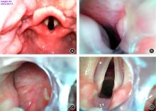
目的 比较可视管芯和可视喉镜在口腔科手术经鼻气管插管时的效果。 方法 选择择期全麻下口腔科手术患者80例,年龄18 ~ 70岁,ASA Ⅰ或Ⅱ级,随机分为可视管芯组(N组)、可视喉镜组(C组),每组40例。N组使用可视管芯将气管导管塑形90°,塑形位置为喉结至鼻孔的垂直距离,可视下将导管从鼻腔插入咽喉部,见声门后,置入导管。C组先将不带管芯的气管导管盲插入鼻腔,当导管到达咽喉部时,使用可视喉镜从口腔进入挑起会厌并暴露声门,借助插管钳或套囊充气法置入导管。主要观察指标为插管时间。记录鼻腔通过时间、声门暴露时间、声门暴露情况。记录插管次数、助手协助情况。记录入室平静休息5 min 时(T0)、暴露声门时(T1)、导管过声门时(T2)、导管进入气管后1 min(T3)时MAP、HR。记录鼻出血、口腔黏膜出血、门齿松动、术后咽喉痛等插管并发症。 结果 N组插管时间、鼻腔通过时间均明显短于C组(P < 0.05)。N组套囊充气、插管钳辅助例数明显少于C组(P < 0.05)。两组患者在声门暴露时间、首次插管成功次数、C-L声门分级、托举下颌辅助插管均无明显差异(P > 0.05)。N组在T1、T2时刻MAP、HR上升幅度均明显低于C组(P < 0.05)。N组轻度鼻出血例数明显少于C组(P < 0.05)。N组门齿松动、口腔黏膜出血发生例数均明显少于C组(P < 0.05)。 结论 与可视喉镜相比,可视管芯引导下经鼻气管插管的插管时间更短,对口鼻咽部损伤更小,不需要借助插管钳,并能够减轻患者插管时的心血管应激反应。

关键词: 可视管芯, 可视喉镜, 经鼻气管插管, 鼻咽腔护理

Abstract:

Objective This study aims to compare the efficacy of video stylets and video laryngoscopes in facilitating nasotracheal intubation during oral surgery. Methods A total of 80 patients, aged between 18 and 70 years old, with ASA grade Ⅰ or Ⅱ, scheduled for elective oral surgery under general anesthesia, were randomly assigned to either the video stylet group (Group N) or the video laryngoscope group (Group C), with 40 patients in each group. In Group N, a video stylet was used to shape the tracheal tube at a 90-degree angle, with the shaping position being the vertical distance from the Adam's apple to the nostril. The tube was inserted from the nasal cavity into the throat under direct visualization, and positioned behind the glottis. In Group C, the tube was initially blindly inserted into the nasal cavity without a core. Upon reaching the throat, a video laryngoscope was employed to lift the epiglottis and expose the glottis from the mouth. The tube was then inserted with the aid of intubation forceps or cuff inflation. The primary outcome measure was the intubation time. Additional measures included the time taken for nasal passage, glottis exposure, and the number of intubation attempts and assistant interventions required. Vital signs, including MAP and HR, were recorded at five minutes of quiet rest upon entering the room (T0), during glottis exposure (T1), upon passage of the tube through the glottis (T2), and one minute after the tube entered the trachea (T3). Complications such as epistaxis, oral mucosal bleeding, loose incisors, and postoperative sore throat were also documented. Results The intubation time and nasal passage time in Group N were significantly shorter than those in Group C (P < 0.05). The number of cuff inflations and intubation forceps assisted cases in Group N was significantly lower than in Group C (P < 0.05). There were no significant differences between the two groups in terms of glottis exposure time, first successful intubation times, C-L glottis classification, and mandibular lift-assisted intubation (P > 0.05). The increase in MAP and HR in Group N at T1 and T2 was significantly less than in Group C (P < 0.05). The number of cases with mild epistaxis in Group N was significantly lower than in Group C (P < 0.05). Similarly, the number of cases with loose incisors and oral mucosal bleeding in Group N was significantly less than in Group C (P < 0.05). Conclusion Compared to the video laryngoscope, the video stylet-guided nasotracheal intubation results in a shorter intubation time, less damage to the oronasopharynx, eliminates the need for intubation forceps, and reduces the patient's stress and vascular stress response during intubation.

Key words: video stylet, video laryngoscope, nasotracheal intubation, nasopharyngeal cavity care

中图分类号: