

实用医学杂志 ›› 2024, Vol. 40 ›› Issue (23): 3306-3316.doi: 10.3969/j.issn.1006-5725.2024.23.005
• 基础研究 • 上一篇
贾祥1,2,徐田杰1,2,樊佳欣1,2,郭小玲1,2,刘凯楠3,张辉4,王永生1,2,王茜1,2( )
)
收稿日期:2024-08-23
出版日期:2024-12-10
发布日期:2024-12-16
通讯作者:
王茜
E-mail:tswqxx008@126.com
基金资助:
Xiang JIA1,2,Tianjie XU1,2,Jiaxin FAN1,2,Xiaoling GUO1,2,Kainan LIU3,Hui ZHANG4,Yongsheng WANG1,2,Qian. WANG1,2()
Received:2024-08-23
Online:2024-12-10
Published:2024-12-16
Contact:
Qian. WANG
E-mail:tswqxx008@126.com
摘要:
目的 探究二甲双胍对骨关节炎大鼠软骨保护作用机制。 方法 取30只雄性SD大鼠,随机分为3组 (10只/组) 。建立大鼠膝骨关节炎模型,二甲双胍组大鼠接受灌胃治疗[200 mg/(kg·d)],空白组和模型组大鼠给予生理盐水作为对照。4周后,采用形态学染色方法观察关节软骨形态,通过免疫组化染色、免疫荧光染色及Western blot检测SIRT1/p53信号通路因子、炎症及凋亡相关因子的表达。 结果 二甲双胍组大鼠较模型组软骨结构损伤明显减轻,软骨层面趋于平整,软骨细胞数目增多,蛋白多糖含量增加。免疫组化染色、免疫荧光染色和Western blot检测发现,二甲双胍组大鼠软骨组织中SOX9、Aggrecan、Bcl-2、SIRT1蛋白表达较模型组显著升高,而IL-6、TNF-α、BAX、Caspase-9、p53蛋白表达明显降低。TUNEL染色结果显示,与模型组比较,二甲双胍组大鼠软骨细胞凋亡数量明显减少。 结论 二甲双胍可通过SIRT1/p53信号通路的激活减少骨关节炎SD模型大鼠软骨细胞凋亡,抑制软骨细胞外基质降解,进而保护关节软骨。
中图分类号:
贾祥,徐田杰,樊佳欣,郭小玲,刘凯楠,张辉,王永生,王茜. 二甲双胍通过激活SIRT1/p53信号通路对骨关节炎大鼠关节软骨发挥保护作用[J]. 实用医学杂志, 2024, 40(23): 3306-3316.
Xiang JIA,Tianjie XU,Jiaxin FAN,Xiaoling GUO,Kainan LIU,Hui ZHANG,Yongsheng WANG,Qian. WANG. Metformin exerts a protective effect on articular cartilage in osteoarthritis rats by activating the SIRT1/p53 signaling pathway[J]. The Journal of Practical Medicine, 2024, 40(23): 3306-3316.
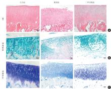
图 1
各组大鼠膝关节软骨形态观察 (× 200)注:A,关节软骨苏木精-伊红染色,空白组软骨表面光滑平整;模型组软骨表面出现破损,细胞数量减少;二甲双胍组软骨表面趋于平整,细胞数量增多;B,阿利新蓝染色,空白组软骨基质均呈蓝色,模型组软骨基质部分区域染色不均,二甲双胍组软骨基质呈蓝色;C,甲苯胺蓝染色,空白组软骨基质呈蓝紫色,模型组软骨基质呈淡蓝色,二甲双胍组软骨基质呈蓝紫色。比例尺=50 μm"
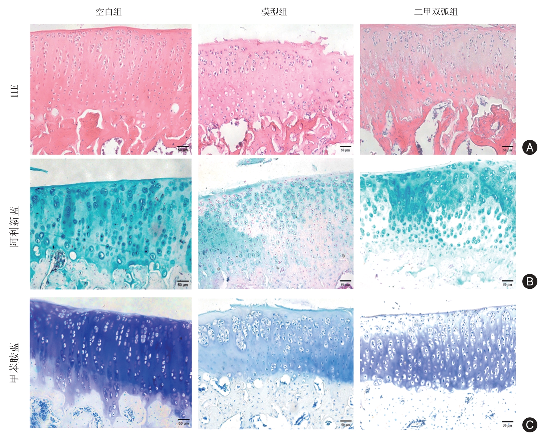
表1
大鼠OARSI评分 (x ± s)"
| 组别 | 例数 | 分数 |
|---|---|---|
| 空白组 | 3 | 0.000 ± 0.000 |
| 模型组 | 3 | 9.667 ± 2.082? |
| 二甲双胍组 | 3 | 3.333 ± 1.528# |
| F值 | 33.650 | |
| P值 | 0.001 |
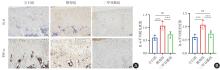
图2
大鼠软骨组织IL-6和TNF-α免疫组化染色结果 (× 200)注:A,IL-6和TNF-α免疫组化染色图,与空白组比较,模型组IL-6和TNF-α阳性细胞数量明显增多且染色较深;与模型组比较,二甲双胍组IL-6和TNF-α阳性细胞数量减少且染色稍浅。比例尺 = 50 μm;B,IL-6和TNF-α 免疫组化染色平均吸光度值统计分析,*P < 0.05,***P < 0.001,nsP > 0.05"

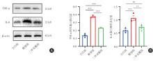
图3
大鼠软骨组织IL-6和TNF-α Western blot检测结果注:A,TNF-α与IL-6蛋白表达免疫印迹图;B,IL-6与TNF-α蛋白比值定量统计分析。*P < 0.05,**P < 0.01,***P < 0.001,nsP > 0.05"
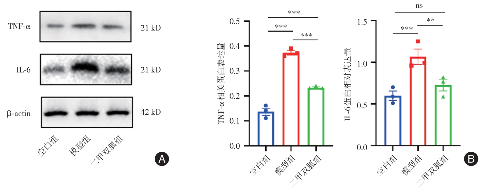
图4
大鼠软骨组织Aggerecan与SOX9免疫组化染色结果 (× 200)注:A,Aggerecan与SOX9免疫组化染色图,与空白组比较,模型组Aggerecan与SOX9阳性细胞数量明显减少且染色变浅;与模型组比较,二甲双胍组Aggerecan与SOX9阳性细胞数量增多且染色较深。比例尺 = 50 μm;B,Aggerecan与SOX9 免疫组化染色平均吸光度值统计分析,**P < 0.01,***P < 0.001,nsP > 0.05"

图5
大鼠软骨Aggerecan与SOX9 Western blot检测结果注:A,Aggrecan与SOX9蛋白表达免疫印迹图;B,Aggrecan与SOX9蛋白比值定量统计分析,*P < 0.05,**P < 0.01,***P < 0.001,nsP > 0.05"
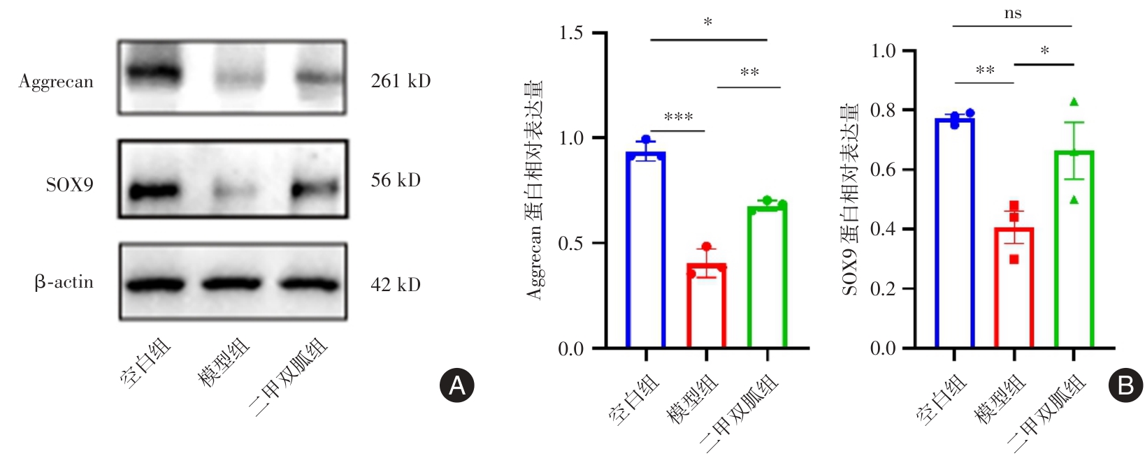
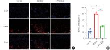
图6
大鼠软骨组织TUNEL荧光染色结果 (× 200)注:A,TUNEL荧光染色图,TUNEL阳性染色为红色荧光,与空白组比较,模型组阳性细胞数量增加;与模型组比较,二甲双胍组阳性细胞数量减少。比例尺 =50 μm;B,TUNEL阳性细胞率,*P < 0.05,**P < 0.01,***P < 0.001"

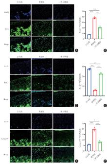
图7
大鼠软骨组织BAX、Bcl-2与Caspase-9免疫荧光染色结果 (× 200)注:A,BAX免疫荧光染色图,阳性表达可见绿色荧光分布于细胞质,与空白组比较,模型组平均荧光强度较强;与模型组比较,二甲双胍组平均荧光强度较弱。比例尺 =50 μm;B,BAX免疫荧光染色图平均荧光强度值分析;C,Bcl-2免疫荧光染色图,阳性表达可见绿色荧光分布于细胞质,与空白组比较,模型组平均荧光强度较弱;与模型组比较,二甲双胍组平均荧光强度较强。比例尺 = 50 μm;D,Bcl-2免疫荧光染色图平均荧光强度值分析;E,Caspase-9免疫荧光染色图,阳性表达可见绿色荧光分布于细胞质,与空白组比较,模型组平均荧光强度较弱;与模型组比较,二甲双胍组平均荧光强度较强。比例尺 = 50 μm;F,Caspase-9免疫荧光染色图平均荧光强度值分析,*P < 0.05,**P < 0.01,***P < 0.001"
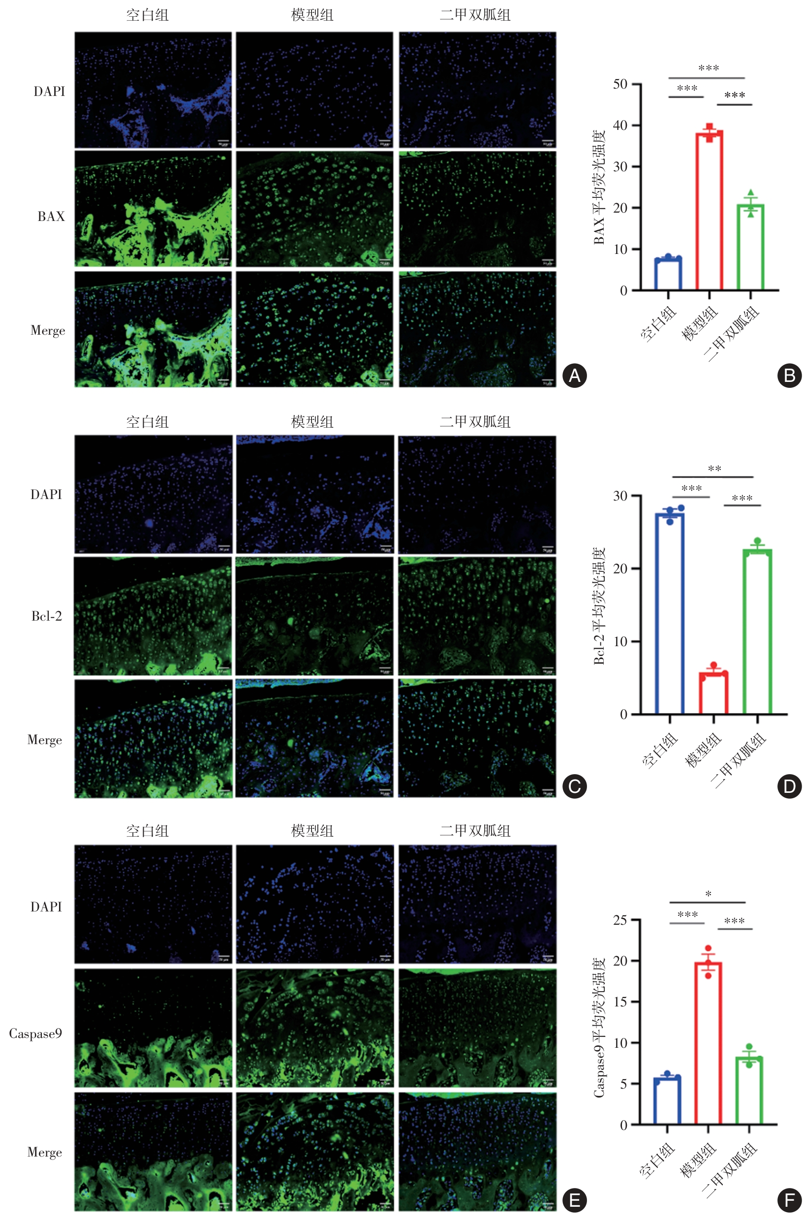
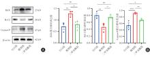
图8
大鼠软骨组织BAX、Bcl-2与Caspase-9 Western blot检测结果注:A,BAX、Bcl-2与Caspase-9蛋白表达免疫印迹图;B,BAX、Bcl-2与Caspase-9蛋白比值定量统计分析,*P < 0.05,**P < 0.01,***P < 0.001,nsP > 0.05"
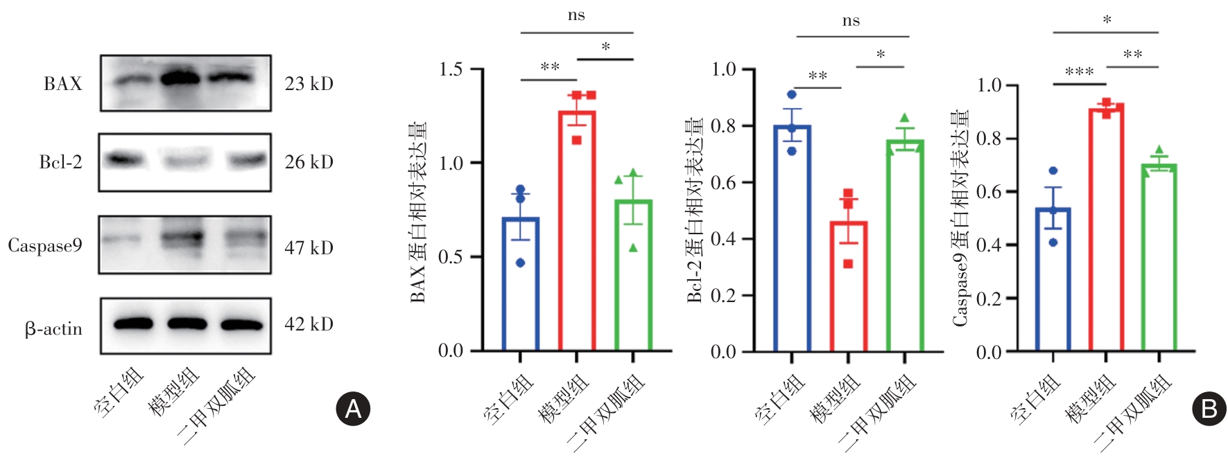
图9
大鼠软骨组织SIRT1与p53免疫组化染色结果 (× 200)注:A,SIRT1与p53免疫组化染色图,与空白组比较,模型组SIRT1阳性细胞数量明显减少且染色较浅;与模型组比较,二甲双胍组SIRT1阳性细胞数量增加且染色较深。与空白组比较,模型组 p53阳性细胞数量明显增加且染色较深;与模型组比较,二甲双胍组p53阳性细胞数量减少且染色稍浅。比例尺 =50 μm。B,SIRT1与p53免疫组化染色平均吸光度值统计分析,*P < 0.05,**P < 0.01,***P < 0.001,nsP > 0.05"
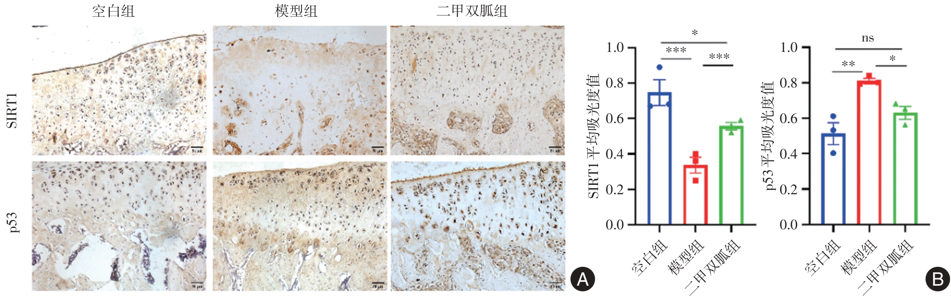
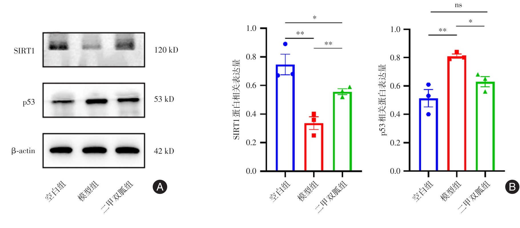
图10
大鼠软骨组织SIRT1与p53 Western blot检测结果注:A,SIRT1与p53蛋白表达免疫印迹图;B,SIRT1与p53蛋白比值定量统计分析,*P < 0.05,**P < 0.01,nsP > 0.05"
| 1 |
DIAMOND L E, GRANT T, UHLRICH S D. Osteoarthritis year in review 2023: Biomechanics[J]. Osteoarthritis Cartilage, 2024, 32(2): 138-147. doi:10.1016/j.joca.2023.11.015
doi: 10.1016/j.joca.2023.11.015 |
| 2 |
MINNIG M C C, GOLIGHTLY Y M, NELSON A E. Epidemiology of osteoarthritis: Literature update 2022-2023[J]. Curr Opin Rheumatol, 2024, 36(2): 108-112. doi:10.1097/bor.0000000000000985
doi: 10.1097/bor.0000000000000985 |
| 3 | PERRUCCIO A V, YOUNG J J, WILFONG J M, 等. Osteoarthritis year in review 2023: Epidemiology & therapy[J]. Osteoarthritis Cartilage, 2024, 32(2): 159-165. |
| 4 |
XU X, SUN Y, CEN X, et al. Metformin activates chaperone-mediated autophagy and improves disease pathologies in an Alzheimer disease mouse model[J]. Protein Cell, 2021, 12(10): 769-787. doi:10.1007/s13238-021-00858-3
doi: 10.1007/s13238-021-00858-3 |
| 5 |
FORETZ M, GUIGAS B, VIOLLET B. Metformin: Update on mechanisms of action and repurposing potential[J]. Nat Rev Endocrinol, 2023, 19(8): 460-476. doi:10.1038/s41574-023-00833-4
doi: 10.1038/s41574-023-00833-4 |
| 6 | 田珂, 冷秋枫, 吕晶, 等. 二甲双胍通过NLRP3炎症小体通路对皮肤角质形成细胞增殖和凋亡的双向调节研究[J]. 中国全科医学, 2025,28(6):742-750. |
| 7 |
KUSWANTO W, BAKER M C. Repurposing drugs for the treatment of osteoarthritis[J]. Osteoarthritis Cartilage, 2024, 32(8): 886-895. doi:10.1016/j.joca.2024.05.008
doi: 10.1016/j.joca.2024.05.008 |
| 8 |
HE M, LU B, OPOKU M, et al. Metformin Prevents or Delays the Development and Progression of Osteoarthritis: New Insight and Mechanism of Action[J]. Cells, 2022, 11(19): 3012. doi:10.3390/cells11193012
doi: 10.3390/cells11193012 |
| 9 |
CHEN C, ZHOU M, GE Y, et al. SIRT1 and aging related signaling pathways[J]. Mech Ageing Dev, 2020, 187: 111215. doi:10.1016/j.mad.2020.111215
doi: 10.1016/j.mad.2020.111215 |
| 10 |
CHEN L, HUADONG Z, YANQING L, et al. Novel Role of the SIRT1 in Endocrine and Metabolic Diseases[J]. Int J Biol Sci, 2023, 19(2):484-501. doi:10.7150/ijbs.78654
doi: 10.7150/ijbs.78654 |
| 11 | 贲莹, 张天雅, 田佳鑫, 等. 基于SIRT1/p53介导的细胞凋亡途径探讨补阳还五汤对糖尿病周围神经病变的治疗作用及方中黄芪用量[J]. 中国实验方剂学杂志, 2022, 28(2): 1-10. |
| 12 |
ZHOU M, LIU B, YE H M, et al. ROS-induced imbalance of the miR-34a-5p/SIRT1/p53 axis triggers chronic chondrocyte injury and inflammation[J]. Heliyon, 2024, 10(11): e31654. doi:10.1016/j.heliyon.2024.e31654
doi: 10.1016/j.heliyon.2024.e31654 |
| 13 | 冯晓峰, 张荣凯, 祁伟仲, 等. 二甲双胍干预骨关节炎模型小鼠早期骨关节炎软骨及软骨下骨变化[J]. 中国组织工程研究, 2019, 23(19): 3031-3036. |
| 14 |
LI J, ZHANG B, LIU W X, et al. Metformin limits osteoarthritis development and progression through activation of AMPK signalling[J]. Ann Rheum Dis, 2020, 79(5): 635-645. doi:10.1136/annrheumdis-2019-216713corr1
doi: 10.1136/annrheumdis-2019-216713corr1 |
| 15 | 徐田杰, 樊佳欣, 郭小玲, 等. 二甲双胍抑制PI3K/AKT/mTOR信号通路保护骨关节炎模型大鼠关节软骨[J]. 中国组织工程研究, 2025, 29(5): 1003-1012. |
| 16 |
LI D, RUAN G, ZHANG Y, et al. Metformin attenuates osteoarthritis by targeting chondrocytes, synovial macrophages and adipocytes[J]. Rheumatology(Oxford), 2023, 62(4): 1652-1661. doi:10.1093/rheumatology/keac467
doi: 10.1093/rheumatology/keac467 |
| 17 | 许学猛, 刘文刚, 许树柴, 等. 膝骨关节炎(膝痹)中西医结合临床实践指南[J]. 实用医学杂志, 2021, 37(22): 2827-2833. |
| 18 |
YAO Q, WU X, TAO C, et al. Osteoarthritis: Pathogenic signaling pathways and therapeutic targets[J]. Signal Transduct Target Ther, 2023, 8(1):56. doi:10.1038/s41392-023-01330-w
doi: 10.1038/s41392-023-01330-w |
| 19 |
TONG L, YU H, HUANG X, et al. Current understanding of osteoarthritis pathogenesis and relevant new approaches[J]. Bone Res, 2022, 10(1):60. doi:10.1038/s41413-022-00226-9
doi: 10.1038/s41413-022-00226-9 |
| 20 |
JONES I A, TOGASHI R, WILSON M L, et al. Intra-articular treatment options for knee osteoarthritis[J]. Nat Rev Rheumatol, 2019, 15(2): 77-90. doi:10.1038/s41584-018-0123-4
doi: 10.1038/s41584-018-0123-4 |
| 21 |
LI D, RUAN G, ZHANG Y, et al. Metformin attenuates osteoarthritis by targeting chondrocytes, synovial macrophages and adipocytes[J]. Rheumatology (Oxford), 2023, 62(4): 1652-1661. doi:10.1093/rheumatology/keac467
doi: 10.1093/rheumatology/keac467 |
| 22 |
ZAKI S, BLAKER C L, LITTLE C B. OA foundations-Exper-imental models of osteoarthritis[J]. Osteoarthritis Cartilage, 2022, 30(3): 357-380. doi:10.1016/j.joca.2021.03.024
doi: 10.1016/j.joca.2021.03.024 |
| 23 |
SZYMCZAK-PAJOR I, WENCLEWSKA S, ŚLIWIŃSKA A. Metabolic Action of Metformin[J]. Pharmaceuticals(Basel), 2022, 15(7): 810. doi:10.3390/ph15070810
doi: 10.3390/ph15070810 |
| 24 |
FENG X, PAN J, LI J, et al. Metformin attenuates cartilage degeneration in an experimental osteoarthritis model by regulating AMPK/mTOR[J]. Aging(Alany NY), 2020, 12(2): 1087-1103. doi:10.18632/aging.102635
doi: 10.18632/aging.102635 |
| 25 | 黄夏荣, 周君, 孙光华, 等. 电针对老年大鼠关节软骨及软骨下骨极化相关蛋白表达的影响[J]. 实用医学杂志, 2023, 39(12): 1473-1479. |
| 26 |
FUJII Y, LIU L, YAGASAKI L, et al. Cartilage Homeostasis and Osteoarthritis[J]. In J Mol Scis, 2022, 23(11): 6316. doi:10.3390/ijms23116316
doi: 10.3390/ijms23116316 |
| 27 |
CHEN Y, QIU F, YU B, et al. Metformin, an AMPK Activator, Inhibits Activation of FLSs but Promotes HAPLN1 Secretion[J]. Mol Ther Methods Clin Dev, 2020, 17: 1202-1214. doi:10.1016/j.omtm.2020.05.008
doi: 10.1016/j.omtm.2020.05.008 |
| 28 |
TYLUTKA A, WALAS Ł, ZEMBRON-LACNY A. Level of IL-6, TNF, and IL-1β and age-related diseases: A systematic review and meta-analysis[J]. Front Immunol, 2024, 15: 1330386. doi:10.3389/fimmu.2024.1330386
doi: 10.3389/fimmu.2024.1330386 |
| 29 |
NEGISHI Y, ADILI A, DE VEGA S, et al. IL-6 Reduces Spheroid Sizes of Osteophytic Cells Derived from Osteoarthritis Knee Joint via Induction of Apoptosis[J]. Am J Pathol, 2024, 194(1): 135-149. doi:10.1016/j.ajpath.2023.10.005
doi: 10.1016/j.ajpath.2023.10.005 |
| 30 |
WANG L, HE C. Nrf2-mediated anti-inflammatory polarization of macrophages as therapeutic targets for osteoarthritis[J]. Front Immunol, 2022, 13:967193. doi:10.3389/fimmu.2022.967193
doi: 10.3389/fimmu.2022.967193 |
| 31 |
NILSSON N, ALIM M D A, DIETRICH-ZAGONEL F, et al. The Delayed Presentation of Achilles Tendon Ruptures Is Associated With Marked Alterations in the Gene Expression of COL1A1, MMPs, TIMPs, and IL-6[J]. Am J Sports Med, 2024, 52(1): 164-173. doi:10.1177/03635465231212669
doi: 10.1177/03635465231212669 |
| 32 | 刘子歌, 陈德胜. 破骨细胞因子和抗破骨细胞因子在骨代谢调控网络中作用的研究进展[J]. 医学研究杂志, 2024, 53(6): 175-178. |
| 33 |
TANG H, GONG X, DAI J, et al. The IRF1/GBP5 axis promotes osteoarthritis progression by activating chondrocyte pyroptosis[J]. J Orthop Translat, 2024, 44: 47-59. doi:10.1016/j.jot.2023.11.005
doi: 10.1016/j.jot.2023.11.005 |
| 34 |
ZHOU Z, LV C, WANG Y, et al. BuShen JianGu Fang alleviates cartilage degeneration via regulating multiple genes and signaling pathways to activate NF-κB/Sox9 axis[J]. Phytomedicine, 2023, 113: 154742. doi:10.1016/j.phymed.2023.154742
doi: 10.1016/j.phymed.2023.154742 |
| 35 | 谭清梅, 杨诚, 廖坚文, 等. 二甲双胍通过AMPK信号通路对小鼠骨关节炎保护作用的研究[J]. 中国临床解剖学杂志, 2017, 35(4): 413-418. |
| 36 | 丁丽宏, 耿世佳, 王玉杰. 蟛蜞菊内酯对肺炎链球菌感染的肺泡上皮细胞凋亡及炎症因子分泌的调节作用[J]. 实用医学杂志, 2024, 40(3): 316-320. |
| 37 | 尹路, 蒋川锋, 陈俊杰, 等. 沙苑子苷A对关节软骨细胞凋亡的影响[J]. 中国组织工程研究, 2025, 29(8): 1541-1547. |
| 38 |
DADSENA S, CUEVAS ARENAS R, VIEIRA G, et al. Lipid unsaturation promotes BAX and BAK pore activity during apoptosis[J]. Nat Commun, 2024, 15(1): 4700. doi:10.1038/s41467-024-49067-6
doi: 10.1038/s41467-024-49067-6 |
| 39 |
VANDENABEELE P, BULTYNCK G, SAVVIDES S N. Pore-forming proteins as drivers of membrane permeabilization in cell death pathways[J]. Nat Rev Mol Cell Biol, 2023, 24(5): 312-333. doi:10.1038/s41580-022-00564-w
doi: 10.1038/s41580-022-00564-w |
| 40 | 李田洋, 高小凤, 王宝娟, 等. 骨炎消巴布剂对膝骨关节炎家兔软骨细胞凋亡及Bcl-2、Bax表达的影响[J]. 中国免疫学杂志, 2023, 39(8): 1647-1652. |
| 41 |
AI Y, MENG Y, YAN B, et al. The biochemical pathways of apoptotic, necroptotic, pyroptotic, and ferroptotic cell death[J]. Mol Cell, 2024, 84(1): 170-179. doi:10.1016/j.molcel.2023.11.040
doi: 10.1016/j.molcel.2023.11.040 |
| 42 |
NOGALES C, MAMDOUH Z M, LIST M, et al. Network pharmacology: Curing causal mechanisms instead of treating symptoms[J]. Trends Pharmacol Sci, 2022, 43(2): 136-150. doi:10.1016/j.tips.2021.11.004
doi: 10.1016/j.tips.2021.11.004 |
| 43 | 曾红玉, 叶贵珊, 武琦, 等. OXSR1活性对p53依赖和非依赖途径介导的犬肾细胞凋亡的影响[J]. 中国畜牧兽医, 2024,51(10): 4222-4234. |
| 44 |
YANG Y, LIU Y, WANG Y, et al. Regulation of SIRT1 and Its Roles in Inflammation[J]. Front Immunol, 2022, 13: 831168. doi:10.3389/fimmu.2022.831168
doi: 10.3389/fimmu.2022.831168 |
| 45 |
XU Y, WAN W. Acetylation in the regulation of autophagy[J]. Autophagy, 2023, 19(2): 379-387. doi:10.1080/15548627.2022.2062112
doi: 10.1080/15548627.2022.2062112 |
| 46 |
LI M, HU J, ZHOU J, et al. Grass carp (Ctenopharyngodon idella) deacetylase SIRT1 targets p53 to suppress apoptosis in a KAT8 dependent or independent manner[J]. Fish Shellfish Immunol, 2024, 144: 109264. doi:10.1016/j.fsi.2023.109264
doi: 10.1016/j.fsi.2023.109264 |
| [1] | 罗珊,冯莹,范丹丹,郑雯鑫,郭兴荣,阮绪芝. 血管生成素样蛋白8敲除减轻脂多糖诱导的肝脏脂质沉积[J]. 实用医学杂志, 2024, 40(9): 1197-1203. |
| [2] | 何伟,刘丽萍,卓静薇,张小冬,杨通,冯巨滨. 拮抗CC趋化因子受体5信号诱导肿瘤细胞凋亡并调节肿瘤微环境抑制肿瘤生长[J]. 实用医学杂志, 2024, 40(9): 1204-1210. |
| [3] | 胡玉莹,朱锦明,刘洁,潘亚文,张倩,冯锦秋. 未足月胎膜早破孕妇血清中miR-223-3p的表达水平及临床意义[J]. 实用医学杂志, 2024, 40(9): 1275-1279. |
| [4] | 李文昕,卢敏君,林莉,刘月琴,朱小兰. circRAF1调节人卵巢颗粒细胞的增殖与凋亡[J]. 实用医学杂志, 2024, 40(7): 910-917. |
| [5] | 杨贞,江少如,陈小燕,陈晓琳,邓伟民,郭新宇. 经后增殖方对控制性超促排卵大鼠卵巢GDF9分泌及颗粒细胞凋亡的影响[J]. 实用医学杂志, 2024, 40(7): 918-923. |
| [6] | 肖凌,高春蕾,郭伟,王宁,张萱,刘明. 党参多糖通过调控MAPK/NF-κB信号通路对脂多糖诱导的急性肺损伤小鼠肺组织的保护作用[J]. 实用医学杂志, 2024, 40(7): 948-954. |
| [7] | 周颖,蒋大军,田勇,古雍翔,杨国辉. 抑制TRAF6调节炎症和自噬改善脓毒症小鼠的心肌损伤和心功能[J]. 实用医学杂志, 2024, 40(5): 608-614. |
| [8] | 蔡康林,张婧恺,冉亮弟,胡大军,冯知涛,黄慧莲. 柴胡-白芍配伍抗抑郁药理作用机制研究进展[J]. 实用医学杂志, 2024, 40(4): 447-452. |
| [9] | 王晓燕,邹小义,祝翔,王婷,强叶涛,周思源,张鹏,张平. 铁超载调控氧化性低密度脂蛋白诱导泡沫细胞促动脉粥样硬化活化的作用[J]. 实用医学杂志, 2024, 40(3): 295-301. |
| [10] | 马润伟,穆纯杰,桂雯婷,邓瑶,赵敏章,柳民,宋怡. LncRNA SENCR靶向miR⁃206调控人主动脉夹层血管平滑肌细胞增殖和凋亡[J]. 实用医学杂志, 2024, 40(3): 302-308. |
| [11] | 丁丽宏,耿世佳,王玉杰. 蟛蜞菊内酯对肺炎链球菌感染的肺泡上皮细胞凋亡及炎症因子分泌的调节作用[J]. 实用医学杂志, 2024, 40(3): 316-320. |
| [12] | 邹波,朱龙川,甘达凯,张鑫垚,姚雪兵. 低置换量血浆置换术联合双重血浆分子吸附术治疗慢加急性肝衰竭患者短期预后的结果[J]. 实用医学杂志, 2024, 40(3): 348-352. |
| [13] | 孔春芳,李安娜,柯波,丁伟荣,刘婷婷,符环,张婷婷,金成豪,吴美. 6-姜辣素对人多发性骨髓瘤细胞的抑制作用及分子机制[J]. 实用医学杂志, 2024, 40(23): 3291-3297. |
| [14] | 吴冰儿,李清,杨可容,张健,余逸,雷蕾,胡博. 亚精胺对弥漫大B细胞淋巴瘤细胞株增殖和凋亡的影响[J]. 实用医学杂志, 2024, 40(22): 3130-3137. |
| [15] | 石喆,左夏林,彭林辉,卢志伟,李孔平. M1型小胶质细胞极化在大脑皮层梗死后继发丘脑损伤中的作用[J]. 实用医学杂志, 2024, 40(22): 3138-3145. |
| 阅读次数 | ||||||
|
全文 |
|
|||||
|
摘要 |
|
|||||