

The Journal of Practical Medicine ›› 2026, Vol. 42 ›› Issue (10): 1720-1728.doi: 10.3969/j.issn.1006-5725.2026.10.005
• Chronic Disease Control • Previous Articles
Jie DU1,2,Yanlin GUO2,3,Hanchi YU4,Anqi LIU1,2,Linfeng XI5,Shuai ZHANG5,Wanmu XIE5,Yong CHENG6,Min LIU2( )
)
Received:2026-02-07
Online:2026-05-25
Published:2026-05-27
Contact:
Min LIU
E-mail:mikie0763@126.com
CLC Number:
Jie DU,Yanlin GUO,Hanchi YU,Anqi LIU,Linfeng XI,Shuai ZHANG,Wanmu XIE,Yong CHENG,Min LIU. The value of cardiac magnetic resonance imaging-derived perimeter-area ratio in ventricular remodeling of chronic pulmonary thromboembolism[J]. The Journal of Practical Medicine, 2026, 42(10): 1720-1728.
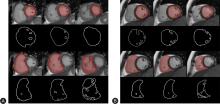
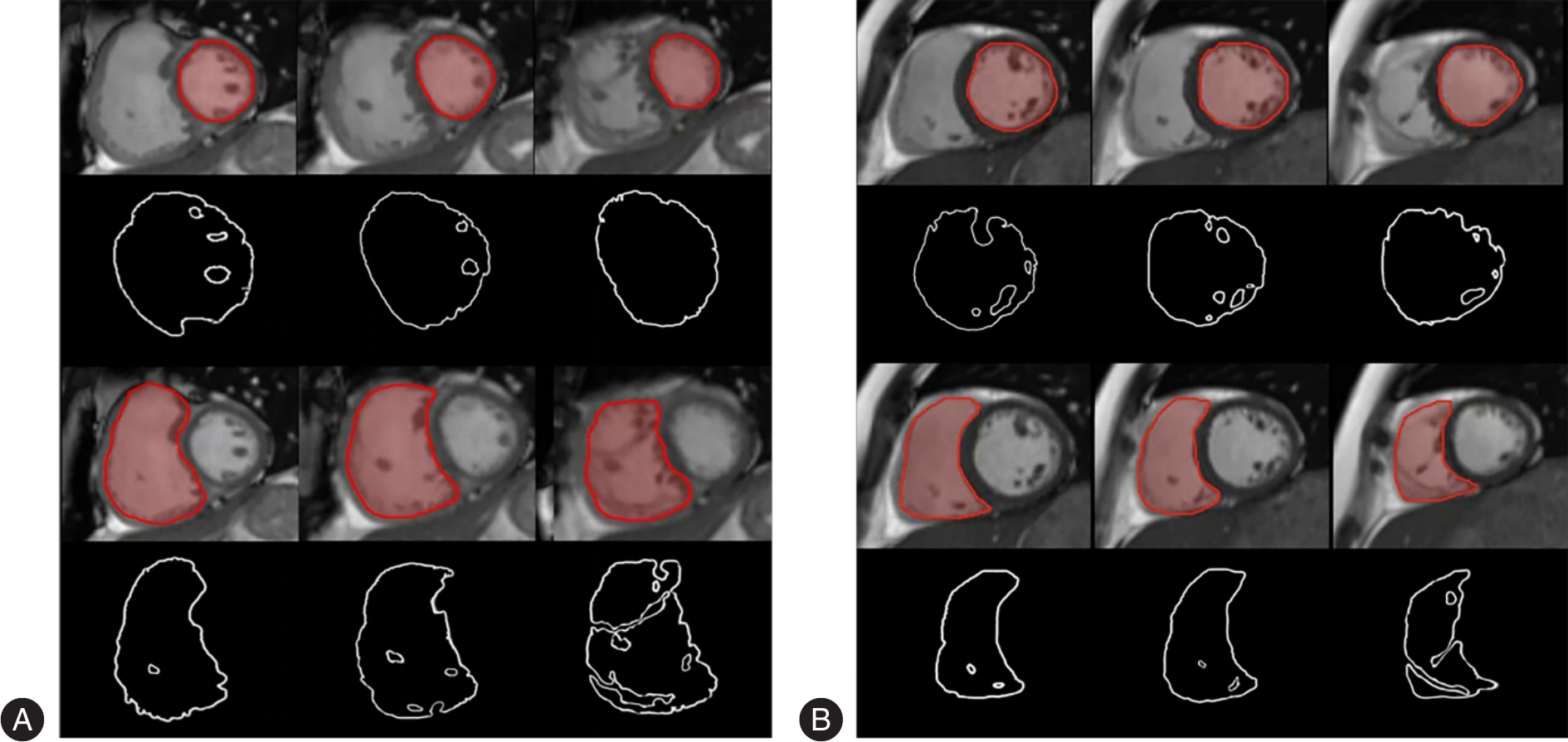
Fig.1
Segmentation of biventricular endocardial contours"
Tab.1
Baseline characteristics of CPTE and healthy controls"
| 项目 | 对照组(n = 53) | CPTE组(n = 53) | t/χ2/Z值 | P值 |
|---|---|---|---|---|
| 年龄/岁 | 52.89 ± 7.86 | 53.30 ± 15.99 | -0.173 | 0.866 |
| 性别/例 | 0.000 | 1.000 | ||
| 男 | 29 | 29 | ||
| 女 | 24 | 24 | ||
| 身高/m | 1.67 ± 0.07 | 1.67 ± 0.07 | 0.171 | 0.864 |
| 体质量/kg | 74.81 ± 10.66 | 69.13 ± 12.53 | 2.512 | 0.014 |
| 体质量指数/(kg/m2) | 26.64 ± 2.64 | 24.54 ± 3.50 | 3.490 | < 0.001 |
| 体表面积/m2 | 1.84 ± 0.15 | 1.77 ± 0.18 | 1.915 | 0.058 |
| 收缩压/mmHg | 130.81 ± 13.83 | 117.00 ± 15.49 | 4.840 | < 0.001 |
| 舒张压/mmHg | 88.60 ± 10.71 | 76.06 ± 12.64 | 5.513 | < 0.001 |
| NT-proBNP[M(P25,P75 )]/(pg/mL) | 29.50(10.00,37.75) | 181.00(34.50,907.00) | -5.274 | < 0.001 |
| D-二聚体[M(P25,P75 )]/(mg/L) | 0.16(0.15,0.23) | 0.25(0.14,0.75) | -2.482 | 0.013 |
Tab.2
Baseline characteristics among three groups"
| 项目 | 对照组(n = 53) | CTEPH组(n = 37) | CTEPD组(n = 16) | F/χ2/Z值 | P值 | t/χ2/Z值 | |||
|---|---|---|---|---|---|---|---|---|---|
CTEPH- CTEPD | CTEPH- 对照组 | CTEPD- 对照组 | |||||||
| 年龄/岁 | 52.89 ± 7.86 | 56.65 ± 14.49 | 45.56 ± 17.05 | 4.686 | 0.011 | 11.086 | 3.762 | -7.324 | |
| 性别/例 | 1.113 | 0.573 | 1.113 | 0.200 | 0.592 | ||||
| 男 | 29 | 22 | 7 | ||||||
| 女 | 24 | 15 | 9 | ||||||
| 身高/m | 1.67 ± 0.07 | 1.67 ± 0.08 | 1.68 ± 0.06 | 0.076 | 0.927 | -0.007 | 0.001 | 0.007 | |
| 体质量/kg | 74.81 ± 10.66 | 66.48 ± 11.15 | 75.25 ± 13.73 | 6.667 | 0.002 | -8.763* | -8.324* | 0.438 | |
| 体质量指数/(kg/m2) | 26.64 ± 2.64 | 23.70 ± 3.19 | 26.48 ± 3.51 | 11.460 | < 0.001 | -2.780* | -2.943* | -0.163 | |
| 体表面积/m2 | 1.84 ± 0.15 | 1.74 ± 0.17 | 1.85 ± 0.18 | 4.156 | 0.018 | -0.106* | -0.095* | 0.011 | |
| 收缩压/mmHg | 130.81 ± 13.83 | 113.91 ± 15.72 | 124.12 ± 12.70 | 15.050 | < 0.001 | -10.206* | -16.892* | -6.686 | |
| 舒张压/mmHg | 88.60 ± 10.71 | 74.21 ± 13.26 | 80.31 ± 10.20 | 17.041 | < 0.001 | -6.096 | -14.387* | -8.291* | |
| NT-proBNP/(pg/mL) | 29.50(10.00,37.75)# | 446.00(57.00,1388.00)# | 36.50(14.50,97.50)# | 40.302 | < 0.001 | 3.534* | 6.242* | 1.239 | |
| D-二聚体/(mg/L) | 0.16(0.15,0.23)# | 0.45(0.21,1.06)# | 0.13(0.10,0.16) | 32.376 | < 0.001 | 5.120* | 4.306* | -1.859 | |
Tab.3
Hemodynamic characteristics of CTEPH and CTEPD"
| 项目 | CTEPH组(n = 37) | CTEPD组(n = 16) | t/χ2/Z值 | P值 |
|---|---|---|---|---|
| 肺动脉收缩压/mmHg | 69.89 ± 20.81 | 26.75 ± 5.10 | 11.757 | < 0.001 |
| 肺动脉舒张压/mmHg | 23.11 ± 8.53 | 10.44 ± 2.70 | 8.249 | < 0.001 |
| 平均肺动脉压/mmHg | 40.00 ± 12.06 | 16.88 ± 3.34 | 10.719 | < 0.001 |
| 肺动脉楔压/mmHg | 10.03 ± 3.26 | 9.69 ± 2.44 | 0.372 | 0.711 |
| 心输出量[M(P25,P75 )]/(L/min) | 3.40(2.84,3.96) | 5.03(4.41,6.08) | -3.991 | < 0.001 |
| 心指数[M(P25,P75 )]/[L/(min·m2)] | 2.00(1.66,2.26) | 2.83(2.29,3.23) | -3.934 | < 0.001 |
| 肺血管阻力[M(P25,P75 )]/(wood units) | 8.14(5.76,13.02) | 1.40(1.05,2.00) | 5.028 | < 0.001 |
| 肺血管阻力指数[M(P25,P75 )]/(wood units·m2) | 14.10(10.27,21.60) | 2.50(1.94,3.77) | 4.960 | < 0.001 |
| 平均右房压[M(P25,P75 )]/mmHg | 4.00(2.00,6.00) | 3.00(2.25,5.75) | 0.176 | 0.860 |
| 右心室收缩压/mmHg | 69.92 ± 23.38 | 27.56 ± 5.91 | 10.285 | < 0.001 |
| 右心室舒张压[M(P25,P75 )]/mmHg | 2.00(1.00,4.00) | 2.00(0.25,4.00) | 0.596 | 0.551 |
| 平均右心室压/mmHg | 25.14 ± 9.46 | 11.88 ± 2.41 | 8.035 | < 0.001 |
| 混合静脉血氧饱和度/% | 67.79 ± 7.08 | 77.30 ± 3.95 | -5.096 | < 0.001 |
Tab.4
Perimeter-area ratio ICC analysis"
| 项目 | 组内相关系数(2,1) | 95%CI |
|---|---|---|
| RVPAR | ||
| D1 | 0.976 | 0.957 ~ 0.987 |
| D2 | 0.969 | 0.946 ~ 0.984 |
| D3 | 0.962 | 0.933 ~ 0.980 |
| S1 | 0.945 | 0.900 ~ 0.972 |
| S2 | 0.949 | 0.910 ~ 0.973 |
| S3 | 0.970 | 0.947 ~ 0.984 |
| LVPAR | ||
| D1 | 0.893 | 0.808 ~ 0.944 |
| D2 | 0.876 | 0.790 ~ 0.933 |
| D3 | 0.914 | 0.852 ~ 0.954 |
| S1 | 0.860 | 0.766 ~ 0.923 |
| S2 | 0.968 | 0.944 ~ 0.983 |
| S3 | 0.951 | 0.914 ~ 0.974 |
Tab.5
Comparison of biventricular PAR among three groups"
| 项目 | 对照组(n = 53) | CTEPH组(n = 37) | CTEPD组(n = 16) | H值 | P值 | Z值 | ||
|---|---|---|---|---|---|---|---|---|
CTEPH- CTEPD | CTEPH- 对照组 | CTEPD- 对照组 | ||||||
| RVPAR | ||||||||
| D1 | 0.185(0.179,0.208) | 0.138(0.127,0.158) | 0.149(0.141,0.154) | 68.932 | < 0.001 | -0.838 | -7.843* | -5.012* |
| D2 | 0.229(0.212,0.270) | 0.145(0.126,0.163) | 0.163(0.153,0.170) | 74.468 | < 0.001 | -1.740 | -8.398* | -4.482* |
| D3 | 0.298(0.252,0.355) | 0.150(0.142,0.180) | 0.196(0.179,0.229) | 74.669 | < 0.001 | -2.795* | -8.593* | -3.522* |
| S1 | 0.234(0.211,0.258) | 0.172(0.151,0.189) | 0.211(0.183,0.228) | 51.155 | < 0.001 | -2.851* | -7.150* | -2.380 |
| S2 | 0.267(0.229,0.296) | 0.157(0.140,0.191) | 0.214(0.194,0.234) | 66.450 | < 0.001 | -3.272* | -8.150* | -2.688* |
| S3 | 0.355(0.283,0.446) | 0.172(0.145,0.193) | 0.223(0.213,0.305) | 71.386 | < 0.001 | -2.897* | -8.418* | -3.283* |
| LVPAR | ||||||||
| D1 | 0.256(0.219,0.306) | 0.194(0.165,0.254) | 0.203(0.171,0.281) | 17.894 | < 0.001 | -0.457 | -4.006* | -2.530* |
| D2 | 0.288(0.261,0.342) | 0.255(0.215,0.293) | 0.252(0.233,0.302) | 12.441 | 0.002 | 0.227 | -3.096* | -2.563* |
| D3 | 0.293(0.260,0.332) | 0.251(0.239,0.275) | 0.261(0.237,0.318) | 16.978 | < 0.001 | -0.963 | -4.039* | -2.023* |
| S1 | 0.332(0.287,0.369) | 0.287(0.238,0.339) | 0.245(0.224,0.283) | 15.591 | < 0.001 | 1.553 | -2.637* | -3.610* |
| S2 | 0.377(0.310,0.460) | 0.412(0.302,0.491) | 0.325(0.291,0.372) | 2.480 | 0.289 | 1.414 | 0.353 | -1.450 |
| S3 | 0.407(0.316,0.574) | 0.438(0.389,0.539) | 0.398(0.354,0.487) | 1.253 | 0.535 | 1.104 | 0.824 | -0.327 |
Tab.6
Receiver operating characteristic curves among three groups"
| P | ||||||
|---|---|---|---|---|---|---|
| < | < | |||||
| < | < | |||||
| < | < | |||||
| < | < | |||||
| < | < | |||||
| < | < | |||||
| < | ||||||
| < |
Tab.7
Correlation analysis between right ventricular perimeter-area ratio and hemodynamic parameters"
| 项目 | sPAP | dPAP | mPAP | PAWP | CO | PVR | mRAP | sRVP | dRVP | mRVP | SvO2 | CI | PVRI | |
|---|---|---|---|---|---|---|---|---|---|---|---|---|---|---|
| D1 | ρ值 | -0.181 | -0.235 | -0.219 | 0.100 | 0.200 | -0.314 | -0.109 | -0.222 | -0.276 | -0.205 | 0.260 | 0.325 | -0.346 |
| P值 | 0.213 | 0.104 | 0.131 | 0.493 | 0.169 | 0.028 | 0.457 | 0.125 | 0.055 | 0.158 | 0.071 | 0.023 | 0.015 | |
| D2 | ρ值 | -0.305 | -0.287 | -0.310 | 0.096 | 0.316 | -0.375 | 0.024 | -0.322 | -0.179 | -0.289 | 0.440 | 0.437 | -0.413 |
| P值 | 0.033 | 0.046 | 0.030 | 0.510 | 0.027 | 0.008 | 0.868 | 0.024 | 0.219 | 0.044 | 0.002 | 0.002 | 0.003 | |
| D3 | ρ值 | -0.428 | -0.400 | -0.433 | 0.062 | 0.312 | -0.434 | 0.005 | -0.414 | -0.157 | -0.381 | 0.485 | 0.423 | -0.471 |
| P值 | 0.002 | 0.004 | 0.002 | 0.671 | 0.029 | 0.002 | 0.972 | 0.003 | 0.281 | 0.007 | < 0.001 | 0.002 | 0.001 | |
| S1 | ρ值 | -0.486 | -0.540 | -0.540 | 0.052 | 0.260 | -0.494 | -0.190 | -0.509 | -0.446 | -0.454 | 0.378 | 0.340 | -0.526 |
| P值 | < 0.001 | < 0.001 | < 0.001 | 0.725 | 0.071 | < 0.001 | 0.191 | < 0.001 | 0.001 | 0.001 | 0.007 | 0.017 | < 0.001 | |
| S2 | ρ值 | -0.521 | -0.531 | -0.567 | 0.075 | 0.436 | -0.566 | -0.180 | -0.550 | -0.355 | -0.488 | 0.564 | 0.531 | -0.602 |
| P值 | < 0.001 | < 0.001 | < 0.001 | 0.610 | 0.002 | < 0.001 | 0.216 | < 0.001 | 0.012 | < 0.001 | < 0.001 | < 0.001 | < 0.001 | |
| S3 | ρ值 | -0.546 | -0.572 | -0.575 | 0.168 | 0.408 | -0.541 | -0.021 | -0.540 | -0.171 | -0.493 | 0.557 | 0.489 | -0.587 |
| P值 | < 0.001 | < 0.001 | < 0.001 | 0.248 | 0.004 | < 0.001 | 0.886 | < 0.001 | 0.241 | < 0.001 | < 0.001 | < 0.001 | < 0.001 |
| [1] |
HUMBERT M, KOVACS G, HOEPER MM,et al. 2022 ESC/ERS Guidelines for the diagnosis and treatment of pulmonary hypertension[J]. Eur Heart J,2022,43(38):3618-3731. doi:10.1093/eurheartj/ehac237 .
doi: 10.1093/eurheartj/ehac237 |
| [2] |
中华医学会呼吸病学分会肺栓塞与肺血管病学组,中国医师协会呼吸医师分会肺栓塞与肺血管病工作委员会,全国肺栓塞与肺血管病防治协作组. 肺血栓栓塞症诊治与预防指南[J]. 中华医学杂志,2018,98(14):1060-1087. doi:10.3760/cma.j. issn.0376-2491.2018.14.007 .
doi: 10.3760/cma.j. issn.0376-2491.2018.14.007 |
| [3] |
TEERAPUNCHAROEN K,BAG R. Chronic thromboembolic pulmonary hypertension[J]. Lung,2022,200(3):283-299. doi:10.1007/s00408-022-00539-w .
doi: 10.1007/s00408-022-00539-w |
| [4] |
杨宗江,舒向阳.微创介入介导联合重组组织纤溶酶原激活剂治疗大面积肺栓塞相关患者的效果[J].实用医学杂志,2026,42(4):639-645. doi:10.3969/j.issn.1006-5725.2026.04.014 .
doi: 10.3969/j.issn.1006-5725.2026.04.014 |
| [5] |
CAPONE C, VALENTINI A, SPINILLO S L,et al. Radiological differences between chronic thromboembolic pulmonary disease (CTEPD) and chronic thromboembolic pulmonary hypertension (CTEPH)[J]. Eur Radiol,2021,31(8):6230-6238. doi:10.1007/s00330-020-07556-4 .
doi: 10.1007/s00330-020-07556-4 |
| [6] |
KIM N H, D'ARMINI AM, DELCROIX M,et al. Chronic thromboembolic pulmonary disease[J]. Eur Respir J,2024,64(4):2401294. doi:10.1183/13993003.01294-2024 .
doi: 10.1183/13993003.01294-2024 |
| [7] |
GHANI H, WEIR-MCCALL J R, RUGGIERO A,et al. Imaging in chronic thromboembolic pulmonary disease:Current practice and advances[J]. Int J Cardiol Congenit Heart Dis,2024,17:100536. doi:10.1016/j.ijcchd.2024.100536 .
doi: 10.1016/j.ijcchd.2024.100536 |
| [8] |
FAIRLEY J L, O'ROURKE R, PURANIK R,et al. Cardiac magnetic resonance imaging in systemic sclerosis:Heart involvement in high-resolution[J]. Rheumatol Immunol Res,2024,5(2):83-92. doi:10.1515/rir-2024-0011 .
doi: 10.1515/rir-2024-0011 |
| [9] |
ZHANG L, DAI J, ZHANG P,et al. Right ventricular end-systolic remodeling index on cardiac magnetic resonance imaging:comparison with other functional markers in patients with chronic thromboembolic pulmonary hypertension[J]. Quant Imaging Med Surg,2022,12(2):894-905. doi:10.21037/qims-21-385 .
doi: 10.21037/qims-21-385 |
| [10] |
DENG M, LIU A, XU W,et al. Right and left ventricular blood pool T2 ratio on cardiac magnetic resonance imaging correlates with hemodynamics in patients with pulmonary hypertension[J]. Insights Imaging,2023,14(1):66. doi:10.1186/s13244-023-01406-9 .
doi: 10.1186/s13244-023-01406-9 |
| [11] |
YANG F, YAN Y, JIANG W,et al. Impaired right atrial function preceding right ventricular systolic dysfunction:clinical utility and long-term prognostic value in pulmonary hypertension[J]. Insights Imaging,2025,16(1):115. doi:10.1186/s13244-025-01996-6 .
doi: 10.1186/s13244-025-01996-6 |
| [12] |
任雯,吴倩,杨振文,等. 右心室-肺动脉耦联对慢性血栓栓塞性肺动脉高压风险分层评估的价值[J]. 放射学实践,2025,40(8):982-988. doi:10.13609/j.cnki.1000-0313.2025.08.006 .
doi: 10.13609/j.cnki.1000-0313.2025.08.006 |
| [13] |
DAWES T J W, CAI J, QUINLAN M,et al. Fractal analysis of right ventricular trabeculae in pulmonary hypertension[J]. Radiology,2018,288(2):386-395. doi:10.1148/radiol.2018172821 .
doi: 10.1148/radiol.2018172821 |
| [14] |
孟夏培,孙学彪,许文清,等. 慢性血栓栓塞性肺疾病与慢性血栓栓塞性肺动脉高压肺血管分形维数和弯曲度对比研究[J]. 中华结核和呼吸杂志,2023,46(8):774-780. doi:10.3760/cma.j.cn112147-20230630-00352 .
doi: 10.3760/cma.j.cn112147-20230630-00352 |
| [15] |
VILADES D, GARCIA-MOLL X, GOMEZ-LLORENTE M,et al. Differentiation of athlete's heart and hypertrophic cardiomyopathy by the fractal dimension of left ventricular trabeculae[J]. Int J Cardiol,2021,330:232-237. doi:10.1016/j.ijcard.2021.02.042 .
doi: 10.1016/j.ijcard.2021.02.042 |
| [16] |
FIKRLE T, PIZINGER K. Digital computer analysis of dermatoscopical images of 260 melanocytic skin lesions; perimeter/area ratio for the differentiation between malignant melanomas and melanocytic nevi[J]. J Eur Acad Dermatol Venereol,2007,21(1):48-55. doi:10.1111/j.1468-3083.2006.01864.x .
doi: 10.1111/j.1468-3083.2006.01864.x |
| [17] |
PATEL N R, SETYA K, PRADHAN S,et al. Microarchitectural Changes of Cardiovascular Calcification in Response to In Vivo Interventions Using Deep-Learning Segmentation and Computed Tomography Radiomics[J]. Arterioscler Thromb Vasc Biol,2022,42(8):e228-e241. doi:10.1161/atvbaha.122.317761 .
doi: 10.1161/atvbaha.122.317761 |
| [18] |
中华医学会呼吸病学分会肺栓塞与肺血管病学组,中国医师协会呼吸医师分会肺栓塞与肺血管病工作组,全国肺栓塞与肺血管病防治协作组,等. 慢性血栓栓塞性肺动脉高压诊断与治疗指南(2024版)[J]. 中华医学杂志,2024,104(24):2200-2221.doi:10.3760/cma.j.cn112137-20240116-00117 .
doi: 10.3760/cma.j.cn112137-20240116-00117 |
| [19] |
KOVACS G, BARTOLOME S, DENTON C P,et al. Definition,classification and diagnosis of pulmonary hypertension[J]. Eur Respir J,2024,64(4):2401324. doi:10.1183/13993003.01324-2024 .
doi: 10.1183/13993003.01324-2024 |
| [20] |
CAPTUR G, SYRRIS P, OBIANYO C,et al. Formation and malformation of cardiac trabeculae:Biological basis,clinical significance,and special yield of magnetic resonance imaging in assessment[J]. Can J Cardiol,2015,31(11):1325-1337. doi:10.1016/j.cjca.2015.07.003 .
doi: 10.1016/j.cjca.2015.07.003 |
| [21] |
CAPTUR G, MUTHURANGU V, COOK C,et al. Quantification of left ventricular trabeculae using fractal analysis[J]. J Cardiovasc Magn Reson,2013,15(1):36. doi:10.1186/1532-429x-15-36 .
doi: 10.1186/1532-429x-15-36 |
| [22] |
VAN DE VEERDONK M C, DUSOSWA S A, MARCUS J T,et al. The importance of trabecular hypertrophy in right ventricular adaptation to chronic pressure overload[J]. Int J Cardiovasc Imaging,2014,30(2):357-365. doi:10.1007/s10554-013-0338-z .
doi: 10.1007/s10554-013-0338-z |
| [23] |
CAPTUR G, WILSON R, BENNETT M F,et al. Morphogenesis of myocardial trabeculae in the mouse embryo[J]. J Anat,2016,229(2):314-325. doi:10.1111/joa.12465 .
doi: 10.1111/joa.12465 |
| [24] |
SACCO F, PAUN B, LEHMKUHL O,et al. Evaluating the roles of detailed endocardial structures on right ventricular haemodynamics by means of CFD simulations[J]. Int J Numer Method Biomed Eng,2018,34(9):e3115. doi:10.1002/cnm.3115 .
doi: 10.1002/cnm.3115 |
| [25] |
KAISER R, LIU D, ARIAS-LOZA P,et al. Right ventricular pressure overload directly affects left ventricular torsion mechanics in patients with precapillary pulmonary hypertension[J]. PLoS One,2020,15(5):e0232544. doi:10.1371/journal.pone.0232544 .
doi: 10.1371/journal.pone.0232544 |
| Viewed | ||||||
|
Full text |
|
|||||
|
Abstract |
|
|||||