| 1 |
张索, 刘冬舟. 系统性红斑狼疮脑病的研究进展[J]. 实用医学杂志, 2020, 36(3): 414-419.
|
| 2 |
SCHWARTZ N, STOCK A D, PUTTERMAN C. Neuropsychiatric lupus: New mechanistic insights and future treatment directions[J]. Nat Rev Rheumatol, 2019,15(3): 137-152. doi:10.1038/s41584-018-0156-8
doi: 10.1038/s41584-018-0156-8
|
| 3 |
申杰, 徐桂华. 阿尔茨海默病与血脑屏障的相关性研究进展[J]. 实用医学杂志, 2024, 40(11): 1602-1606.
|
| 4 |
SANTOS G S P, PRAZERES P H D M, MINTZ A, et al. Role of pericytes in the retina[J]. Eye (Lond), 2018,32(3): 483-486. doi:10.1038/eye.2017.220
doi: 10.1038/eye.2017.220
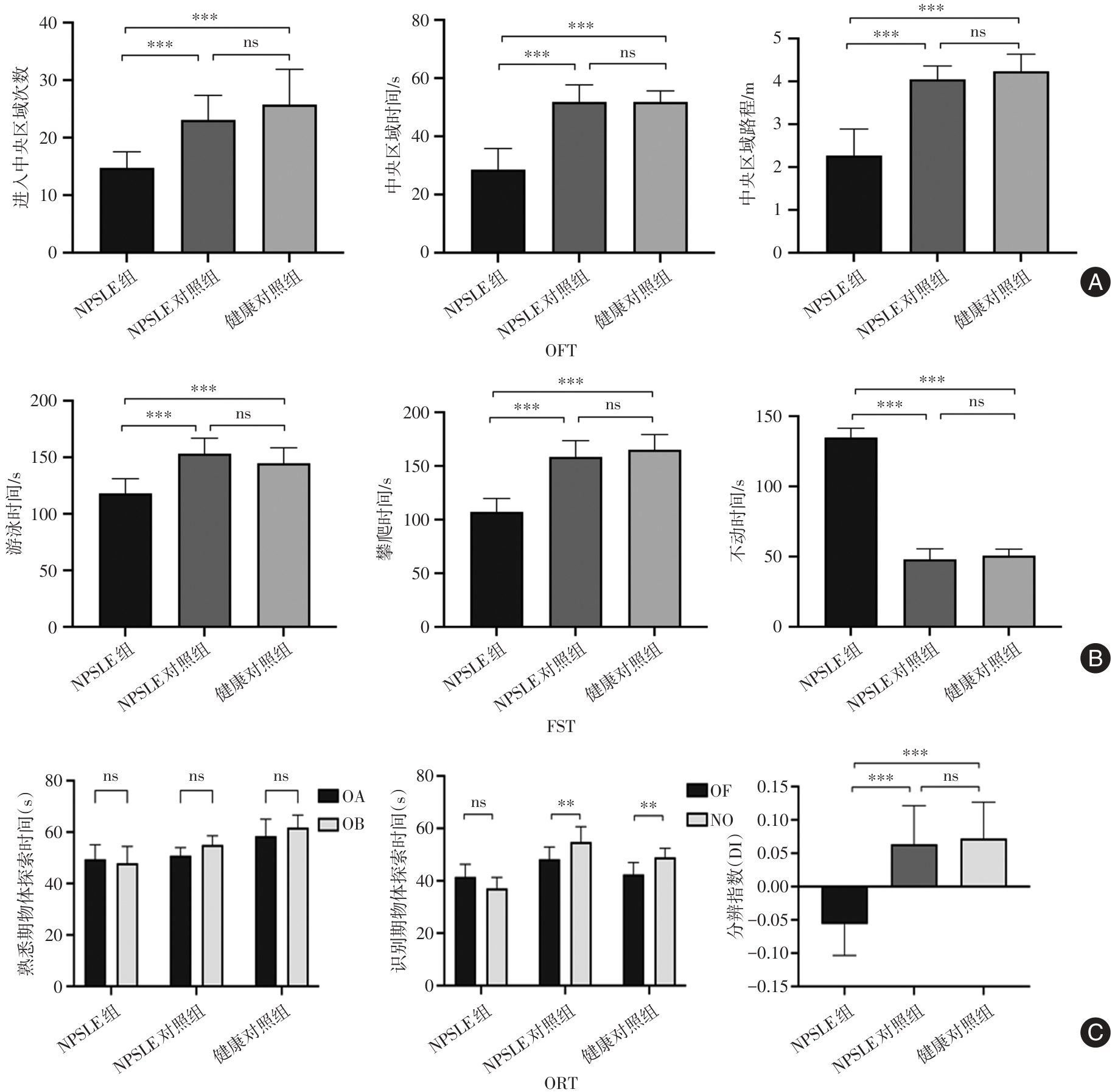
|
| 5 |
KURELI G, YILMAZ-OZCAN S, ERDENER S E, et al. F-actin polymerization contributes to pericyte contractility in retinal capillaries[J]. Exp Neurol, 2020, 332: 113392. doi:10.1016/j.expneurol.2020.113392
doi: 10.1016/j.expneurol.2020.113392
|
| 6 |
HUANG H. Pericyte-Endothelial Interactions in the Retinal Microvasculature[J]. Int J Mol Sci, 2020, 21(19): 7413. doi:10.3390/ijms21197413
doi: 10.3390/ijms21197413
|
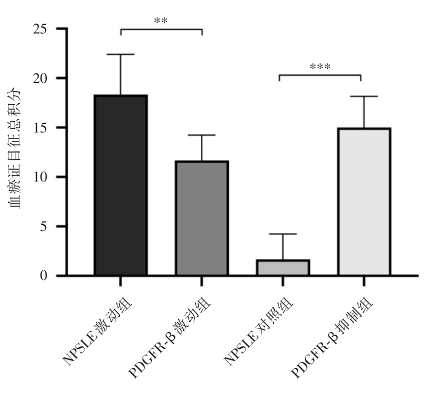
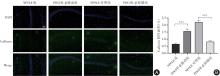
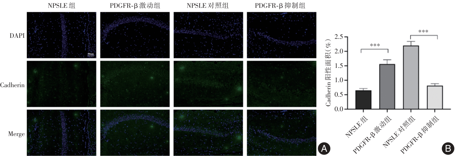
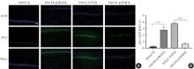
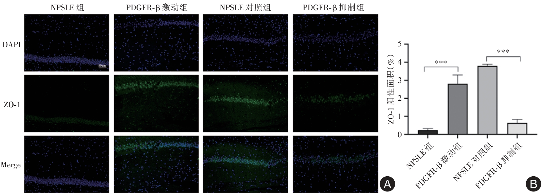
| 7 |
NIKOLAKOPOULOU A, MONTAGNE A, et al. Pericyte loss leads to circulatory failure and pleiotrophin depletion causing neuron loss[J]. Nat Neurosci, 2019, 22: 1089-1098. doi:10.1038/s41593-019-0434-z
doi: 10.1038/s41593-019-0434-z
|
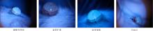
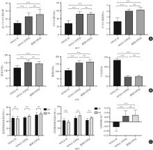
| 8 |
SHI H, KORONYO Y, RENTSENDORJ A, et al. Identification of early pericyte loss and vascular amyloidosis in Alzheimer disease retina[J]. Acta Neuropathol, 2020, 139: 813-836. doi:10.1007/s00401-020-02134-w
doi: 10.1007/s00401-020-02134-w
|
| 9 |
SWEENEY M D, AYYADURAI S, ZLOKOVIC B V. Pericytes of the neurovascular unit: Key functions and signaling pathways[J]. Nat Neurosci, 2016, 19(6): 771-783. doi:10.1038/nn.4288
doi: 10.1038/nn.4288
|
| 10 |
HIRUNPATTARASILP C, ATTWELL D, FREITAS F. The role of pericytes in brain disorders: From the periphery to the brain[J]. J Neurochem, 2019, 150: 648-665. doi:10.1111/jnc.14725
doi: 10.1111/jnc.14725
|
| 11 |
HAN X, XU T, DING C, et al. Neuronal NR4A1 deficiency drives complement-coordinated synaptic stripping by microglia in a mouse model of lupus[J]. Signal Transduct Target Ther, 2022, 7(1): 47-56. doi:10.1038/s41392-021-00867-y
doi: 10.1038/s41392-021-00867-y
|
| 12 |
赖琴, 郭雪, 王梅英. 系统性红斑狼疮动物模型及研究进展[J]. 中国实验动物学报, 2024, 32(11): 1493-1504.
|
| 13 |
TOMALLA V, SCHMEISSER M J, WEINMANN-MENKE J. Mouse models, antibodies, and neuroimaging: Current knowledge and future perspectives in neuropsychiatric systemic lupus erythematosus (NPSLE)[J]. Front Psychiatry, 2023, 14: 1078607. doi:10.3389/fpsyt.2023.1078607
doi: 10.3389/fpsyt.2023.1078607
|
| 14 |
WU C, YANG L, LI Y, et al. Effects of exercise training on anxious-depressive-like behavior in Alzheimer rat[J]. Med Sci Sports Exerc, 2020, 52(7): 1456-1469. doi:10.1249/mss.0000000000002294
doi: 10.1249/mss.0000000000002294
|
| 15 |
GERANMAYEH M H, RAHBARGHAZI R, FARHOUDI M. Targeting pericytes for neurovascular regeneration[J]. Cell Commun Signal, 2019, 17(1): 26. doi:10.1186/s12964-019-0340-8
doi: 10.1186/s12964-019-0340-8
|
| 16 |
OTA Y, SRINIVASAN A, CAPIZZANO A A, et al. Central nervous system systemic lupus erythematosus: Pathophysiologic, clinical, and imaging features[J]. Radiographics, 2022, 42(1): 212-232. doi:10.1148/rg.210045
doi: 10.1148/rg.210045
|
| 17 |
PERSIDSKY Y, HILL J, ZHANG M, et al. Dysfunction of brain pericytes in chronic neuroinflammation[J]. J Cereb Blood Flow Metab, 2016, 36(4): 794-807. doi:10.1177/0271678x15606149
doi: 10.1177/0271678x15606149
|
| 18 |
SMYTH L C, RUSTENHOVEN J, PARK T I H, et al. Unique and shared inflammatory profiles of human brain endothelia and pericytes[J]. J Neuroinflammation, 2018, 15: 138. doi:10.1186/s12974-018-1167-8
doi: 10.1186/s12974-018-1167-8
|
| 19 |
KIM Y, LEE S, ZHANG H, et al. CLEC14A deficiency exacerbates neuronal loss by increasing blood-brain barrier permeability and inflammation[J]. J Neuroinflammation, 2020, 17(1): 48. doi:10.1186/s12974-020-1727-6
doi: 10.1186/s12974-020-1727-6
|
| 20 |
CHEN X, XUE J, ZOU J, et al. Resveratrol alleviated neuroinflammation induced by pseudorabies virus infection through regulating microglial M1/M2 polarization[J]. Biomed Pharmacother, 2023, 160: 114271. doi:10.1016/j.biopha.2023.114271
doi: 10.1016/j.biopha.2023.114271
|
| 21 |
LI W, NIU X, DAI Y, et al. Rnf-213 knockout induces pericyte reduction and blood-brain barrier impairment in mouse[J]. Mol Neurobiol, 2023, 60(11): 6188-6200. doi:10.1007/s12035-023-03480-y
doi: 10.1007/s12035-023-03480-y
|
| 22 |
LIU G, WANG J, WEI Z, et al. Elevated PDGF-BB from bone impairs hippocampal vasculature by inducing PDGFRβ shedding from pericytes[J]. Adv Sci (Weinh), 2023, 10(20): e2206938. doi:10.1002/advs.202206938
doi: 10.1002/advs.202206938
|
| 23 |
SHI H, KORONYO Y, RENTSENDORJ A, et al. Identification of early pericyte loss and vascular amyloidosis in Alzheimer's disease retina[J]. Acta Neuropathol, 2020, 139(5): 813-836. doi:10.1007/s00401-020-02134-w
doi: 10.1007/s00401-020-02134-w
|
| 24 |
KVERNEBO A K, MIYAMOTO T, SPORAST A H, et al. Quantification of ocular surface microcirculation by computer-assisted video microscopy and diffuse reflectance spectroscopy[J]. Exp Eye Res, 2020, 201: 108312. doi:10.1016/j.exer.2020.108312
doi: 10.1016/j.exer.2020.108312
|
| 25 |
ZHAO S, YANG Z, SUN P, et al. Conjunctival microcirculation is associated with cerebral cortex microcirculation in post-resuscitation mild hypothermia: A rat model[J]. Microcirculation, 2020, 27(3): e12604. doi:10.1111/micc.12604
doi: 10.1111/micc.12604
|
| 26 |
SHI L H, LIU Z Y, YU S J, et al. Performance of eye sign combined with increased interleukin-6 in cerebrospinal fluid in patients with neuropsychiatric lupus erythematosus[J]. Int J Rheum Dis, 2023, 26(8): 1464-1473. doi:10.1111/1756-185x.14731
doi: 10.1111/1756-185x.14731
|


 )
)