

The Journal of Practical Medicine ›› 2025, Vol. 41 ›› Issue (22): 3510-3519.doi: 10.3969/j.issn.1006-5725.2025.22.007
• Basic Research • Previous Articles
Jingyu LIU1,Chenlu DAI1,Min JI2,Liping SU2,Min LIANG3,Ming CHENG1,Xuanming LIU1,Linlin ZHANG1,Yujie GAO1,Shaoshuai CHEN1,Hongwei PU4,5( )
)
Received:2025-08-17
Online:2025-11-25
Published:2025-11-26
Contact:
Hongwei PU
E-mail:576250630@qq.com
CLC Number:
Jingyu LIU,Chenlu DAI,Min JI,Liping SU,Min LIANG,Ming CHENG,Xuanming LIU,Linlin ZHANG,Yujie GAO,Shaoshuai CHEN,Hongwei PU. Role of SPP1 and MYD88 in diacetylmorphine⁃induced apoptosis in cardiomyocytes[J]. The Journal of Practical Medicine, 2025, 41(22): 3510-3519.
Tab.1
Peimer sequence"
| 引物 | 序列(5′— 3′) |
|---|---|
| SPP1 | 上游ACCATGCAGAGAGCGAGGAT |
| 下游TGCTGGCAGTGAAGGACTCA | |
| MYD88 | 上游ACGCAACCAGCAGAAACAGGAG |
| 下游GGTGATGCCTCCCAGTTCCTTTG | |
| Bax | 上游GACGCATCCACCAAGAAGCTGAG |
| 下游GCTGCCACACGGAAGAAGACC | |
| Bcl2 | 上游TGGAGAGCGTCAACAGGGAGATG |
| 下游GGTGTGCAGATGCCGGTTCAG | |
| Caspase?9 | 上游CATCTCAGACTGCCTTGGGAAAC |
| 下游GCTCTGCCAGAACCAATGTCCAC | |
| Caspase?3 | 上游GCTGGACTGCGGTATTGAGACAG |
| 下游GATGGACCATGACCCGTCCCTTG | |
| GAPDH | 上游ACGGCAAGTTCAACGGCACAG |
| 下游TCGCTCCTGGAAGATGGTGATGG |
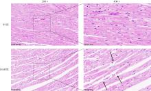
Fig.1
Morphological changes in the myocardium of SD rats in each group detected by HE staining"


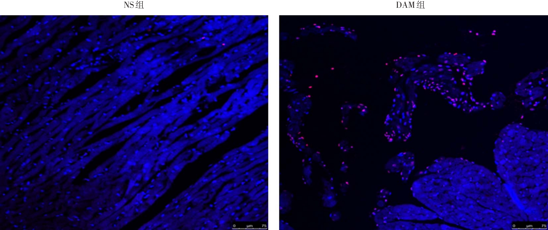
Fig. 2
TUNEL detection of apoptosis in myocardial tissue cells of each group"
Tab.2
Effect of DAM on the expression levels of SPP1, MYD88, Bax, Bcl2, Caspase?9, and Caspase?3 mRNAin myocardial tissues of SD rats"
| 组别 | SPP1/GAPDH | MYD88/GAPDH | Bax/GAPDH | Bcl2/GAPDH | Caspase?9/GAPDH | Caspase?3/GAPDH |
|---|---|---|---|---|---|---|
| NS | 1.01 ± 0.14 | 1.01 ± 0.13 | 1.01 ± 0.16 | 1.00 ± 0.03 | 1.00 ± 0.10 | 1.00 ± 0.08 |
| DAM | 2.74 ± 0.32# | 1.69 ± 0.16# | 2.49 ± 0.21& | 0.25 ± 0.01∧ | 1.57 ± 0.23* | 1.65 ± 0.03& |
Fig.3
Immunohistochemical detection of SPP1 and MYD88 expression in myocardial tissues of each group (400 ×)"
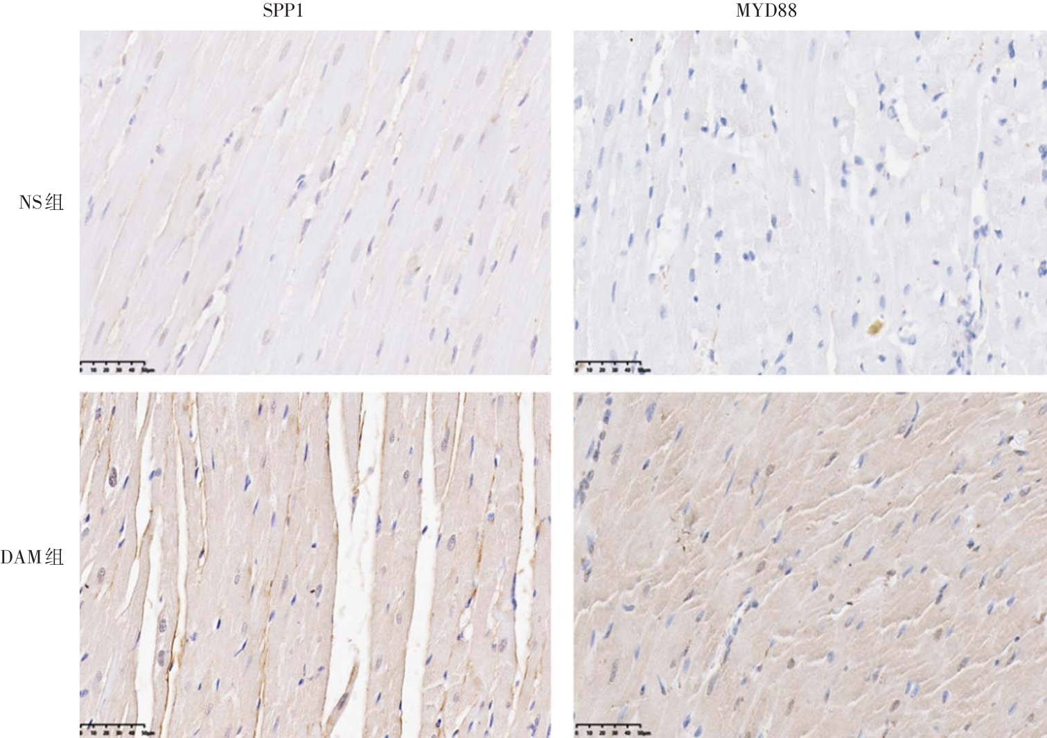
Tab.3
Effect of DAM on the percentage of mean positive area of SPP1 and MYD88 in myocardial tissue of SD rats"
| 组别 | SPP1 | MYD88 |
|---|---|---|
| NS组 | 0.096 ± 0.002 | 0.031 ± 0.009 |
| DAM组 | 0.264 ± 0.005* | 0.311 ± 0.005* |
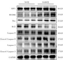
Fig.4
Western blot detection of SPP1, MYD88 and apoptosis-related protein expression in each group"
Tab.4
Effects of DAM on the expression levels of SPP1, MYD88, Bax, Bcl2, Caspase-9, Caspase-3, Cleaved Caspase-9,and Cleaved Caspase-3 proteins in myocardial tissues of SD rats (n = 3)"
| 组别 | SPP1/ GAPDH | MYD88/ GAPDH | Bax/ GAPDH | Bcl2/ GAPDH | Caspase?9/ GAPDH | Caspase?3/ GAPDH | Cleaved Caspase9/ GAPDH | Cleaved Caspase3/ GAPDH |
|---|---|---|---|---|---|---|---|---|
| NS组 | 1.01 ± 0.14 | 1.01 ± 0.13 | 1.01 ± 0.16 | 1.00 ± 0.03 | 1.00 ± 0.10 | 1.00 ± 0.08 | 1.00 ± 0.05 | 1.00 ± 0.06 |
| DAM组 | 2.74 ± 0.32# | 1.69 ± 0.16# | 2.49 ± 0.21& | 0.25 ± 0.01∧ | 1.57 ± 0.23? | 1.65 ± 0.03& | 1.23 ± 0.09# | 1.48 ± 0.16? |
Fig. 5
Laser confocal identification of NRCMs"
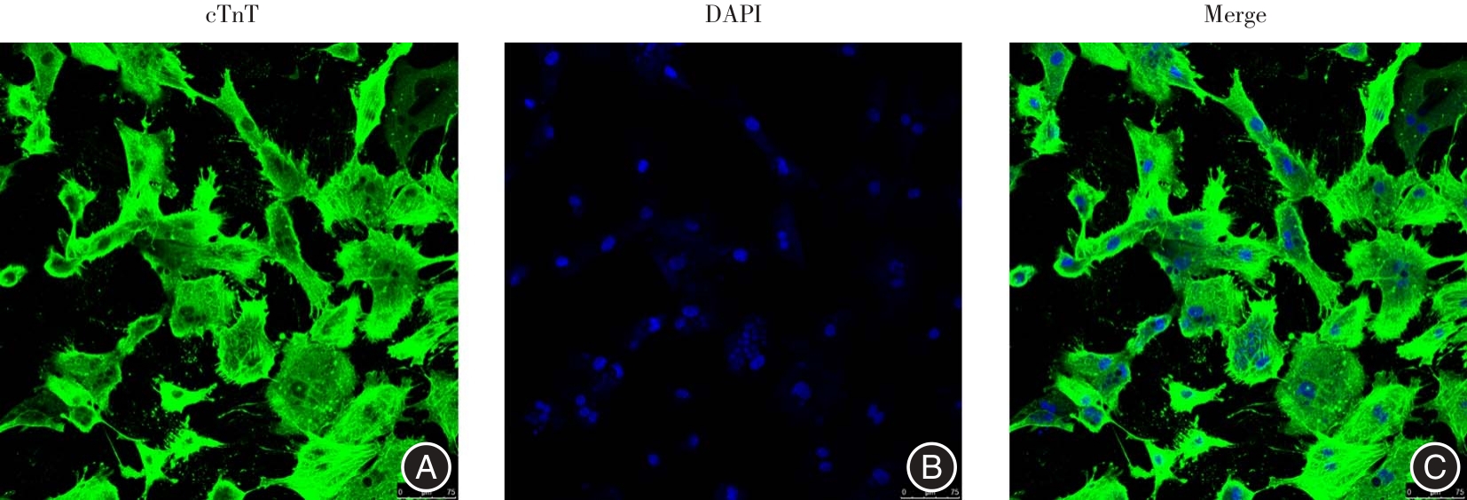
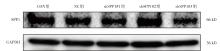
Fig.6
Western blot detection of silencing SPP1 transfection efficiency in each group"
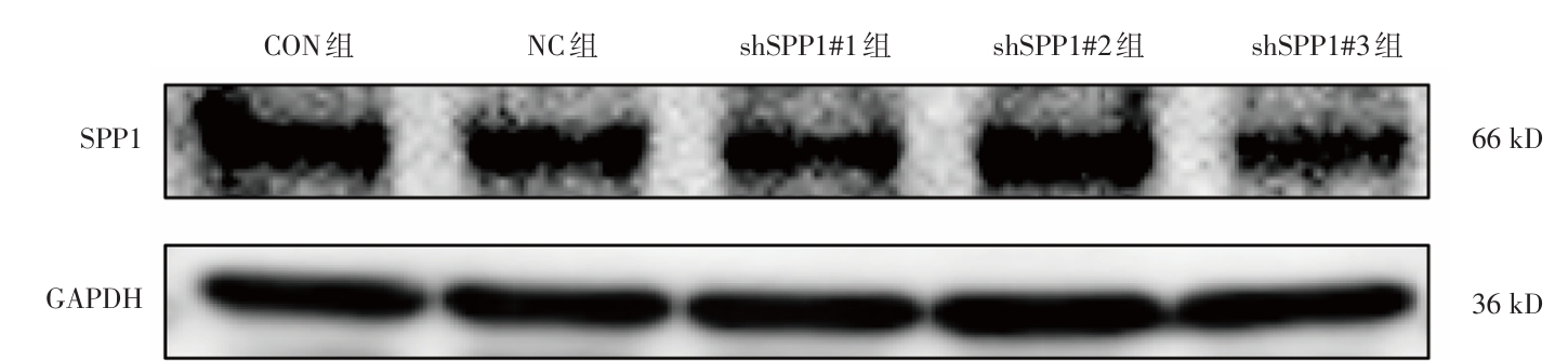
Tab.5
Effect of transfection of NRCMs with different lentiviral sequences on SPP1 protein expression"
| 组别 | SPP1/GAPDH |
|---|---|
| CON组 | 0.99 ± 0.14 |
| NC组 | 0.87 ± 0.14 |
| shSPP1#1组 | 0.77 ± 0.05 |
| shSPP1#2组 | 0.64 ± 0.16 |
| shSPP1#3组 | 0.39 ± 0.25? |
Tab.6
Effect of transfection of NRCMs with differentlentiviral sequences on SPP1 mRNA expression"
| 组别 | SPP1/GAPDH |
|---|---|
| CON组 | 1.07 ± 0.09 |
| NC组 | 0.98 ± 0.18 |
| shSPP1#1组 | 0.46 ± 0.06? |
| shSPP1#2组 | 0.33 ± 0.09# |
| shSPP1#3组 | 0.04 ± 0.01# |
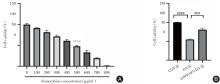
Fig.7
Vitality of NRCMs in each group detected by CCK-8 method"
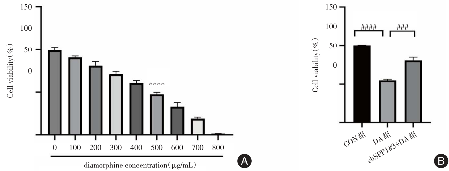
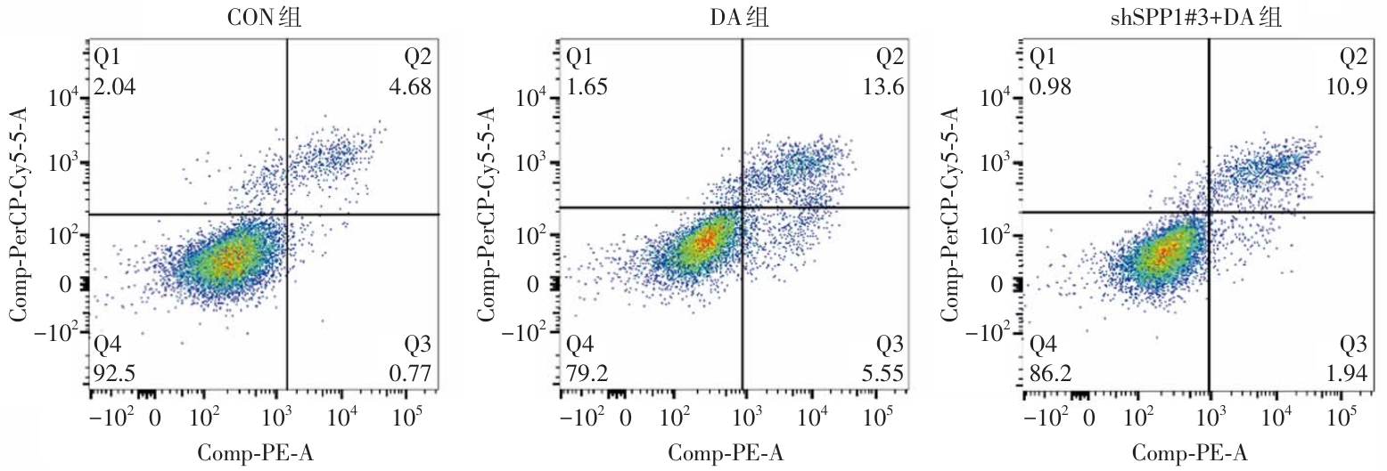
Fig. 8
Effect of DAM on apoptosis rate of NRCMs detected by flow cytometry"
Tab.7
SPP1, MYD88, Bax, Bcl2, Caspase?9, Caspase?3 mRNA changes in NRCMs after DAM intervention"
| 组别 | SPP1/GAPDH | MYD88/GAPDH | Bax/GAPDH | Bcl2/GAPDH | Caspase?9/GAPDH | Caspase?3/GAPDH |
|---|---|---|---|---|---|---|
| CON组 | 1.00 ± 0.04 | 1.00 ± 0.07 | 1.01 ± 0.17 | 1.00 ± 0.17 | 1.00 ± 0.07 | 1.01 ± 0.13 |
| DA组 | 1.94 ± 0.23& | 2.26 ± 0.09∧ | 1.46 ± 0.18? | 0.41 ± 0.02& | 2.92 ± 0.19∧ | 1.63 ± 0.16# |
| shSPP1#3+DA组 | 1.37 ± 0.11△ | 1.56 ± 0.14▽ | 0.73 ± 0.09△ | 0.69 ± 0.02○ | 1.52 ± 0.09▽ | 0.81 ± 0.06▽ |
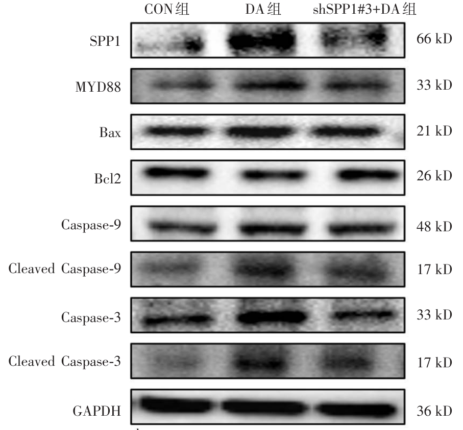
Fig.9
Expression changes of SPP1, MYD88, Bax, Bcl2,Caspase-9, Cleaved-Caspase-9, Caspase-3, and Cleaved-Caspase-3 protein expression in each group of NRCMs detected by Western blot"
Tab.8
Protein changes of SPP1, MYD88, Bax, Bcl2, Caspase-9, Caspase-3, Cleaved Caspase-9, and Cleaved Caspase-3in NRCMs after DAM intervention (n = 3)"
| 组别 | SPP1/ GAPDH | MYD88/ GAPDH | Bax/ GAPDH | Bcl2/GAPDH | Caspase?9/ GAPDH | Caspase?3/ GAPDH | Cleaved Caspase?9/ GAPDH | Cleaved Caspase?3/ GAPDH |
|---|---|---|---|---|---|---|---|---|
| CON组 | 1.00 ± 0.10 | 1.00 ± 0.67 | 1.00 ± 0.13 | 1.00 ± 0.03 | 1.00 ± 0.06 | 1.00 ± 0.12 | 1.00 ± 0.17 | 1.00 ± 0.11 |
| DA组 | 1.64 ± 0.04& | 1.37 ± 0.15? | 1.45 ± 0.06# | 0.60 ± 0.14# | 1.27 ± 0.05? | 1.47 ± 0.08# | 1.75 ± 0.18? | 1.95 ± 0.23# |
| shSPP1#3+DA组 | 1.44 ± 0.02○ | 1.04 ± 0.10○ | 1.20 ± 0.03○ | 0.90 ± 0.08○ | 0.86 ± 0.16△ | 1.11 ± 0.09△ | 1.16 ± 0.33○ | 1.24 ± 0.35○ |
| [1] | 中国国家禁毒委员会办公室. 2023年中国毒情形势报告[N].中国禁毒报,2024-06-28(003). |
| [2] |
HAN X, LIU X, ZHAO X, et al. Dapagliflozin ameliorates sepsis-induced heart injury by inhibiting cardiomyocyte apoptosis and electrical remodeling through the PI3K/Akt pathway[J]. Eur J Pharmacol, 2023, 955: 175930. doi:10.1016/j.ejphar.2023.175930
doi: 10.1016/j.ejphar.2023.175930 |
| [3] | 蔡文臣, 张诗汇, 魏中秋, 等. Ac-SDKP抑制SPP1/TGF-β1信号拮抗实验性矽肺的机制[J]. 实用医学杂志, 2021, 37(2): 159-163. |
| [4] |
ZHAO Y, HUANG Z, GAO L, et al. Osteopontin/SPP1: A potential mediator between immune cells and vascular calcification[J]. Front Immunol, 2024, 15: 1395596. doi:10.3389/fimmu.2024.1395596
doi: 10.3389/fimmu.2024.1395596 |
| [5] |
SONG Z, CHEN W, ATHAVALE D, et al. Osteopontin Takes Center Stage in Chronic Liver Disease[J]. Hepatology, 2021, 73(4): 1594-1608. doi:10.1002/hep.31582
doi: 10.1002/hep.31582 |
| [6] |
SHIRAKAWA K, SANO M. Osteopontin in Cardiovascular Diseases[J]. Biomolecules, 2021, 11(7):1047. doi:10.3390/biom11071047
doi: 10.3390/biom11071047 |
| [7] | 范晓芳. AMI患者血清VEGF-B、OPN水平变化对预后的影响[J]. 湖北科技学院学报(医学版), 2021, 35(3): 221-223,277. |
| [8] |
YANG X, WANG X, LI K, et al. Osteopontin regulates right ventricular failure through integrin ανβ3/PERK/CHOP-dependent inflammatory and apoptotic pathways[J]. Front Immunol, 2025, 16: 1569210. doi:10.3389/fimmu.2025.1569210
doi: 10.3389/fimmu.2025.1569210 |
| [9] |
BAYER A L, ALCAIDE P. MyD88: At the heart of inflammatory signaling and cardiovascular disease[J]. J Mol Cell Cardiol, 2021, 161: 75-85. doi:10.1016/j.yjmcc.2021.08.001
doi: 10.1016/j.yjmcc.2021.08.001 |
| [10] | 梁旭阳,徐萍,吕胜祥,等. TLR4和MyD88在食管鳞癌组织中的表达及临床意义[J]. 中山大学学报(医学科学版),2021,42(3):475-481. |
| [11] |
YANG F, JIANG X, CAO H, et al. Daphnetin Preconditioning Decreases Cardiac Injury and Susceptibility to Ventricular Arrhythmia following Ischaemia-Reperfusion through the TLR4/MyD88/NF-Κb Signalling Pathway[J]. Pharmacology, 2021, 106(7-8): 369-383. doi:10.1159/000513631
doi: 10.1159/000513631 |
| [12] |
GAO F, WANG X, FAN T, et al. LncRNA LINC00461 exacerbates myocardial ischemia-reperfusion injury via microRNA-185-3p/Myd88[J]. Mol Med, 2022, 28(1): 33. doi:10.1186/s10020-022-00452-1
doi: 10.1186/s10020-022-00452-1 |
| [13] | 季敏, 苏丽萍, 刘丽, 等. 阿片受体调节CaMKⅡ磷酸化在二乙酰吗啡致心肌细胞节律异常中的作用[J]. 毒理学杂志, 2023, 37(4): 297-302. |
| [14] | OELHAF R C, AZADFARD M. Heroin Toxicity[M]. Treasure Island (FL): StatPearls Publishing, 2025. |
| [15] |
YILDIRIM E, SELCUK M, SAYLIK F, et al. Effect of Heroin on Electrocardiographic Parameters[J]. Arq Bras Cardiol, 2020, 115(6): 1135-1141. doi:10.36660/abc.20190296
doi: 10.36660/abc.20190296 |
| [16] | 朱森森, 苏丽萍, 庄梦婕, 等. NEFL、NRN1在二乙酰吗啡致神经元凋亡中的作用[J]. 新疆医科大学学报, 2024, 47(6): 765-771. |
| [17] |
COLBENSON G A, KUBBARA A, COX C W, et al. Excipient lung disease secondary to intravenous heroin use[J]. BMJ Case Rep, 2022, 15(4):e247763. doi:10.1136/bcr-2021-247763
doi: 10.1136/bcr-2021-247763 |
| [18] | 卢钰芬, 郑孝明, 韦少娟, 等. miR-483-3p对乏氧/复氧诱导的心肌细胞凋亡和焦亡的作用研究[J]. 实用医学杂志, 2025, 41(3): 339-346. |
| [19] |
NEWTON K, STRASSER A, KAYAGAKI N, et al. Cell death[J]. Cell, 2024, 187(2): 235-256. doi:10.1016/j.cell.2023.11.044
doi: 10.1016/j.cell.2023.11.044 |
| [20] | 肖锦玲, 管雅玲, 朱森森, 等. HK2和VDAC1在二乙酰吗啡致心肌细胞凋亡中的作用[J]. 昆明医科大学学报, 2024, 45(2): 7-13. |
| [21] |
LIN E Y, XI W, AGGARWAL N, et al. Osteopontin (OPN)/SPP1: From its biochemistry to biological functions in the innate immune system and the central nervous system (CNS)[J]. Int Immunol, 2023, 35(4): 171-180. doi:10.1093/intimm/dxac060
doi: 10.1093/intimm/dxac060 |
| [22] |
DALAL S, ZHA Q, DANIELS C R, et al. Osteopontin stimulates apoptosis in adult cardiac myocytes via the involvement of CD44 receptors, mitochondrial death pathway, and endoplasmic reticulum stress[J]. Am J Physiol Heart Circ Physiol, 2014, 306(8): H1182-H1191. doi:10.1152/ajpheart.00954.2013
doi: 10.1152/ajpheart.00954.2013 |
| [23] |
CHEN B, LIANG S, GUO H, et al. OPN Promotes Cell Proliferation and Invasion through NF-κB in Human Esophageal Squamous Cell Carcinoma[J]. Genet Res (Camb), 2022, 2022: 3154827. doi:10.1155/2022/3154827
doi: 10.1155/2022/3154827 |
| [24] |
ZHANG Q, WANG L, WANG S, et al. Signaling pathways and targeted therapy for myocardial infarction[J]. Signal Transduct Target Ther, 2022, 7(1): 78. doi:10.1038/s41392-022-00925-z
doi: 10.1038/s41392-022-00925-z |
| [25] |
LIU Z W, WANG J K, QIU C, et al. Matrine pretreatment improves cardiac function in rats with diabetic cardiomyopathy via suppressing ROS/TLR-4 signaling pathway[J]. Acta Pharmacol Sin, 2015, 36(3): 323-333. doi:10.1038/aps.2014.127
doi: 10.1038/aps.2014.127 |
| [1] | Zhiwei GUAN,Qiong ZHAO,Jianli QIU,Yan XU,Qinwan HUANG,Hongyun ZHOU,Junqi ZHAO,Yinghui WU. Exploring the effect of modified Renshen Wumei Decoction on intestinal mucosal barrier in diarrhea rats based on TLR4/MyD88/pNF⁃κBp65 signaling pathway [J]. The Journal of Practical Medicine, 2025, 41(7): 944-952. |
| [2] | Linsha KONG,Gaigai HE,Ting LI,Kun FANG,Wei HE,Li WEI. Experimental study of doxorubicin coated by magnetic drug⁃carrying gel beads in the treatment of colorectal cancer [J]. The Journal of Practical Medicine, 2025, 41(7): 953-959. |
| [3] | Meiyu GAN,Chunjiao WU,Jingyi QIN,Zuojie. LUO. The effect of vanadyl bis(acetylacetonato) on the proliferation and invasion of human adrenocortical carcinoma cells [J]. The Journal of Practical Medicine, 2025, 41(6): 781-789. |
| [4] | Wei WANG,Min WANG,Minmin CHENG,Tingting. ZHANG. Impacts of safflower polysaccharide on tumor growth and PI3K/Akt/mTOR signal pathway in mice with colorectal cancer [J]. The Journal of Practical Medicine, 2025, 41(5): 670-675. |
| [5] | Hongyan BIAN,Shu ZHANG,Shanshan MENG,Ying WEI. Effect of pomegranate peel polyphenols on the malignant biological behavior of colon cancer cells by regulating the miR⁃138⁃5p/HIF⁃1α pathway [J]. The Journal of Practical Medicine, 2025, 41(5): 676-682. |
| [6] | Huixia YANG,Ning DING,Runqiu MA,Guizhong LI,Yinju HAO,Shengchao MA,Yideng JIANG,Zhigang. BAI. Role and mechanism of circular RNA mmu_circ_0000818 in dexamethasone⁃induced apoptosis of MC3T3⁃E1 cells [J]. The Journal of Practical Medicine, 2025, 41(4): 478-489. |
| [7] | Zonglin LI,Chunlin FENG,Xin LIU,Xingming SHU,Min. SONG. CCCTC⁃binding factors promote the formation of oxaliplatin related gastric cancer drug-tolerant cells by resisting apoptosis [J]. The Journal of Practical Medicine, 2025, 41(4): 490-499. |
| [8] | Zhizhou XIAO,Ying HUANG,Huawei. BIAN. To investigate the effect of aloperine on bone metabolism in osteoporotic mice based on autophagy and apoptosis mediated by Wnt/β⁃catenin signaling pathway [J]. The Journal of Practical Medicine, 2025, 41(4): 500-508. |
| [9] | Yufen LU,Xiaoming ZHENG,Shaojuan WEI,Liqin CHEN,Tongtong XU,Xiangwei. LÜ. Effects of miR⁃483⁃3p on hypoxia/reoxygenation⁃induced apoptosis and pyroptosis in cardiomyocytes [J]. The Journal of Practical Medicine, 2025, 41(3): 339-346. |
| [10] | Li MAI,Yongting PAN,Wenkai HE,Xiaoqian WU,Mingyan LI. Effect of Ad⁃HIF⁃1α⁃Trip on apoptosis of H9c2 cardiomyocytes under high glucose and hypoxic conditions [J]. The Journal of Practical Medicine, 2025, 41(17): 2617-2623. |
| [11] | Jingshuo LI,Shoushi LIU,Hongwei. GUO. Advances in the mechanism and therapeutic potential of Erianin⁃induced apoptosis in breast cancer cells [J]. The Journal of Practical Medicine, 2025, 41(14): 2132-2137. |
| [12] | Chang ZHANG,Chunshan LIU,Huaying LIAO,Yuchao WU,Yunhong TIAN. The effect of pseudouracil modifying enzyme 3 activating AKT pathway on malignant progression of glioblastoma [J]. The Journal of Practical Medicine, 2025, 41(12): 1825-1834. |
| [13] | Shan LUO,Ying FENG,Dandan FAN,Wenxin ZHENG,Xingrong GUO,Xuzhi. RUAN. ANGPTL8 knockout reduces lipopolysaccharide⁃induced hepatic lipid deposition [J]. The Journal of Practical Medicine, 2024, 40(9): 1197-1203. |
| [14] | Wei HE,Liping LIU,Jingwei ZHUO,Xiaodong ZHANG,Tong YANG,Jubin. FENG. CCR5 blockade reduces tumor growth by inducing apoptosis and impairing immunosuppression of tumor microenvironment [J]. The Journal of Practical Medicine, 2024, 40(9): 1204-1210. |
| [15] | Wenxin LI,Minjun LU,Li LIN,Yueqin LIU,Xiaolan. ZHU. circRAF1 regulates the proliferation and apoptosis of human ovarian granulosa cells [J]. The Journal of Practical Medicine, 2024, 40(7): 910-917. |
| Viewed | ||||||
|
Full text |
|
|||||
|
Abstract |
|
|||||