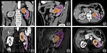
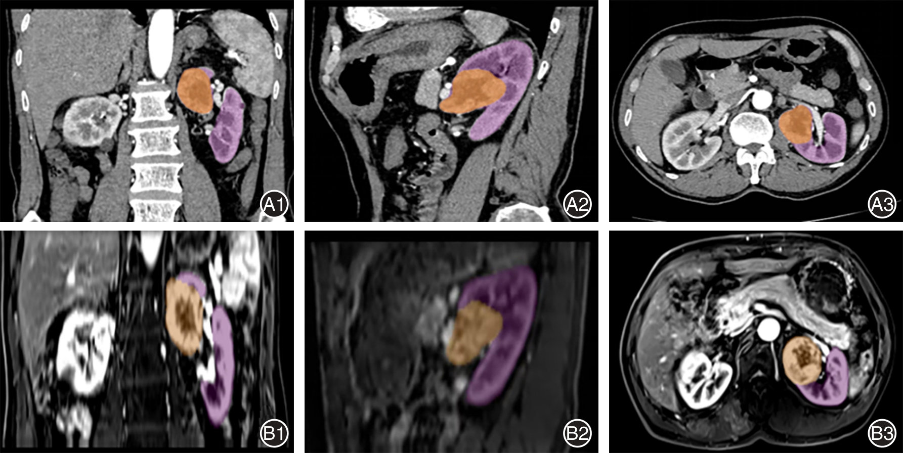
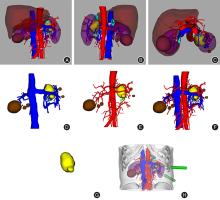
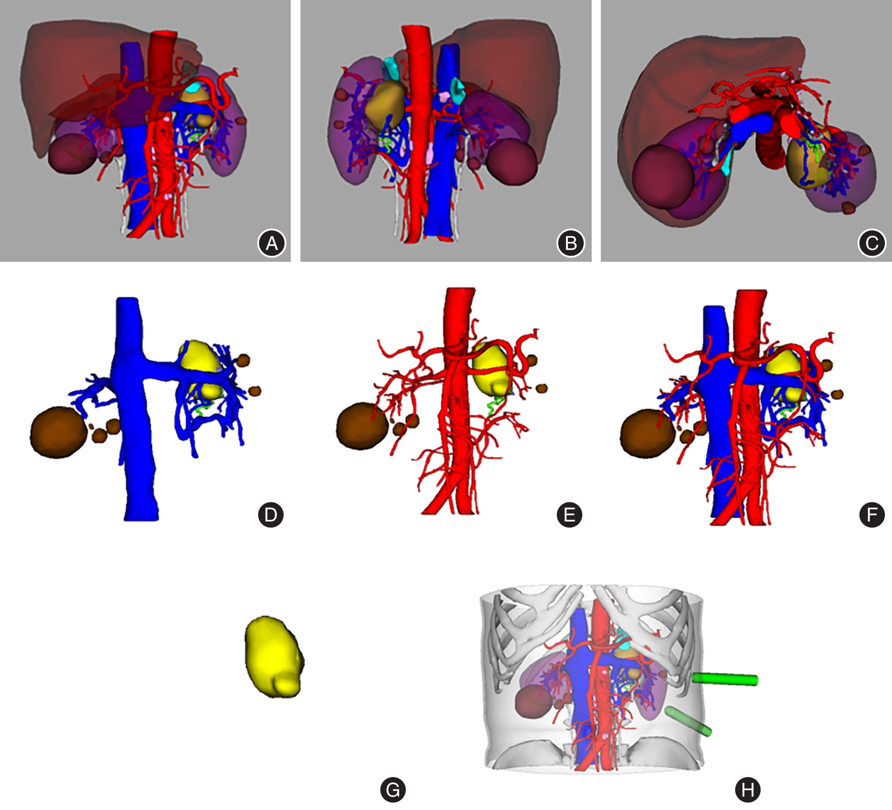
| [1] |
BUKAVINA L, BENSALAH K, BRAY F, et al. Epidemiology of renal cell carcinoma: 2022 update[J]. Eur Urol, 2022, 82(5): 529-542. doi:10.1016/j.eururo.2022.08.019
doi: 10.1016/j.eururo.2022.08.019
|
| [2] |
BAHADORAM S, DAVOODI M, HASSANZADEH S, et al. Renal cell carcinoma: An overview of the epidemiology, diagnosis, and treatment[J]. G Ital Nefrol, 2022, 39(3): 2022.
|
| [3] |
WEI L, WANG C, FU J, et al. Holographic 3D renal segments reconstruction protects renal function by promote choice of selective renal artery clamping during robot-assisted partial nephrectomy[J]. World J Urol, 2023, 41(11): 2975-2983. doi:10.1007/s00345-023-04599-2
doi: 10.1007/s00345-023-04599-2
|
| [4] |
GAO Y, LI H, YAO Y, et al. Vessel and tension-free reconstruction during robot-assisted partial nephrectomy for hilar tumors:“garland” technique and midterm outcomes[J]. J Endourol, 2020, 34(4): 469-474. doi:10.1089/end.2019.0792
doi: 10.1089/end.2019.0792
|
| [5] |
DESAI S, RAC G, PATEL H D, et al. Imaging features of renal masses to select optimal candidates for tumor enucleation partial nephrectomy[J]. Curr Urol Rep, 2022, 23(12): 345-353. doi:10.1007/s11934-022-01121-w
doi: 10.1007/s11934-022-01121-w
|
| [6] |
D'AIELLO A F, BOGNONI L, BEVILACQUA F, et al. Holographic Techniques as a Novel Method for Intervention Planning: A Tertiary Centres Experience[J]. Curr Health Sci J, 2023, 49(4): 584.
|
| [7] |
陈建新,沈海平,袁燕文,等. 三维可视化技术在腹腔镜结直肠癌D3根治术中应用价值[J]. 实用医学杂志,2022,38(12):1533-1540.
|
| [8] |
陈梓键, 王峻峰, 余闫宏, 等. 混合现实技术对腹腔镜下肿瘤动脉阻断肾部分切除手术的指导价值[J]. 临床泌尿外科杂志, 2021, 36(4): 310-313.
|
| [9] |
何跃,张海梁,秦晓健,等. 保留肾单位手术治疗完全内生性中央型肾肿瘤20例报告[J]. 临床泌尿外科杂志,2018,33(4):325-326,330.
|
| [10] |
陆智强,张艳斌,席俊华,等. 男性盆腔三维数字化重建在腹腔镜前列腺癌根治术中的应用[J]. 实用临床医药杂志,2022,26(5):1-5.
|
| [11] |
PHUNG M C, LEE B R. Recent advancements of robotic surgery for kidney cancer[J]. Asian J Endosc Surg, 2018, 11(4): 300-307. doi:10.1111/ases.12635
doi: 10.1111/ases.12635
|
| [12] |
LJUNGBERG B, ALBIGES L, ABU-GHANEM Y, et al. European association of urology guidelines on renal cell carcinoma: The 2019 update[J]. Eur Urol, 2019, 75(5): 799-810.
|
| [13] |
HUNG A J, CAI J, SIMMONS M N, et al. "Trifecta" in partial nephrectomy[J]. J Urol, 2013, 189(1): 36-42. doi:10.1016/j.juro.2012.09.042
doi: 10.1016/j.juro.2012.09.042
|
| [14] |
刘高,王祥宇,方先林,等. 基于CT的可视化三维影像重建技术在腹腔镜肾部分切除手术中的临床应用研究[J]. 世界复合医学,2022,8(10):6-10.
|
| [15] |
张童鑫,艾龙龙,张玉江,等. 三维可视化技术与二维影像比较辅助肝切除术临床效果的Meta分析[J]. 中国循证医学杂志,2018,18(8):850-857
|
| [16] |
付坚,王聪,何鹏,等. 全息影像CT三维重建技术在机器人辅助腹腔镜肾部分切除术中的应用研究[J]. 临床泌尿外科杂志,2022,37(9):698-701,707.
|
| [17] |
吕建敏,潘秀武,干思舜,等. 三维智能定性定量分析系统在双肾肿瘤精准手术规划、模拟及实施中的应用效果分析[J]. 中华泌尿外科杂志,2019,40(5):356-360.
|
| [18] |
李新飞,彭意吉,余霄腾,等. 肾部分切除术前CT三维可视化评估标准的初步探究[J]. 北京大学学报(医学版),2021,53(3):613-622.
|
| [19] |
LIANG C, ZHU J, MIAO C, et al. Protective Effects of the Segmental Renal Artery Clamping Technique on Ischemia‐Reperfusion Injury in db/db Diabetic Mice[J]. Biomed Res Int, 2017, 2017(1): 4763828. doi:10.1155/2017/4763828
doi: 10.1155/2017/4763828
|
| [20] |
NGUYEN M M, GILL I S. Halving ischemia time during laparoscopic partial nephrectomy[J]. J Urol, 2008, 179(2): 627-632. doi:10.1016/j.juro.2007.09.086
doi: 10.1016/j.juro.2007.09.086
|
| [21] |
WU X, LIU R, YU J, et al. Mixed reality technology–assisted orthopedics surgery navigation[J]. Surg Innov, 2018, 25(3): 304-305. doi:10.1177/1553350618771413
doi: 10.1177/1553350618771413
|
| [22] |
张凯,朱刚,李鸿波,等. 三维影像重建在泌尿外科机器人手术中的应用[J]. 中华泌尿外科杂志,2018,39(9): 690-693.
|
| [23] |
冯超,申玉兰,陈磊,等. 多模态三维影像重建技术在尿道狭窄诊断中的应用[J]. 中华泌尿外科杂志,2018,39(5):367-371
|
| [24] |
王振龙,李晓会,李和程,等. 3D打印模型或CT三维重建指导下的肿瘤四点定位法在完全内生型肾癌腹腔镜下肾部分切除术中的应用[J]. 中华泌尿外科杂志, 2016, 37(10): 735-739.
|
| [25] |
燕荣帅,李翔,肖晶晶,等. 混合现实技术在整形外科教学中的应用探索[J]. 中国美容医学, 2018, 27(2): 140-142.
|
| [26] |
朱凯,罗建斌. 全息影像腹腔镜融合技术在肾肿瘤切除术中的临床效果观察[J]. 中国现代药物应用,2023,17(22):61-63
|


 )
)