

The Journal of Practical Medicine ›› 2025, Vol. 41 ›› Issue (1): 23-29.doi: 10.3969/j.issn.1006-5725.2025.01.004
• Basic Research • Previous Articles Next Articles
Chunyan LI1,Ting XIAO2,Bangcui WU1,Yong CHEN1,Mei TIAN1( )
)
Received:2024-05-27
Online:2025-01-10
Published:2025-01-14
Contact:
Mei TIAN
E-mail:348820517@qq.com
CLC Number:
Chunyan LI,Ting XIAO,Bangcui WU,Yong CHEN,Mei TIAN. PKCβ inhibitor modulates macrophage phenotype and affects kidney ischemia-reperfusion injury during transplantation[J]. The Journal of Practical Medicine, 2025, 41(1): 23-29.
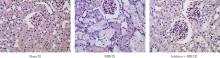
Fig. 1
PAS staining in each group(× 400)"
Tab.1
Renal tubular injury score、Cr and BUN values in each group"
| 组别 | 肾小管损伤评分 | Cr | BUN |
|---|---|---|---|
| Sham组 | 0.11 ± 0.33 | 32.33 ± 3.51 | 4.20 ± 0.16 |
| RIRI组 | 2.56 ± 0.53* | 239.00 ± 123.45* | 37.79 ± 17.64* |
| Inhibitor + RIRI组 | 1.56 ± 0.53#& | 197.57 ± 101.97*& | 17.65 ± 8.44& |
| F值 | 21.62 | 4.191 | 8.64 |
| P值 | < 0.05 | < 0.05 | < 0.05 |

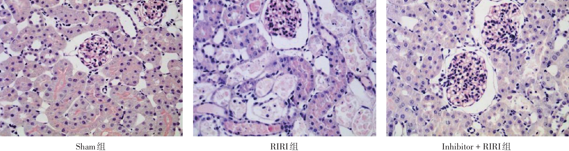
Fig. 2
Expression of KIM-1 and RPA-1 in each group (IHC,× 400)"
Tab.2
KIM-1 and RPA-1 values in each group"
| 组别 | KIM-1 | RPA-1 |
|---|---|---|
| Sham组 | 2.52 ± 1.83 | 11.16 ± 9.83 |
| RIRI组 | 110.79 ± 13.59* | 141.15 ± 9.33* |
| Inhibitor + RIRI组 | 47.46 ± 8.83*& | 54.05 ± 4.47*& |
| F值 | 200.23 | 23.14 |
| P值 | < 0.01 | < 0.01 |

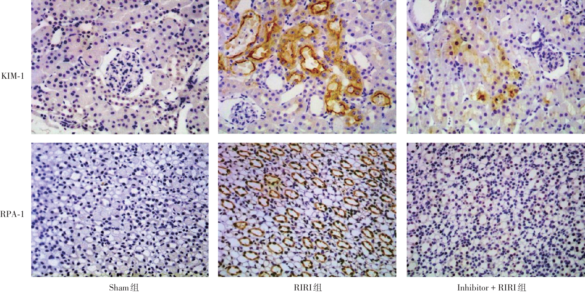
Fig.3
Expression of iNOS、IL-12 and ARG-1 in each group (IHC,× 400)"
Tab.3
The total IOD levels of iNOS、IL-12 and ARG-1 in each group"
| 组别 | iNOS | IL-12 | ARG-1 |
|---|---|---|---|
| Sham组 | 1.76 ± 0.92 | 1.93 ± 1.48 | 10.45 ± 4.23 |
| RIRI组 | 200.44 ± 14.95* | 23.89 ± 1.46* | 26.05 ± 5.37 |
| Inhibitor + RIRI组 | 47.36 ± 2.08*& | 2.8 ± 0.75& | 122.23 ± 22.46#& |
| F值 | 710.58 | 10.50 | 179.55 |
| P值 | < 0.05 | < 0.05 | < 0.05 |
Tab. 4
mRNA expression levels of Dectin-1 and Arg-1 in each group"
| 组别 | Dectin-1 | Arg-1 |
|---|---|---|
| Sham组 | 0.08 ± 0.04 | 0.19 ± 0.11 |
| RIRI组 | 2.39 ± 1.96 | 2.85 ± 1.78 |
| Inhibitor + RIRI组 | 18.40 ± 12.94*& | 12.27 ± 7.83*& |
| F值 | 8.72 | 7.51 |
| P值 | < 0.05 | < 0.05 |
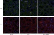
Fig. 4
Expression of CD197 and CD163 in each group ( IF,× 400)"

Tab.5
The fluorescence intensity of CD197 and CD163 in each group"
| 组别 | CD197 | CD163 |
|---|---|---|
| Sham组 | 21.04 ± 1.20 | 23.36 ± 1.15 |
| RIRI组 | 32.67 ± 0.38* | 20.10 ± 3.62 |
| Inhibitor + RIRI组 | 26.40 ± 1.49*& | 35.00 ± 1.46*& |
| F值 | 7.20 | 33.43 |
| P值 | < 0.05 | < 0.05 |
| 1 |
MARYAM B, SMITH M E, MILLER S J, et al. Macrophage Ontogeny, Phenotype, and Function in Ischemia Reperfusion-Induced Injury and Repair[J]. Kidney360,2024,5(3):459-470. doi:10.34067/kid.0000000000000376
doi: 10.34067/kid.0000000000000376 |
| 2 |
ZHANG Y L, TANG T T, WANG B,et al. Identification of a Novel ECM Remodeling Macrophage Subset in AKl to CKD Transition by Integrative Spatial and Single-Cell Analysis[J].Adv Sci (Weinh),2024,11(38):e2309752. doi:10.1002/advs.202470228
doi: 10.1002/advs.202470228 |
| 3 |
KAWANO T, INOKUCHI J, ETO M, et al. Activators and Inhibitors of Protein Kinase C (PKC):Their Applications in Clinical Trials[J]. Pharmaceutics,2021,13(11):1748. doi:10.3390/pharmaceutics13111748
doi: 10.3390/pharmaceutics13111748 |
| 4 |
ALLEBOINA S, WONG T, SINGH M V, et al. Inhibition of protein kinase C beta phosphorylation activates nuclear factor-kappa B and improves postischemic recovery in type 1 diabetes[J]. Exp Biol Med (Maywood),2020,245(9):785-796. doi:10.1177/1535370220920832
doi: 10.1177/1535370220920832 |
| 5 |
LIN Y F, LEU S J, HUANG H M,et al. Selective activation of specific PKC isoforms dictating the fate of CD14(+) monocytes towards differentiation or apoptosis [J]. J Cell Physiol,2011,226(1):122-131. doi:10.1002/jcp.22312
doi: 10.1002/jcp.22312 |
| 6 |
LIN Y F, LEE H M, LEU S J, et al. The essentiality of PKC alpha and PKC betaI translocation for CD14+monocyte differentiation towards macrophages and dendritic cells, respectively [J]. J Cell Biochem,2007,102(2):429-441. doi:10.1002/jcb.21305
doi: 10.1002/jcb.21305 |
| 7 |
LIU H L, HUANG Z, LI Q Z, et al.Schisandrin A alleviates renal fibrosis by inhibiting PKC beta and oxidative stress[J]. Phytomedicine,2024,126:155372. doi:10.1016/j.phymed.2024.155372
doi: 10.1016/j.phymed.2024.155372 |
| 8 |
WANG J, CASIMIRO-GARCIA A, JOHNSON B G, et al. A protein kinase C alpha and beta inhibitor blunts hyperphagia to halt renal function decline and reduces adiposity in a rat model of obesity-driven type 2 diabetes[J]. Sci Rep,2023,13(1):16919. doi:10.1038/s41598-023-43759-7
doi: 10.1038/s41598-023-43759-7 |
| 9 |
RONG S, HUEPER K, KIRSCH T, et al. Renal PKC-epsilon deficiency attenuates acute kidney injury and ischemic allograft injury via TNF-alpha-dependent inhibition of apoptosis and inflammation [J]. Am J Physiol Renal Physiol,2014, 307(6):F718-F726. doi:10.1152/ajprenal.00372.2013
doi: 10.1152/ajprenal.00372.2013 |
| 10 | 杨旸, 杨亦斌, 梁国标, 等. 蛋白激酶C抑制剂对小鼠肾移植后炎性细胞浸润的影响[J]. 中华实验外科杂志,2015, 32(1): 204. |
| 11 | 樊雪梅, 容松. 蛋白激酶C β抑制剂对肾缺血-再灌注损伤模型及其巨噬细胞亚型表达的影响 [J]. 器官移植,2019,10(4):423-427. |
| 12 |
RONG S, PARK J K, KIRSCH T, et al. The TIM-1:TIM-4 pathway enhances renal ischemia-reperfusion injury[J]. J Am Soc Nephrol,2011,22(3):484-495. doi:10.1681/asn.2010030321
doi: 10.1681/asn.2010030321 |
| 13 |
TSENG W C, LEE P Y, TSAI M T, et al. Hypoxic mesenchymal stem cells ameliorate acute kidney ischemia reperfusion injury via enhancing renal tubular autophagy[J]. Stem Cell Res Ther,2021,12(1):367. doi:10.1186/s13287-021-02374-x
doi: 10.1186/s13287-021-02374-x |
| 14 |
YAO W, CHEN Y, LI Z, et al. Single Cell RNA Sequencing Identifies a Unique Inflammatory Macrophage Subset as a Druggable Target for Alleviating Acute Kidney Injury[J]. Adv Sci (Weinh),2022,9(12):e2103675. doi:10.1002/advs.202270079
doi: 10.1002/advs.202270079 |
| 15 |
ZHANG J, LI Q, ZOU Y R, et al. HMGB1-TLR4-IL-23-IL-17A axis accelerates renal ischemia-reperfusion injury via the recruitment and migration of neutrophils[J]. Int Immunopharmacol, 2021,94:107433. doi:10.1016/j.intimp.2021.108461
doi: 10.1016/j.intimp.2021.108461 |
| 16 |
MA T, LI X, ZHU Y, et al. Excessive Activation of Notch Signaling in Macrophages Promote Kidney Inflammation, Fibrosis, and Necroptosis[J]. Front Immunol,2022,13:835879. doi:10.3389/fimmu.2022.835879
doi: 10.3389/fimmu.2022.835879 |
| 17 |
SUN Z, ZHANG F, GAO Z, et al. Liraglutide alleviates ferroptosis in renal ischemia reperfusion injury via inhibiting macrophage extracellular trap formation[J]. Int Immunopharmacol,2024,142(Pt B):113258. doi:10.1016/j.intimp.2024.113258
doi: 10.1016/j.intimp.2024.113258 |
| 18 |
KADIR R R A, ALWJWAJ M, BAYRAKTUTAN U. Protein kinase C-beta distinctly regulates blood-brain barrier-forming capacity of Brain Microvascular endothelial cells and outgrowth endothelial cells[J]. Metab Brain Dis,2022,37(6):1815-1827. doi:10.1007/s11011-022-01041-1
doi: 10.1007/s11011-022-01041-1 |
| 19 |
TANG P M, NIKOLIC-PATERSON D J, LAN H Y. Macrophages: Versatile players in renal inflammation and fibrosis [J].Nat Rev Nephrol,2019,15(3):144-158. doi:10.1038/s41581-019-0110-2
doi: 10.1038/s41581-019-0110-2 |
| 20 |
ZHANG R, QIN C, ZHANG J, et al. DNA hypomethylation of Syk induces oxidative stress and apoptosis via the PKCβ/P66shc signaling pathway in diabetic kidney disease[J]. FASEB J,2024,38(6):e23564. doi:10.1096/fj.202301579r
doi: 10.1096/fj.202301579r |
| 21 |
LI Y, LIANG Q, ZHOU L,et al. An ROS-responsive artesunate prodrug nanosystem co-delivers dexamethasone for rheumatoid arthritis treatment through the HIF-1 α/NF-κB cascade regulation of ROS scavenging and macrophage repolarization[J]. Acta Biomater, 2022,152:406-424. doi:10.1016/j.actbio.2022.08.054
doi: 10.1016/j.actbio.2022.08.054 |
| Viewed | ||||||
|
Full text |
|
|||||
|
Abstract |
|
|||||