

The Journal of Practical Medicine ›› 2026, Vol. 42 ›› Issue (12): 2119-2127.doi: 10.3969/j.issn.1006-5725.2026.12.005
• Oncology: Diagnosis, Treatment and Prevention • Previous Articles
Hongmei YUAN1( ),Dongyuan LIU1,Yajun XU1,Xiuping ZHAO1,Wenjie MA2
Received:2026-01-28
Online:2026-06-25
Published:2026-06-30
Contact:
Hongmei YUAN
E-mail:smxhongmei@163.com
CLC Number:
Hongmei YUAN,Dongyuan LIU,Yajun XU,Xiuping ZHAO,Wenjie MA. The relationship between high-resolution CT features of pulmonary ground-glass nodules and pathological subtypes of lung adenocarcinoma[J]. The Journal of Practical Medicine, 2026, 42(12): 2119-2127.
Tab.1
Comparison of baseline data and clinical indicators among patients with different pathological types"
| 指标 | IAC组(n = 67) | MIA组(n = 14) | AIS组(n = 13) | AAH组(n = 10) | F/χ2值 | P值 |
|---|---|---|---|---|---|---|
| 性别 | 0.404 | 0.939 | ||||
| 男 | 35(52.24) | 7(50.00) | 7(53.85) | 6(60.00) | ||
| 女 | 32(47.76) | 7(50.00) | 6(46.15) | 4(40.00) | ||
| 年龄(x ± s)/岁 | 58.56 ± 15.17 | 59.68 ± 17.24 | 58.11 ± 14.10 | 58.27 ± 15.63 | 0.039 | 0.990 |
| BMI(x ± s)/(kg/m2) | 22.24 ± 2.36 | 22.63 ± 2.70 | 22.43 ± 3.01 | 22.51 ± 2.48 | 0.089 | 0.966 |
| 病灶位置 | 4.529 | 0.972 | ||||
| 左肺上叶 | 12(17.91) | 2(14.29) | 2(15.38) | 2(20.00) | ||
| 左肺下叶 | 12(17.91) | 4(28.57) | 3(23.08) | 2(20.00) | ||
| 右肺上叶 | 19(28.36) | 5(35.71) | 5(38.46) | 2(20.00) | ||
| 右肺中叶 | 12(17.91) | 2(14.29) | 1(7.69) | 3(30.00) | ||
| 右肺下叶 | 12(17.91) | 1(7.14) | 2(15.38) | 1(10.00) |
Tab.2
Comparison of HRCT features among patients with different pathological types"
| 指标 | IAC组(n = 67) | MIA组(n = 14) | AIS组(n = 13) | AAH组(n = 10) | χ2值 | P值 |
|---|---|---|---|---|---|---|
| 形态 | 1.674 | 0.643 | ||||
| 圆形/类圆 | 20(29.85) | 6(42.86) | 3(23.08) | 4(40.00) | ||
| 不规则 | 47(70.15) | 8(57.14) | 10(76.92) | 6(60.00) | ||
| 分叶征 | 46(68.66) | 5(35.71)* | 5(38.46)* | 3(30.00)* | 11.080 | 0.011 |
| 毛刺征 | 50(74.63) | 7(50.00)* | 6(46.15)* | 4(40.00)* | 8.810 | 0.032 |
| 界面 | 10.500 | 0.015 | ||||
| 清楚光滑 | 10(14.93) | 5(35.71)* | 4(30.77)* | 3(30.00)* | ||
| 清楚毛糙 | 45(67.16) | 5(35.71)* | 4(30.77)* | 3(30.00)* | ||
| 模糊 | 12(17.91) | 4(28.57)* | 5(38.46)* | 4(40.00)* | ||
| 空泡征 | 30(44.78) | 1(7.14)* | 0(0.00)* | 0(0.00)* | 20.379 | <0.001 |
| 支气管截断征 | 26(38.81) | 0(0.00)* | 0(0.00)* | 0(0.00)* | 19.144 | <0.001 |
| 胸膜凹陷征 | 31(46.27) | 2(14.29)* | 1(7.69)* | 0(0.00)* | 16.317 | <0.001 |
| 血管集束征 | 25(37.31) | 1(7.14)* | 0(0.00)* | 0(0.00)* | 15.466 | 0.001 |
Tab.3
Comparison of HRCT quantitative parameters among patients with different pathological types"
| 指标 | IAC组(n = 67) | MIA组(n = 14) | AIS组(n = 13) | AAH组(n = 10) | F值 | P值 |
|---|---|---|---|---|---|---|
| 结节最大径/mm | 12.50 ± 3.12 | 6.82 ± 2.06* | 6.61 ± 1.83* | 6.34 ± 1.70* | 44.729 | < 0.001 |
| CT值/HU | -520.18 ± 68.01 | -569.38 ± 74.16* | -575.83 ± 80.23* | -581.28 ± 87.41* | 4.887 | 0.003 |
| 相对CT值 | 0.60 ± 0.12 | 0.66 ± 0.09* | 0.67 ± 0.10* | 0.67 ± 0.09* | 3.370 | 0.021 |
| 体积/mm3 | 1 005.45 ± 247.45 | 572.12 ± 108.144* | 593.72 ± 120.17* | 580.42 ± 131.89* | 54.801 | < 0.001 |
| 质量/mg | 623.65 ± 173.19 | 289.67 ± 82.55* | 300.06 ± 91.76* | 293.65 ± 93.61* | 38.325 | < 0.001 |
Tab.4
Comparison of HRCT qualitative features among IAC subtypes"
| 指标 | 附壁型组(n = 35) | 腺泡型组(n = 21) | 乳头型组(n = 11) | χ2值 | P值 |
|---|---|---|---|---|---|
| 形态 | 1.210 | 0.546 | |||
| 圆形/类圆 | 11(31.43) | 6(28.57) | 3(27.27) | ||
| 不规则 | 24(68.57) | 15(71.43) | 8(72.73) | ||
| 分叶征 | 24(68.57) | 15(71.43) | 7(63.64) | 0.204 | 0.903 |
| 毛刺征 | 26(74.29) | 15(71.43) | 9(81.82) | 0.416 | 0.812 |
| 界面 | 1.042 | 0.903 | |||
| 清楚光滑 | 4(11.43) | 3(14.29) | 3(27.27) | ||
| 清楚毛糙 | 25(71.43) | 15(71.43) | 5(45.45)?# | ||
| 模糊 | 6(17.14) | 3(14.29) | 3(27.27) | ||
| 空泡征 | 17(48.57) | 11(52.38) | 2(18.18)?# | 3.841 | 0.147 |
| 支气管截断征 | 14(40.00) | 8(38.10) | 4(36.36) | 0.053 | 0.974 |
| 胸膜凹陷征 | 16(45.71) | 11(52.38) | 4(36.36) | 0.754 | 0.686 |
| 血管集束征 | 15(42.86) | 9(42.86) | 1(9.09)?# | 4.482 | 0.106 |
Tab.5
Comparison of HRCT quantitative parameters among IAC subtypes"
| 指标 | 附壁型组(n = 35) | 腺泡型组(n = 21) | 乳头型组(n = 11) | F值 | P值 |
|---|---|---|---|---|---|
| 结节最大径/mm | 11.95 ± 3.06 | 12.90 ± 3.20 | 13.50 ± 3.30 | 1.924 | 0.154 |
| CT值/HU | -540.13 ± 69.10 | -510.38 ± 67.80* | -475.42 ± 66.20*& | 8.736 | < 0.001 |
| 相对CT值 | 0.63 ± 0.12 | 0.58 ± 0.12* | 0.55 ± 0.13* | 3.512 | 0.036 |
| 体积/mm3 | 890.32 ± 233.54 | 1 105.80 ± 255.40* | 1 180.20 ± 260.30* | 7.843 | < 0.001 |
| 质量/mg | 558.52 ± 167.42 | 689.14 ± 187.21* | 705.87 ± 192.04* | 6.724 | 0.002 |
Tab.6
Multivariate binary logistic regression analysis of influencing factors of IAC"
| 指标 | β值 | SE | Wald χ2 | OR(95%CI) | P值 |
|---|---|---|---|---|---|
| 结节最大径 | 0.239 | 0.105 | 5.181 | 1.270(1.034 ~ 1.560) | 0.023 |
| CT值 | 0.291 | 0.061 | 22.903 | 1.338(1.187 ~ 1.508) | < 0.001 |
| 相对CT值 | -0.571 | 0.334 | 2.923 | 0.565(0.294 ~ 1.087) | 0.088 |
| 体积 | 0.113 | 0.037 | 9.327 | 1.120(1.041 ~ 1.204) | 0.002 |
| 质量 | 2.117 | 1.027 | 4.249 | 1.658(1.110 ~ 62.13) | 0.059 |
| 分叶征 | 0.133 | 0.060 | 4.194 | 1.142(1.016 ~ 1.285) | 0.027 |
| 毛刺征 | 0.271 | 0.206 | 1.731 | 1.311(0.876 ~ 1.964) | 0.189 |
| 空泡征 | 0.394 | 0.245 | 2.586 | 1.483(0.917 ~ 2.397) | 0.109 |
| 支气管截断征 | 0.457 | 0.387 | 1.394 | 1.579(0.740 ~ 3.372) | 0.238 |
| 胸膜凹陷征 | 0.592 | 0.405 | 2.137 | 1.808(0.817 ~ 3.998) | 0.145 |
| 血管集束征 | 0.333 | 0.177 | 3.540 | 1.395(0.986 ~ 1.974) | 0.061 |
Tab.7
ROC curve analysis of diagnostic value of HRCT features on IAC"
| 指标 | AUC | SE | P值 | 95%CI | 截断值 | 约登指数 | 敏感度/% | 特异度/% | |
|---|---|---|---|---|---|---|---|---|---|
| 下限 | 上限 | ||||||||
| 结节最大径 | 0.929 | 0.027 | < 0.001 | 0.876 | 0.982 | 9.48 mm | 0.706 | 89.55 | 81.08 |
| CT值 | 0.827 | 0.045 | < 0.001 | 0.739 | 0.915 | -550.24 HU | 0.605 | 82.09 | 78.38 |
| 体积 | 0.909 | 0.033 | < 0.001 | 0.844 | 0.973 | 803.67 mm3 | 0.664 | 88.06 | 78.38 |
| 分叶征 | 0.668 | 0.056 | 0.005 | 0.557 | 0.778 | / | 0.518 | 76.12 | 75.68 |
| 联合预测 | 0.992 | 0.006 | < 0.001 | 0.981 | 1.000 | / | 0.727 | 97.01 | 75.68 |
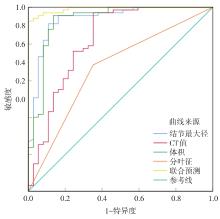
Fig.1
ROC curve analysis of diagnostic value of HRCT features on IAC"
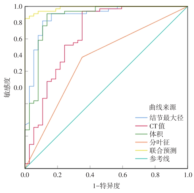
Fig.2
CT plain scan and HRCT examination images of a 28-year-old female patient with microinvasive adenocarcinoma"
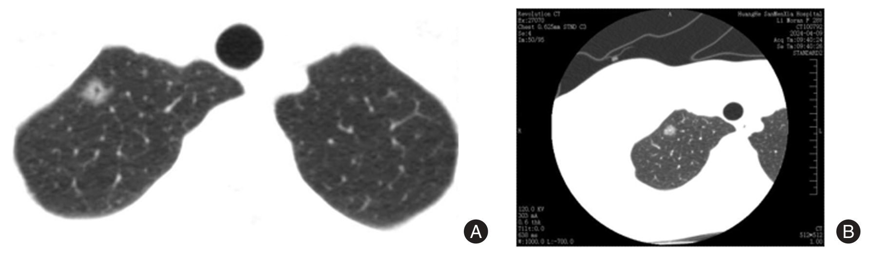
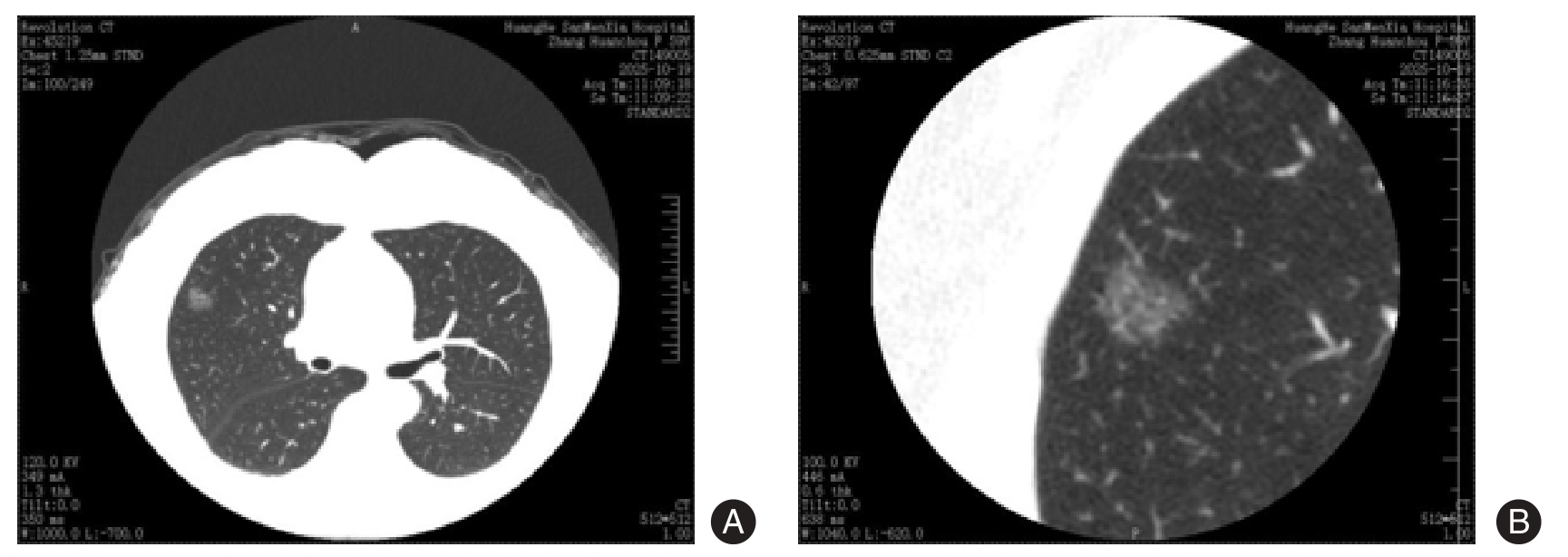
Fig.3
CT plain scan and HRCT examination images of a 59-year-old female patient with invasive adenocarcinoma"
| [1] |
李祥天,温小晖,王若琳,等. 机械拉伸载荷调控肺癌细胞行为的分子机制[J]. 中国临床解剖学杂志,2025,43(4):444-449.doi:10.13418/j.issn.1001-165x.2025.4.13 .
doi: 10.13418/j.issn.1001-165x.2025.4.13 |
| [2] |
KANZAKI R, REID S, BOLIVAR P,et al. FHL2 expression by cancer-associated fibroblasts promotes metastasis and angiogenesis in lung adenocarcinoma[J]. Int J Cancer,2025,156(2):431-446.doi: 10.1002/ijc.35174 .
doi: 10.1002/ijc.35174 |
| [3] |
YIN X, LU Y, CUI Y,et al. CT-based radiomics-deep learning model predicts occult lymph node metastasis in early-stage lung adenocarcinoma patients: A multicenter study[J]. Chin J Cancer Res,2025,37(1):12-27.doi: 10.21147/j.issn.1000-9604.2025. 01.02 .
doi: 10.21147/j.issn.1000-9604.2025. 01.02 |
| [4] |
LIU J, LI Y, LONG Y,et al. Predicting High-risk Lung Adenocarcinoma in Solid and Part-solid Nodules on Low-dose CT: A Multicenter Study[J]. Acad Radiol,2025,32(5): 2966-2976.doi: 10.1016/j.acra.2024.11.059 .
doi: 10.1016/j.acra.2024.11.059 |
| [5] |
ZOU P L, MA C H, LI X,et al. Early Lung Adenocarcinoma Manifesting as Irregular Subsolid Nodules: Clinical and CT Characteristics[J]. Acad Radiol,2025,32(4): 2320- 2329.doi: 10.1016/j.acra.2024.12.010 .
doi: 10.1016/j.acra.2024.12.010 |
| [6] |
石琴,张依凡,杨易,等. 18F-FDG PET/CT预测浸润性肺腺癌WHO(2021)组织学分级[J]. 中国医学影像学杂志,2025,33(2):171-176.doi: 10.3969/j.issn.1005-5185. 2025.02.012 .
doi: 10.3969/j.issn.1005-5185. 2025.02.012 |
| [7] |
WU L H, CHEN L, WANG Q Y,et al. Correlation between HRCT signs and levels of CA125, SCCA, and NSE for different pathological types of lung cancer[J]. Eur Rev Med Pharmacol Sci,2023,27(9):4162-4168.doi: 10.26355/eurrev_202305_32325 .
doi: 10.26355/eurrev_202305_32325 |
| [8] |
ZHANG G, SHANG L, LI S,et al. Non-enhanced CT deep learning model for differentiating lung adenocarcinoma from tuberculoma: A multicenter diagnostic study[J]. Eur Radiol,2025,35(12):8116-8125.doi: 10.1007/s00330-025-11721-y .
doi: 10.1007/s00330-025-11721-y |
| [9] |
AZUMA Y, SAKAMOTO S, HOMMA S,et al. Impact of accurate diagnosis of interstitial lung diseases on postoperative outcomes in lung cancer[J]. Gen Thorac Cardiovasc Surg,2023,71(2):129-137.doi: 10.1007/s11748-022-01868-6 .
doi: 10.1007/s11748-022-01868-6 |
| [10] |
中国医疗保健国际交流促进会肿瘤内科学分会,中国医师协会肿瘤医师分会. Ⅳ期原发性肺癌中国治疗指南(2024版)[J].中华肿瘤杂志,2024,46(7):595-636. doi:10.3760/cma.j.cn112152-20240311-00104 .
doi: 10.3760/cma.j.cn112152-20240311-00104 |
| [11] |
SASAKI T, KUNO H, HIYAMA T,et al. 2021 WHO Classification of Lung Cancer: Molecular Biology Research and Radiologic-Pathologic Correlation[J]. Radiographics, 2024,44(3):1-36.doi: 10.1148/rg.230136 .
doi: 10.1148/rg.230136 |
| [12] |
王春,王晓娣,张海涛,等. 人工智能量化参数联合256层螺旋CT扫描对肺磨玻璃结节浸润程度的预测[J]. 实用医学杂志,2025,41(19):3106-3111. doi:10.3969/j.issn.1006-5725.2025. 19.022 .
doi: 10.3969/j.issn.1006-5725.2025. 19.022 |
| [13] |
CHEN L, SU Y, HUANG Y,et al. Predicting lymphovascular invasion in stage IA lung adenocarcinoma: A CT-based classification and regression tree model[J]. Eur Radiol,2025,35(10):6357-6368.doi: 10.1007/s00330-025-11593-2 .
doi: 10.1007/s00330-025-11593-2 |
| [14] |
古明宇, 郑翔匀, 马吉尔, 等. 干细胞治疗在肺移植领域中的应用现状[J]. 器官移植, 2026, 17(2): 311-318. doi: 10.12464/ j.issn.1674-7445.2025233 .
doi: 10.12464/ j.issn.1674-7445.2025233 |
| [15] |
XU P, YAO F, XU Y,et al. Habitat Radiomics and Deep Learning Features Based on CT for Predicting Lymphovascular Invasion in T1-stage Lung Adenocarcinoma: A Multicenter Study[J]. Acad Radiol,2025,32(8):4860-4870.doi: 10.1016/j.acra.2025. 04.005 .
doi: 10.1016/j.acra.2025. 04.005 |
| [16] |
HUANG L, XU L, WANG X,et al. Prediction of EGFR Mutations in Lung Adenocarcinoma via CT Images: A Comparative Study of Intratumoral and Peritumoral Radiomics, Deep Learning, and Fusion Models[J]. Acad Radiol, 2025,32(8):4880-4892.doi: 10.1016/j.acra.2025.04.029 .
doi: 10.1016/j.acra.2025.04.029 |
| [17] |
汪琼,王之悦,雍千叶,等. 肺结节圆度在HRCT多平面重建图像上对纯磨玻璃结节的浸润性预测优于常规横断位[J]. 中国CT和MRI杂志,2025,23(9): 44-46, 93.doi: 10.3969/j.issn.1 672-5131.2025.09.013 .
doi: 10.3969/j.issn.1 672-5131.2025.09.013 |
| [18] |
武国华,伏平友,邢璐,宋方. 51例单发炎性肺磨玻璃结节患者HRCT特异征象分析[J]. 医学影像学杂志,2025,35(9):65-67.doi: 10.20258/j.cnki.1006-9011.2025. 09.015 .
doi: 10.20258/j.cnki.1006-9011.2025. 09.015 |
| [19] |
张利霞,刘海霞,陈玮,等. 纯磨玻璃结节的HRCT征象对非附壁型浸润性肺腺癌的诊断价值[J]. 河北医学,2023,29(1):112-115.doi: 10.3969/j.issn.1006-6233. 2023. 01.021 .
doi: 10.3969/j.issn.1006-6233. 2023. 01.021 |
| [20] |
刘江勇,黄文才,薛阳,等. 基于HRCT浸润性肺腺癌异质性分析及其与肿瘤倍增时间的相关性[J]. 放射学实践,2023,38(8):990-995.doi: 10.13609/j.cnki.1000-0313. 2023.08.006 .
doi: 10.13609/j.cnki.1000-0313. 2023.08.006 |
| [21] |
CHEN X, QI H, ZOU Y,et al. Predicting the spread through air spaces in lung adenocarcinoma from preoperative 18 F-FDG PET/CT radiomics[J]. Nucl Med Commun,2025,46(6):570-578.doi: 10.1097/MNM.0000000000001975 .
doi: 10.1097/MNM.0000000000001975 |
| [22] |
SATO J, YANAGAWA M, NISHIGAKI D,et al. Radiologists Versus AI-Based Software: Predicting Lymph Node Metastasis and Prognosis in Lung Adenocarcinoma From CT Under Various Image Display Conditions[J]. Clin Lung Cancer,2025,26(1): 58-71.doi: 10.1016/j.cllc.2024.10.015 .
doi: 10.1016/j.cllc.2024.10.015 |
| [23] |
DING X, ZHOU W, ZHONG G,et al. One-year mortality risk prediction model for patients with interstitial lung disease and lung cancer[J]. Transl Lung Cancer Res,2025, 14(5):1786-1803.doi: 10.21037/tlcr-2025-235 .
doi: 10.21037/tlcr-2025-235 |
| [24] |
KUDO Y, SAITO A, HORIUCHI T,et al. Preoperative evaluation of visceral pleural invasion in peripheral lung cancer utilizing deep learning technology[J]. Surg Today, 2025,55(1):18-28.doi: 10.1007/s00595-024-02869-z .
doi: 10.1007/s00595-024-02869-z |
| [25] |
CHEN S, WANG X, LIN X,et al. CT-based radiomics predictive model for spread through air space of IA stage lung adenocarcinoma[J]. Acta Radiol,2025,66(5): 477- 486.doi: 10.1177/02841851241305737 .
doi: 10.1177/02841851241305737 |
| [26] |
MORI S, HASEGAWA M, SAKAI F,et al. Incidence of and predictive factors for lung cancer in patients with rheumatoid arthritis: A retrospective long-term follow-up study[J]. Mod Rheumatol,2025,35(2):240-248.doi: 10.1093/mr/roae084 .
doi: 10.1093/mr/roae084 |
| [27] |
邓琦,徐志锋,周涛,等. 基于人工智能CT定量分析对长径≤10 mm肺磨玻璃结节浸润程度的预测价值[J].实用放射学杂志,2023,39(7):1088-1092.doi: 10.3969/j.issn.1002-1671. 2023.07.010 .
doi: 10.3969/j.issn.1002-1671. 2023.07.010 |
| [1] | Xiaoyue LI,Na WANG,Xianni LIU,Tingli DAI,Haiwen CHEN,Jianguo XIN,Wei WANG,Menglan ZHANG. Analysis of the relationship between stromal fibrosis degree and targeted therapy resistance and prognosis in EGFR mutant lung adenocarcinoma [J]. The Journal of Practical Medicine, 2025, 41(15): 2381-2387. |
| [2] | Ying LIU,Yun LIU,Jiahui LIU,Lu WANG,Wenrui HOU,Xiaoli LI,Junxin XIANG,Dianming. LI. An analysis on clinical characteristics in patients with lung adenocarcinomas tested positive for EGFR mutation in pleural effusion [J]. The Journal of Practical Medicine, 2024, 40(1): 43-47. |
| [3] |
HUANG Hancong, CHEN Qianhua, YANG Xiaorui..
Effects of cisplatin and pemetrexed combined with recombinant human endostatin injection in elderly patients with lung adenocarcinoma [J]. The Journal of Practical Medicine, 2023, 39(8): 1009-1014. |
| [4] |
LI Xing, MA Li′na, CHENG Xiaozhen, CUI Ronghua, ZHANG Shubo.
Effect and safety of chemotherapy⁃based PD⁃1 inhibitors in the treatment of advanced lung adenocarcino⁃ma
[J]. The Journal of Practical Medicine, 2021, 37(3): 365-368.
|
| [5] |
ZHANG Caixia, WANG Xinwei, LIU Xinnian..
The value of evaluating the invasion of lung adenocarcinoma using autoantibodies combined with HRCT in pure ground ⁃glass nodule [J]. The Journal of Practical Medicine, 2021, 37(16): 2142-2146. |
| [6] | CHE Siyu, JIANG Yining, HAN Guangq⁃ ing, ZHAO Wenjing, LI Guosheng, LI Zhiyong.. A neural network model based on CT image used to differentiate minimally invasive adenocarcinoma and invasive adenocarcinoma presenting as pure ground⁃glass nodules [J]. The Journal of Practical Medicine, 2020, 36(23): 3273-3278. |
| [7] | LIN Zhongyuan, WANG Yimin, ZHANG Hui, LIN Shiqing, LIU Decheng, TANG Kejing, ZHANG Zha⁃ ohui, DOU Yunling. Transcriptome profiling of peripheral blood cells in patients with metastatic bone painfrom lung adenocar⁃cinoma [J]. The Journal of Practical Medicine, 2020, 36(21): 2953-2957. |
| Viewed | ||||||
|
Full text |
|
|||||
|
Abstract |
|
|||||
