实用医学杂志 ›› 2025, Vol. 41 ›› Issue (8): 1238-1242.doi: 10.3969/j.issn.1006-5725.2025.08.022

• 医学检查与临床诊断 • 上一篇    

3.0 T磁共振T2 mapping序列联合血清新饱食分子蛋白1水平检测在老年膝关节早期骨关节炎诊断中的应用价值

唐利1,巩玉荣1,曾立叶2,高艳芳2,邓成哲3()   

  1. 1.邢台医学高等专科学校第二附属医院体检中心 ( 河北 邢台 054000 )
    2.河北省第一荣军优抚医院老年病科
    1.河北 邢台 054000),邢台医学院中医系,(河北邢台 054000
  • 收稿日期:2024-12-25 出版日期:2025-04-25 发布日期:2025-04-30
  • 通讯作者: 邓成哲 E-mail:dengazhe@126.com
  • 基金资助:
    河北省医学科学研究课题计划项目(20241818);邢台市重点研发计划自筹项目(2020ZC316)

Application value of 3.0T magnetic resonance imaging T2 mapping sequence combined with serum nesfatin⁃1 level detection in the diagnosis of elderly knee early osteoarthritis

Li TANG1,Yurong GONG1,Liye ZENG2,Yanfang GAO2,Chengzhe. DENG3()   

  1. Physical Examination Center,Xingtai Medical College Second Affiliated Hospital,Xingtai 054000,Hebei,China
  • Received:2024-12-25 Online:2025-04-25 Published:2025-04-30
  • Contact: Chengzhe. DENG E-mail:dengazhe@126.com

摘要:

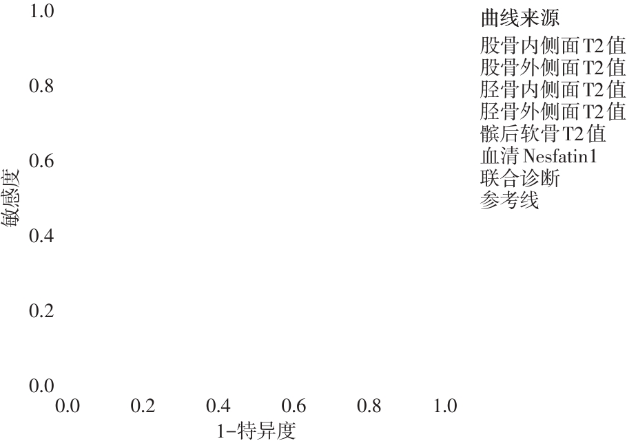
目的 探讨3.0 T磁共振(MRI)T2 mapping序列联合血清新饱食分子蛋白1(nesfatin-1)水平对老年膝关节早期骨关节炎(OA)的诊断价值。 方法 选取2023年5月至2024年5月医院收治的膝关节OA的97例老年患者(OA组)和52例同期老年体检者(对照组),根据X线结果将膝关节OA组分为早期组和非早期组,均接受3.0T MRI T2 mapping序列扫描检测膝关节软骨区域T2值,检测血清nesfatin-1水平,比较上述指标差异并采用ROC曲线分析其对老年膝关节早期OA的诊断价值。 结果 97例老年膝关节OA患者中,早期组35例,非早期组62例,OA组患者膝关节5个软骨区域的T2值及nesfatin-1血清均高于对照组( P < 0.05),早期组均低于非早期组( P < 0.05);膝关节软骨区域T2值和血清nesfatin-1水平单独诊断早期OA的AUC在0.774 ~ 0.871范围,联合诊断的AUC为0.939。 结论 3.0 T磁共振T2 mapping序列联合血清nesfatin-1水平检测对老年膝关节早期OA具有较高的诊断价值。

关键词: 膝关节早期骨关节炎, 老年, 磁共振, T2 mapping序列, 新饱食分子蛋白1

Abstract:

Objective To investigate the diagnostic potential of combining 3.0T magnetic resonance imaging (MRI) T2 mapping sequences with serum nesfatin-1 levels for detecting early osteoarthritis (OA) in elderly knees. Methods 97 elderly patients with knee osteoarthritis (OA group) and 52 elderly individuals undergoing physical examinations (control group) were recruited from a hospital between May 2023 and May 2024. Based on X-ray findings, the OA group was further divided into an early-stage subgroup and a non-early-stage subgroup. All participants underwent 3.0T MRI T2 mapping sequence scans to measure the T2 values in the knee cartilage region, and serum nesfatin-1 levels were assessed. Differences in these indicators were compared between groups. Additionally, a receiver operating characteristic (ROC) curve was constructed to evaluate the diagnostic value of these parameters for early-stage knee OA in elderly individuals. Results Among 97 elderly patients with knee OA, 35 cases were classified into the early-stage group, and 62 cases were categorized into the non-early-stage group. The T2 values in five cartilage regions of the knee and serum nesfatin-1 levels were significantly higher in the OA group compared to the control group ( P < 0.05). Moreover, these values were significantly lower in the early-stage group than in the non-early-stage group ( P < 0.05). The areas under the curve (AUC) for diagnosing early-stage OA using T2 values in knee cartilage regions and serum nesfatin-1 levels ranged from 0.774 to 0.871. Notably, the AUC for combined diagnosis reached 0.939 ( P < 0.05). Conclusion 3.0T MRI using the T2 mapping sequence in combination with serum nesfatin-1 level detection demonstrates high diagnostic value for early OA in elderly patients with knee involvement.

Key words: early knee osteoarthritis, elderly, magnetic resonance imaging, T2 mapping sequence, nesfatin-1

中图分类号: