实用医学杂志 ›› 2023, Vol. 39 ›› Issue (19): 2469-2474.doi: 10.3969/j.issn.1006-5725.2023.19.010

• 基础研究 • 上一篇    下一篇

ADAMTS-5在加速形成中性粒细胞胞外陷阱恶化颅内动脉瘤疾病进展中的作用机制

张俊,王巍,王帮奎,曾杰   

  1. 新疆生产建设兵团医院神经外科 (乌鲁木齐 830002 )
  • 收稿日期:2023-06-09 出版日期:2023-10-10 发布日期:2023-11-22
  • 基金资助:
    新疆维吾尔自治区自然科学基金资助项目(2022D01C412)

Mechanism of ADAMTS⁃5 in accelerating formation of neutrophil extracellular traps and worsening progression of intracranial aneurysm

Jun ZHANG,Wei WANG,Bangkui WANG,Jie. ZENG   

  1. Department of Neurosurgery,Xinjiang Production and Construction Corps Hospital,Urumqi 830002,China
  • Received:2023-06-09 Online:2023-10-10 Published:2023-11-22

摘要:

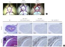
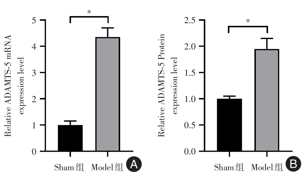
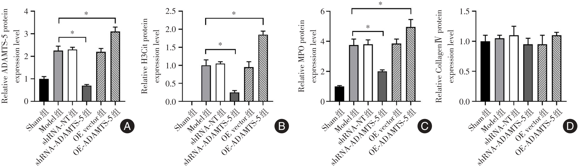
目的 探讨ADAMTS-5在加速形成中性粒细胞胞外陷阱恶化颅内动脉瘤疾病进展中的作用机制。 方法 构建颅内动脉瘤小鼠模型。采用腺病毒介导颅脑右侧脑池Willis环动脉血管壁过表达或敲低ADAMTS-5。实验分组为Sham组、Model组、shRNA-NT组、shRNA-ADAMTS-5组、OE-vector组和OE-ADAMTS-5组,每组10只小鼠。采用HE染色分析小鼠Willis环动脉瘤血管壁免疫细胞浸润情况;采用免疫组化(IHC)对中性粒细胞标志物MPO、中性粒细胞胞外陷阱(Neutrophil extracellular traps,NETs)标志物H3Cit和ADAMTS-5在Willis环动脉瘤血管壁的表达情况进行染色;采用qPCR和Western blot分析Sham组和Model组Willis环动脉瘤血管壁ADAMTS-5 mRNA和蛋白质的表达情况;采用Western blot法对6个分组Willis环动脉瘤血管壁MPO、H3Cit、Collagen Ⅳ和ADAMTS-5的表达情况进行测定。 结果 HE染色显示,在Model组小鼠颅内动脉瘤处血管壁存在大量的免疫细胞浸润。IHC-MPO染色、IHC-H3Cit染色和IHC-ADAMTS-5染色显示,在此处血管壁存在大量中性粒细胞浸润和NETs,且ADAMTS-5的染色阳性区域与H3Cit染色阳性区域存在部分重叠的现象。与Sham组相比,Model组ADAMTS-5 mRNA和蛋白质表达水平均升高(P < 0.05)。Western blot结果显示,与Model组相比,敲低ADAMTS-5/过表达ADAMTS-5降低/增加颅内动脉瘤小鼠模型Willis环动脉瘤血管壁NETs的形成(P < 0.05)。 结论 ADAMTS-5在驱动形成NETs促进颅内动脉瘤小鼠模型疾病进展中发挥重要的作用。

关键词: 颅内动脉瘤, 中性粒细胞胞外陷阱, 金属蛋白酶5

Abstract:

Objective To investigate the mechanism of ADAMTS-5 in accelerating the formation of neutrophil extracellular traps (NETs) and worsening the progression of intracranial aneurysm (IA). Methods IA mouse model was constructed. Adenovirus-mediated overexpression or knockdown ADAMTS-5 in arterial wall of the circle of Willis in the right cisterna of mice brain was conducted. Sixty mice were randomly divided into six groups of sham group, model group, shRNA-NT group, shRNA-ADAMTS-5 group, OE-vector group and OE-ADAMTS-5 group, with ten mice in each. H&E staining was used to analyze the infiltration of immune cells in arterial wall of the circle of Willis; and immunohistochemistry (IHC) to detect the expression of neutrophil markers MPO, neutrophil extracellular trap (NETs) markers H3Cit and ADAMTS-5 protein expression level in arterial wall of the circle of Willis. qPCR and western blot were used to analyze the expression of ADAMTS-5 mRNA and protein in arterial wall of the circle of Willis in sham group and model group. The expression of MPO, H3Cit, ADAMTS-5 and Collagen Ⅳ in arterial wall of the circle of Willis was detected by western blot. Results H&E staining showed that there were a large number of immune cells infiltrated in arterial wall of the circle of Willis in the model group, and IHC-MPO staining, IHC-H3Cit staining and IHC-ADAMTS-5 staining showed a large number of neutrophils infiltrated and NETs formed. ADAMTS-5 staining positive area overlapped with H3Cit staining positive area. Compared with that in the sham group, the expression of ADAMTS-5 mRNA and protein was higher in the model group (P < 0.05). Compared with the model group, knocking down/ overexpression ADAMTS-5 in arterial wall of the circle of Willis decreased/increased NETs formation (P < 0.05). Conclusion ADAMTS-5 plays an important role in driving formation of NETs and promoting disease progression of IA in mouse models.

Key words: intracranial aneurysms, neutrophil extracellular traps, ADAMTS-5

中图分类号: