实用医学杂志 ›› 2026, Vol. 42 ›› Issue (7): 1177-1182.doi: 10.3969/j.issn.1006-5725.2026.07.009

• 肿瘤诊治与预后专栏 • 上一篇    

超声造影肝脏影像报告与数据系统调整分类在≤ 5 cm肝细胞癌中的诊断价值

陈丽红(),陈惠春,冯斯奕   

  1. 福建医科大学孟超肝胆医院超声科 (福建 福州 350025 )
  • 收稿日期:2025-10-12 修回日期:2025-11-25 接受日期:2025-12-02 出版日期:2026-04-10 发布日期:2026-04-13
  • 通讯作者: 陈丽红 E-mail:992550062@qq.com
  • 基金资助:
    福建省自然科学基金项目(2023J011470);福州市科技计划项目(2022-S-034)

Application of modified contrast-enhanced ultrasound Liver Imaging Reporting and Data System in diagnosis of ≤5cm hepatocellular carcinoma

Lihong CHEN(),Huichun CHEN,Siyi FENG   

  1. Department of Ultrasound,Mengchao Hepatobiliary Hospital of Fujian Medical University,Fuzhou 350025,Fujian,China
  • Received:2025-10-12 Revised:2025-11-25 Accepted:2025-12-02 Online:2026-04-10 Published:2026-04-13
  • Contact: Lihong CHEN E-mail:992550062@qq.com

摘要:

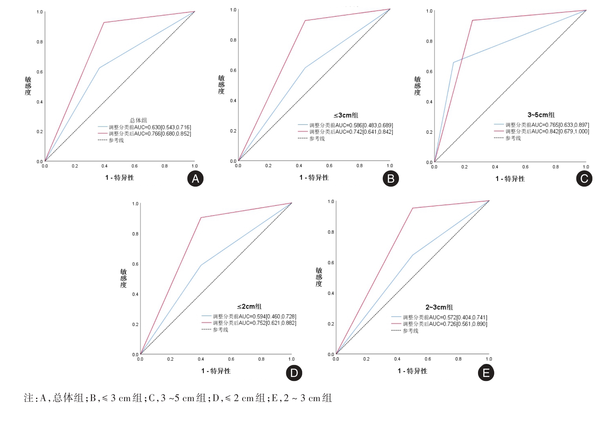
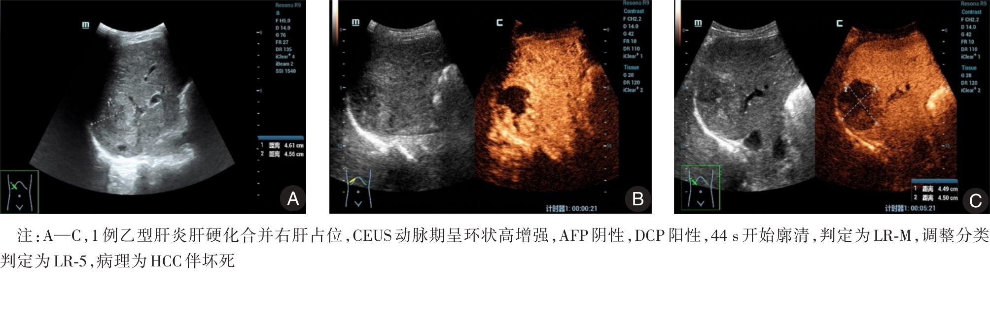
目的 探讨超声造影肝脏影像报告与数据系统(CEUS LI-RADS)调整分类在≤ 5 cm肝细胞癌(hepatocellular carcinoma, HCC)中的诊断价值。 方法 回顾性分析461个肝癌高风险的肝脏结节CEUS资料,以病理结果为金标准。先参照2017版CEUS LI-RADS对结节进行分类,再根据以下两点进行调整:(1)动脉期高增强、开始廓清时间在45 ~ 60 s内的LR-M类结节重新判定为LR-5;(2)LR-4、LR-M类结节若合并AFP/DCP阳性则重新判定为LR-5。比较调整前后LR-5的诊断效能。 结果 379个HCC中,调整后LR-M类HCC占比从32.70%降至4.70%(χ2 = 97.367, P < 0.001),在肿瘤最大径总体组、≤ 3 cm组、3 ~ 5 cm组、0 ~ 2 cm组、2 ~ 3 cm组中,调整分类均显著提高灵敏度[(92.61%、92.41%、93.33%、90.30%、95.04%) vs.(62.27%、61.24%、65.56%、58.79%、64.46%)]、准确度[(90.5%、89.52%、91.84%、87.78%、91.60%) vs. (62.38%、60.38%、67.35%、58.89%、63.36%)]、AUC值[(0.766、0.742、0.842、0.752、0.726) vs.(0.630、0.586、0.765、0.594、0.572)],阳性预测值达96%以上。 结论 CEUS LI-RADS调整分类在不降低阳性预测值的同时,显著提高了≤ 5 cm HCC诊断的灵敏度、准确度、AUC值,在≤ 2 cm病灶中同样适用,可为临床决策提供更可靠的参考依据。

关键词: 超声造影, 肝脏影像报告与数据系统, 肝细胞癌, 调整分类

Abstract:

Objective To explore the application value of the modified Contrast-Enhanced Ultrasound Liver Imaging Reporting and Data System (CEUS LI-RADS) in the diagnosis of hepatocellular carcinoma (HCC) with the diameter of ≤ 5 cm. Methods A retrospective analysis was performed on contrast-enhanced ultrasound (CEUS) data from 461 liver nodules in high-risk HCC patients, with pathological results serving as the gold standard. The nodules were classified according to the 2017 CEUS Liver Imaging Reporting and Data System (LI-RADS) and then modified based on the following two criteria: (1) nodules with arterial phase hyper-enhancement and a washout time of 45-60 s were re-classified as LR-5; (2) If LR-4 and LR-M nodules were combined with positive alpha-fetoprotein (AFP) or des-gamma-carboxy prothrombin (DCP), they were re-judged as LR-5. The diagnostic efficacy of LR-5 before and after modification was compared. Results Among 379 HCC lesions, after modification, the proportion of HCC cases classified as LR-M decreased from 32.70% to 4.70% (χ2 = 97.367, P < 0.001). In the overall group, the ≤ 3 cm group, the 3-5 cm group, the 0-2 cm group, and the 2-3 cm group of the tumor maximum diameter, the modified classification significantly improved the sensitivity [(92.61%, 92.41%, 93.33%, 90.30%, 95.04%) vs. (62.27%, 61.24%, 65.56%, 58.79%, 64.46%)], accuracy [(90.5%, 89.52%, 91.84%, 87.78%, 91.60%) vs. (62.38%, 60.38%, 67.35%, 58.89%, 63.36%)], and the area under the curve value (AUC) [(0.766, 0.742, 0.842, 0.752, 0.726) vs. (0.630, 0.586, 0.765, 0.594, 0.572)]. The positive predictive value (PPV) was more than 96%. Conclusions The modified CEUS LI-RADS classification significantly enhanced the sensitivity, accuracy, and AUC of ≤ 5 cm HCC diagnosis without compromising the PPV. The same applies to ≤ 2 cm HCC diagnosis, which would provide more reliable evidence for clinical decision-making.

Key words: contrast-enhanced ultrasound, liver imaging reporting and data system, hepatocellular carcinoma, modified classification

中图分类号: