实用医学杂志 ›› 2024, Vol. 40 ›› Issue (17): 2435-2439.doi: 10.3969/j.issn.1006-5725.2024.17.014

• 临床研究 • 上一篇    下一篇

体表监测系统在胸部肿瘤调强放疗中的应用

陈莹,陈飞,龚筱钦,黄建,杨武阳,游涛,戴春华,胡静()   

  1. 江苏大学附属医院放疗科 (江苏 镇江 212000 )
  • 收稿日期:2024-03-28 出版日期:2024-09-10 发布日期:2024-09-13
  • 通讯作者: 胡静 E-mail:hujing@ujs.edu.cn
  • 基金资助:
    江苏省卫生健康委医学科研重点项目(ZDB2020022);镇江市重点研发计划(社会发展)项目(SH2023002);宿迁市指导性科技计划项目(Z202323)

Application of surface monitoring system for thoracic tumors treated with intensity modulated radiotherapy

Ying CHEN,Fei CHEN,Xiaoqin GONG,Jian HUANG,Wuyang YANG,Tao YOU,Chunhua DAI,Jing. HU()   

  1. Department of Radiotherapy,Affiliated Hospital of Jiangsu University,Zhenjiang 212000,China
  • Received:2024-03-28 Online:2024-09-10 Published:2024-09-13
  • Contact: Jing. HU E-mail:hujing@ujs.edu.cn

摘要:

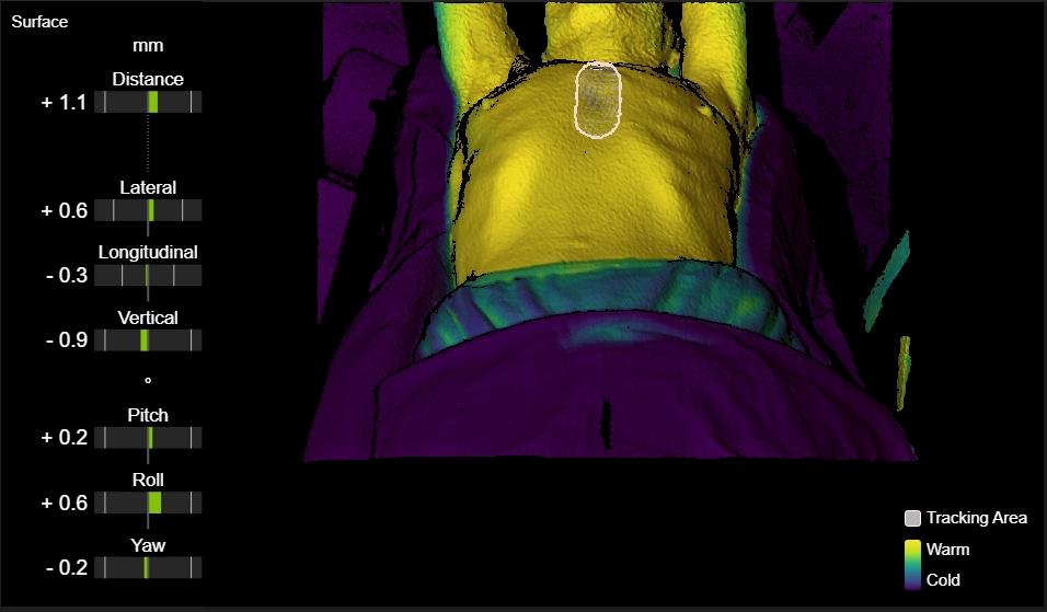
目的 探讨体表监测系统(ExacTracDynamic, ETD)辅助摆位和体表监测功能在胸部肿瘤患者调强放疗中应用的可行性。 方法 选取进行放疗的胸部肿瘤患者,入组患者交替行常规十字标记线摆位(对照组)和体表监测系统辅助摆位(观察组)。治疗前均用ETD的X线影像行摆位校准,在胸骨上勾画感兴趣区并实时体表监测患者治疗中的位置变化,治疗结束后再行X线影像验证。记录左右(X)、头脚(Y)、腹背(Z)、俯仰(Pitch)、横滚(Roll)、旋转(Yaw)方向上的数据并比较分析。 结果 入组60 例患者,对照组放疗754次,观察组718次。观察组在X和Z方向的摆位误差小于对照组(P < 0.05);观察组在X ≤ 0.50 cm、Y ≤ 0.50 cm、Z ≤ 0.50 cm和Roll ≤ 1.00°方向的摆位误差次数均大于对照组(P < 0.05);体表监测和治疗后的位置偏差在Y和Z方向上存在差异性(P < 0.05),但均值在亚毫米级。 结论 体表监测系统辅助摆位可提高胸部肿瘤患者放疗摆位的精度,尤其在X和Z方向。感兴趣区设置在胸骨上时,体表监测能较好地反映患者靶区内部位置的变化。

关键词: 体表监测系统, 胸部肿瘤, 调强放疗, 摆位误差

Abstract:

Objective To assess the feasibility of utilizing the ExacTracDynamic surface monitoring system (ETD) for setup and body surface monitoring in patients with thoracic tumors undergoing intensity?modulated radiotherapy (IMRT). Methods Patients receiving IMRT for thoracic tumors were included in this study. The enrolled patients were alternatively assigned to either conventional cross curve positioning (control group) or surface monitoring system?assisted positioning (experimental group). ETD X?ray images were utilized for calibration purposes prior to radiotherapy, enabling the determination of setup errors. A region of interest (ROI) was delineated on the body surface above the sternum, and real?time body surface monitoring was performed based on this ROI during radiotherapy. Post?radiotherapy X?ray images were obtained to verify patient position. Data regarding left?right (X), head?foot (Y), abdomen?back (Z), pitch, roll, and yaw directions were recorded and analyzed. Results A total of 60 patients were enrolled, with 754 fractions of radiotherapy in the control group and 718 fractions in the experimental group. The setup errors in the X and Z directions were significantly smaller in the experimental group compared to the control group (P < 0.05). Moreover, there was a significant reduction in the number of setup errors ≤ 0.50 cm for X, Y, and Z directions, as well as ≤ 1.00° for Roll angle in the experimental group compared to the control group (P < 0.05). Additionally, differences were observed between surface monitoring and X?ray image verification regarding position deviation along Y and Z directions (P < 0.05), although these deviations remained within submillimeter levels on average. Conclusion Surface monitoring system?assisted positioning can enhance radiotherapy setup accuracy among thoracic tumor patients, particularly along X and Z directions. Furthermore, when setting ROI above sternum on body surface area, surface monitoring provides better reflection of target area's position deviation.

Key words: body surface monitoring system, thoracic tumors, IMRT, setup errors

中图分类号: