

The Journal of Practical Medicine ›› 2025, Vol. 41 ›› Issue (11): 1645-1654.doi: 10.3969/j.issn.1006-5725.2025.11.006
• Basic Research • Previous Articles
Li CUI1,Feiyan MA1,2,3( )
)
Received:2025-03-15
Online:2025-06-10
Published:2025-06-19
Contact:
Feiyan MA
E-mail:f4ma@hebmu.edu.cn
CLC Number:
Li CUI,Feiyan MA. Mechanism of p300 mediated PPAR⁃γ affecting diabetes retinopathy through lipid peroxidation[J]. The Journal of Practical Medicine, 2025, 41(11): 1645-1654.
Tab.1
Primer information for each gene"
| 基因 | 引物序列 | 扩增产物长度/bp |
|---|---|---|
| p300 | 上游:5′-TCCTATGGGAGAACGGCAGA-3′ 下游:5′-TCCTTTGTCCCCTGAGCTTG-3′ | 676 |
| PPAR-γ | 上游:5′-GCTCAGGGGACAAAGGAAGC-3′ 下游:5′-TCGCTCTCTCGTGGCTAGTA-3′ | 814 |
| PKCβ | 上游:5′-GAAGGCTATAGTCACCTCGGG-3′ 下游:5′-ATGGTAATAATGCGGCCGGT-3′ | 143 |
| P66Shc | 上游:5′-AGGGGACAAAGGAAGCGCA-3′ 下游:5′-GGAACCAGACGCTACGATCA-3′ | 511 |
| β-actin | 上游:5′-GGCTTGCCACTCCCAAAGTA-3′ 下游:5′-TCTGCGCTTCCTTTGTCCCC-3′ | 302 |
Tab.2
Comparison of the degree of damage to the blood retinal barrier among different groups of rats"
| 组别 | n | Evans blue浓度/(ng/mg) | 白蛋白渗透/(μL血浆*g视网膜湿重wt-1×hr-1) |
|---|---|---|---|
| 正常组 | 5 | 10.15 ± 2.07 | 6.58 ± 1.03 |
| 模型组 | 5 | 25.57 ± 3.59? | 14.06 ± 2.51? |
| p300上调组 | 5 | 18.10 ± 2.35?&# | 10.20 ± 1.89?&# |
| p300下调组 | 5 | 30.68 ± 4.32?&# | 19.33 ± 3.01?&# |
| PPAR-γ上调组 | 5 | 18.21 ± 2.37?&# | 10.22 ± 1.91?&# |
| PPAR-γ下调组 | 5 | 30.71 ± 4.50?&# | 19.40 ± 3.18?&# |
| 空载对照组 | 5 | 25.60 ± 3.66? | 14.20 ± 2.47? |
| F值 | 145.836 | 158.967 | |
| P值 | < 0.001 | < 0.001 |
Tab.3
Comparison of lipid indicators among different groups of rats (x ± s)/(mmol/L)"
| 组别 | n | TG | TC | LDL-C | HDL-C |
|---|---|---|---|---|---|
| 正常组 | 10 | 1.15 ± 0.21 | 1.09 ± 0.20 | 0.20 ± 0.04 | 1.98 ± 0.31 |
| 模型组 | 10 | 1.80 ± 0.30? | 1.78 ± 0.29? | 0.63 ± 0.10? | 1.35 ± 0.27? |
| p300上调组 | 10 | 1.40 ± 0.24?&# | 1.36 ± 0.23?&# | 0.41 ± 0.06?&# | 1.75 ± 0.30?&# |
| p300下调组 | 10 | 2.05 ± 0.32?&# | 2.08 ± 0.33?&# | 0.89 ± 0.12?&# | 1.11 ± 0.22?&# |
| PPAR-γ上调组 | 10 | 1.41 ± 0.22?&# | 1.35 ± 0.25?&# | 0.43 ± 0.07?&# | 1.76 ± 0.28?&# |
| PPAR-γ下调组 | 10 | 2.07 ± 0.31?&# | 2.10 ± 0.34?&# | 0.90 ± 0.11?&# | 1.13 ± 0.24?&# |
| 空载对照组 | 10 | 1.79 ± 0.28? | 1.80 ± 0.31? | 0.62 ± 0.09? | 1.37 ± 0.29? |
| F值 | 112.315 | 108.743 | 129.546 | 138.735 | |
| P值 | < 0.001 | < 0.001 | < 0.001 | < 0.001 |
Tab.4
Comparison of peroxidation reaction indicators among different groups of rats"
| 组别 | n | SOD活性/(U/mL) | MDA含量/(μmol/L) | ROS水平/(ng/mL) |
|---|---|---|---|---|
| 正常组 | 10 | 151.36 ± 12.58 | 1.52 ± 0.33 | 6.15 ± 1.12 |
| 模型组 | 10 | 112.73 ± 11.55? | 3.26 ± 0.61? | 13.43 ± 2.41? |
| p300上调组 | 10 | 130.07 ± 13.54?&# | 2.57 ± 0.43?&# | 10.14 ± 2.02?&# |
| p300下调组 | 10 | 90.75 ± 10.13?&# | 4.35 ± 0.74?&# | 16.25 ± 2.73?&# |
| PPAR-γ上调组 | 10 | 131.26 ± 13.89?&# | 2.59 ± 0.45?&# | 10.07 ± 2.05?&# |
| PPAR-γ下调组 | 10 | 91.20 ± 10.22?&# | 4.38 ± 0.76?&# | 16.31 ± 2.70?&# |
| 空载对照组 | 10 | 111.86 ± 11.32? | 3.29 ± 0.64? | 13.28 ± 2.27? |
| F值 | 169.706 | 154.689 | 141.655 | |
| P值 | < 0.001 | < 0.001 | < 0.001 |
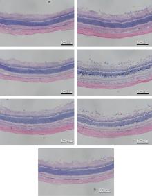
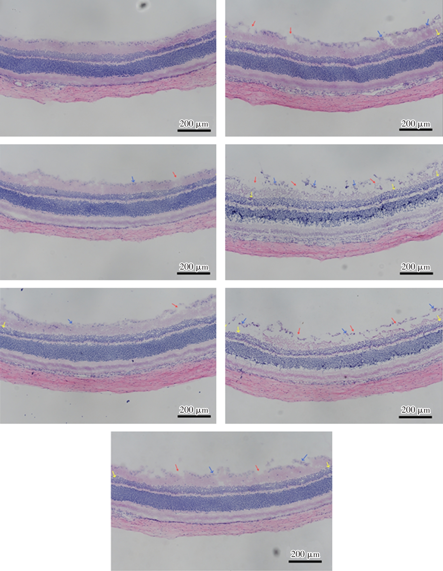
Fig.1
Pathological changes in retinal tissue of rats in each group (HE staining, × 100)"
Tab. 5
Comparison of p300, PPAR - γ, PKC β, P66Shc mRNA expressions in retinal tissues of rats in each group"
| 组别 | n | p300 | PPAR-γ | PKCβ | P66Shc |
|---|---|---|---|---|---|
| 正常组 | 5 | 1.00 | 1.00 | 1.00 | 1.00 |
| 模型组 | 5 | 0.71 ± 0.12? | 0.65 ± 0.10? | 0.54 ± 0.09? | 0.48 ± 0.07? |
| p300上调组 | 5 | 0.95 ± 0.14?&# | 0.83 ± 0.12?&# | 0.80 ± 0.11?&# | 0.85 ± 0.12?&# |
| p300下调组 | 5 | 0.40 ± 0.06?&# | 0.35 ± 0.06?&# | 0.27 ± 0.04?&# | 0.25 ± 0.05?&# |
| PPAR-γ上调组 | 5 | 0.71 ± 0.10?# | 0.85 ± 0.13?&# | 0.81 ± 0.12?&# | 0.84 ± 0.11?&# |
| PPAR-γ下调组 | 5 | 0.72 ± 0.11?# | 0.34 ± 0.05?&# | 0.28 ± 0.05?&# | 0.26 ± 0.05?&# |
| 空载对照组 | 5 | 0.70 ± 0.11? | 0.67 ± 0.12? | 0.55 ± 0.08? | 0.46 ± 0.08? |
| F值 | 196.321 | 201.457 | 178.310 | 186.293 | |
| P值 | < 0.001 | < 0.001 | < 0.001 | < 0.001 |
Tab. 6
Comparison of p300, PPAR-γ, PKCβ, P66Shc protein expressions and P66Shc phosphorylation levels in retinal tissues of rats in each group"
| 组别 | n | p300 | PPAR-γ | PKCβ | P66Shc | P66Shc磷酸化水平 |
|---|---|---|---|---|---|---|
| 正常组 | 5 | 1.78 ± 0.33 | 1.86 ± 0.28 | 1.92 ± 0.29 | 1.95 ± 0.34 | 1.85 ± 0.21 |
| 模型组 | 5 | 0.45 ± 0.11? | 0.72 ± 0.13? | 0.81 ± 0.12? | 0.88 ± 0.14? | 0.43 ± 0.05* |
| p300上调组 | 5 | 1.12 ± 0.24?&# | 1.28 ± 0.22?&# | 1.45 ± 0.27?&# | 1.53 ± 0.22?&# | 0.76 ± 0.13?&# |
| p300下调组 | 5 | 0.18 ± 0.05?&# | 0.47 ± 0.06?&# | 0.60 ± 0.10?&# | 0.61 ± 0.12?&# | 0.15 ± 0.03?&# |
| PPAR-γ上调组 | 5 | 0.45 ± 0.11?# | 1.30 ± 0.24?&# | 1.43 ± 0.29?&# | 1.51 ± 0.21?&# | 0.78 ± 0.15?&# |
| PPAR-γ下调组 | 5 | 0.43 ± 0.10?# | 0.46 ± 0.07?&# | 0.61 ± 0.12?&# | 0.59 ± 0.11?&# | 0.15 ± 0.04?&# |
| 空载对照组 | 5 | 0.42 ± 0.12? | 0.70 ± 0.15? | 0.84 ± 0.15? | 0.90 ± 0.16? | 0.22 ± 0.06? |
| F值 | 222.454 | 218.736 | 220.915 | 239.753 | 241.078 | |
| P值 | < 0.001 | < 0.001 | < 0.001 | < 0.001 | < 0.001 |
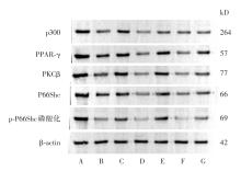
Fig.2
Detection of p300, PPAR-γ, PKC β, P66Shc proteins and P66Shc phosphorylation levels in each group(Western blot method)"

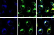
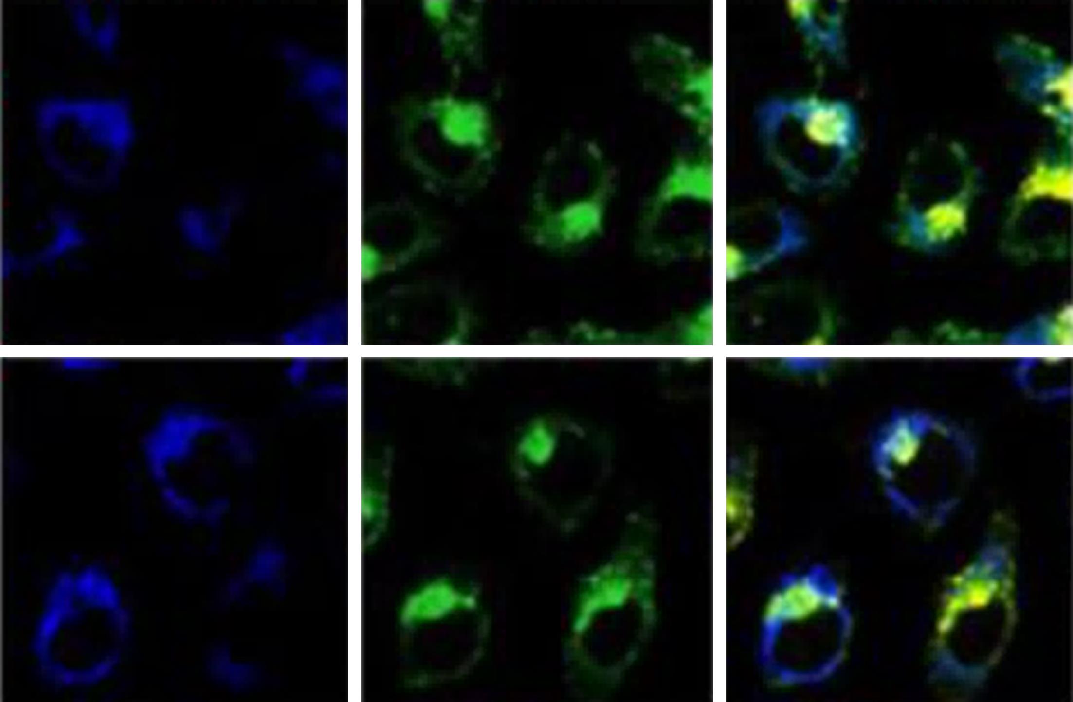
Fig.3
Validation of p300 targeting PPAR-γ and co localization of cells"
| 1 |
FUNG T H, PATEL B, WILMOT E G, et al. Diabetic retinopathy for the non-ophthalmologist[J]. Clin Med (Lond), 2022, 22(2):112-116. doi:10.7861/clinmed.2021-0792
doi: 10.7861/clinmed.2021-0792 |
| 2 |
LI S, GUO B, JIANG Y, et al. Long-term Exposure to Ambient PM2.5 and Its Components Associated With Diabetes: Evidence From a Large Population-Based Cohort From China[J]. Diabetes Care, 2023, 46(1):111-119. doi:10.2337/dc22-1585
doi: 10.2337/dc22-1585 |
| 3 |
HOU X, WANG L, ZHU D, et al. Prevalence of diabetic retinopathy and vision-threatening diabetic retinopathy in adults with diabetes in China[J]. Nat Commun, 2023, 14(1):4296. doi:10.1038/s41467-023-39864-w
doi: 10.1038/s41467-023-39864-w |
| 4 |
SHABALALA S C, JOHNSON R, BASSON A K, et al. Detrimental Effects of Lipid Peroxidation in Type 2 Diabetes: Exploring the Neutralizing Influence of Antioxidants[J]. Antioxidants (Basel), 2022, 11(10):2071. doi:10.3390/antiox11102071
doi: 10.3390/antiox11102071 |
| 5 | 张薇, 陈聪. p300介导组蛋白乙酰化修饰下调PPAR-γ致妊娠期糖尿病小鼠子代心肌细胞糖脂代谢紊乱[J]. 中国病理生理杂志, 2017, 33(12):2222-2226. |
| 6 | 安娜, 郑卫东, 杨丽君. PPAR-γ防治糖尿病视网膜病变的作用机制[J]. 海峡科学, 2016, 32(4):6-8, 30. |
| 7 |
KU Y H, CHO B J, KIM M J, et al. Rosiglitazone increases endothelial cell migration and vascular permeability through Akt phosphorylation[J]. BMC Pharmacol Toxicol, 2017, 18(1):62. doi:10.1186/s40360-017-0169-y
doi: 10.1186/s40360-017-0169-y |
| 8 |
HUANG T S, WU T, WU Y D, et al. Long-term statins administration exacerbates diabetic nephropathy via ectopic fat deposition in diabetic mice[J]. Nat Commun, 2023, 14(1):390. doi:10.1038/s41467-023-35944-z
doi: 10.1038/s41467-023-35944-z |
| 9 | 戴旭锋, 刘晓玲. 大鼠玻璃体内注射二甲基亚砜对视网膜电图的影响[J]. 中华眼视光学与视觉科学杂志, 2007, 9(1):51-53. |
| 10 | 张锐, 周颖, 倪文吉, 等. 人工智能视网膜微血管分析在糖尿病并发症中的应用价值[J]. 实用医学杂志, 2024, 40(8):1142-1147. |
| 11 |
FERNANDES SILVA L, HOKKANEN J, VANGIPURAPU J, et al. Metabolites as Risk Factors for Diabetic Retinopathy in Patients With Type 2 Diabetes: A 12-Year Follow-up Study[J]. J Clin Endocrinol Metab, 2023, 109(1):100-106. doi:10.1210/clinem/dgad452
doi: 10.1210/clinem/dgad452 |
| 12 |
CHEN S N, CHEN S J, WU T T, et al. Refining vitrectomy for proliferative diabetic retinopathy[J]. Graefes Arch Clin Exp Ophthalmol, 2023, 261(12):3659-3670. doi:10.1007/s00417-023-06134-w
doi: 10.1007/s00417-023-06134-w |
| 13 |
STUDNIČKA J, NĚMČANSKÝ J, VYSLOUŽILOVÁ D, et al. Diabetic Retinopathy-Diagnostics and Treatment Guidelines[J]. Cesk Slov Oftalmol, 2023, 79(5):238-247. doi:10.31348/2023/28
doi: 10.31348/2023/28 |
| 14 |
SOLDO A M, SOLDO I, KARAČIĆ A, et al. Lipid Peroxidation in Obesity: Can Bariatric Surgery Help?[J]. Antioxidants (Basel), 2022, 11(8):1537. doi:10.3390/antiox11081537
doi: 10.3390/antiox11081537 |
| 15 |
GAO J, TAO L, JIANG Z. Alleviate oxidative stress in diabetic retinopathy: Antioxidant therapeutic strategies[J]. Redox Rep, 2023, 28(1):2272386. doi:10.1080/13510002.2023.2272386
doi: 10.1080/13510002.2023.2272386 |
| 16 |
PERDICES L, FUENTES-BROTO L, SEGURA F, et al. Systemic epigallocatechin gallate protects against retinal degeneration and hepatic oxidative stress in the P23H-1 rat[J]. Neural Regen Res, 2022, 17(3):625-631. doi:10.4103/1673-5374.320990
doi: 10.4103/1673-5374.320990 |
| 17 |
FAN X, XU M, REN Q, et al. Downregulation of fatty acid binding protein 4 alleviates lipid peroxidation and oxidative stress in diabetic retinopathy by regulating peroxisome proliferator-activated receptor γ-mediated ferroptosis[J]. Bioengineered, 2022, 13(4):10540-10551. doi:10.1080/21655979.2022.2062533
doi: 10.1080/21655979.2022.2062533 |
| 18 |
LIU J, ZHANG Y, SHI D, et al. Vitamin D Alleviates Type 2 Diabetes Mellitus by Mitigating Oxidative Stress-Induced Pancreatic β-Cell Impairment[J]. Exp Clin Endocrinol Diabetes, 2023, 131(12):656-666. doi:10.1055/a-2191-9969
doi: 10.1055/a-2191-9969 |
| 19 |
STOCKWELL B R. Ferroptosis turns 10: Emerging mechanisms, physiological functions, and therapeutic applications[J]. Cell, 2022, 185(14):2401-2421. doi:10.1016/j.cell.2022.06.003
doi: 10.1016/j.cell.2022.06.003 |
| 20 |
HSU C G, CHÁVEZ C L, ZHANG C, et al. The lipid peroxidation product 4-hydroxynonenal inhibits NLRP3 inflammasome activation and macrophage pyroptosis[J]. Cell Death Differ, 2022, 29(9):1790-1803. doi:10.1038/s41418-022-00966-5
doi: 10.1038/s41418-022-00966-5 |
| 21 |
YANG H C, DELEUZE S, ZUO Y, et al. The PPARgamma agonist pioglitazone ameliorates aging-related progressive renal injury[J]. J Am Soc Nephrol, 2009, 20(11):2380-8. doi:10.1681/asn.2008111138
doi: 10.1681/asn.2008111138 |
| 22 |
MATTOS R T, BOSCO A A, NOGUEIRA-MACHADO J A. Rosiglitazone, a PPAR-γ agonist, inhibits VEGF secretion by peripheral blood mononuclear cells and ROS production by human leukocytes[J]. Inflamm Res, 2012, 61(1):37-41. doi:10.1007/s00011-011-0386-6
doi: 10.1007/s00011-011-0386-6 |
| 23 | 蒋玲, 廖洪霞, 吴燕, 等. 过氧化物酶增生体激活受体-γ激动剂对糖尿病大鼠血-视网膜屏障通透性的影响[J]. 眼科研究, 2010, 28(11):1054-1058. |
| 24 |
NIU T, SHI X, LIU X, et al. Porous Se@SiO2 nanospheres alleviate diabetic retinopathy by inhibiting excess lipid peroxidation and inflammation[J]. Mol Med, 2024, 30(1):24. doi:10.1186/s10020-024-00785-z
doi: 10.1186/s10020-024-00785-z |
| 25 |
LIU Q, LIU C Q, YI W Z, et al. Ferroptosis Contributes to Microvascular Dysfunction in Diabetic Retinopathy[J]. Am J Pathol, 2024, 194(6):1078-1089. doi:10.1016/j.ajpath.2024.01.019
doi: 10.1016/j.ajpath.2024.01.019 |
| 26 |
DONG M, ZHANG Y, CHEN M, et al. ASF1A-dependent P300-mediated histone H3 lysine 18 lactylation promotes atherosclerosis by regulating EndMT[J]. Acta Pharm Sin B, 2024, 14(7):3027-3048. doi:10.1016/j.apsb.2024.03.008
doi: 10.1016/j.apsb.2024.03.008 |
| 27 |
RIEMANN A, RAUSCHNER M, REIME S, et al. The Role of microRNAs in gene expression and signaling response of tumor cells to an acidic environment[J]. Int J Mol Sci, 2023, 24(23):16919. doi:10.3390/ijms242316919
doi: 10.3390/ijms242316919 |
| [1] | Rui ZHANG,Ying ZHOU,Wenji NI,Ya HUANG,Dandan LI,Tao JIN,Yong. ZHONG. Application value of artificial intelligence⁃basedretinal microvascular analysis in diagnosis of diabetes complications [J]. The Journal of Practical Medicine, 2024, 40(8): 1142-1147. |
| [2] | Xiaoyan WANG,Xiaoyi ZOU,Xiang ZHU,Ting WANG,Yetao QIANG,Siyuan ZHOU,Peng ZHANG,Ping ZHANG. Iron overload regulates atherosclerotic activity of foam cells induced by oxLDL [J]. The Journal of Practical Medicine, 2024, 40(3): 295-301. |
| [3] |
LIU Yang, SUN Yue, YANG Anning, LIU Zige, LIU Taiyang, HAO Wei, LIU Yaoyang, WANG Qiushi, LIU Zhihong.
Involvement of ferroptosis in atherosclerosis induced by high⁃fat diet in ApoE-/- mouse and formation of ox ⁃LDL ⁃induced foam cell [J]. The Journal of Practical Medicine, 2021, 37(5): 585-590. |
| Viewed | ||||||
|
Full text |
|
|||||
|
Abstract |
|
|||||