实用医学杂志 ›› 2025, Vol. 41 ›› Issue (5): 751-755.doi: 10.3969/j.issn.1006-5725.2025.05.020

• 医学检查与临床诊断 • 上一篇    

超声剪切波频散成像指导肝细胞肝癌患者术前肝纤维化程度评估的价值

吕悦,孟彦娜,李盼盼,陈英红()   

  1. 河南大学淮河医院超声医学科 (河南 开封 475000 )
  • 收稿日期:2024-07-11 出版日期:2025-03-10 发布日期:2025-03-20
  • 通讯作者: 陈英红 E-mail:mu29898@163.com
  • 基金资助:
    河南省科技攻关计划项目(182102311206)

Value of SWD in preoperative assessment of liver fibrosis in patients with hepatocellular carcinoma

Yue LV,Yanna MENG,Panpan LI,Yinghong. CHEN()   

  1. Department of Ultrasound Medicine,Henan University Huaihe Hospital,Kaifeng 475000,Henan,China
  • Received:2024-07-11 Online:2025-03-10 Published:2025-03-20
  • Contact: Yinghong. CHEN E-mail:mu29898@163.com

摘要:

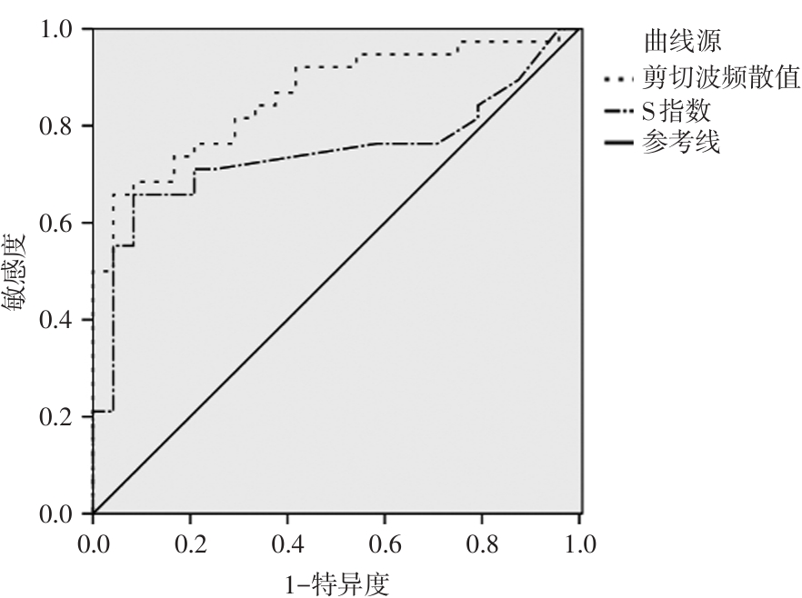
目的 观察超声剪切波频散成像(SWD)指导肝细胞肝癌(HCC)患者术前肝纤维化程度评估的价值。 方法 选取2022年1月至2024年1月我院收治的62例HCC患者,所有患者均于肝切除术前接受SWD检查,根据手术病理结果评估患者肝纤维化程度,并将患者分为低度纤维化组(S0~S2级)与高度纤维化组(S3 ~ S4级),比较两组患者基线资料与肝实质剪切波频散值,采用多因素logistic回归分析HCC患者肝纤维化程度的影响因素,并采用受试者工作特征(ROC)曲线分析SWD对HCC患者术前肝纤维化程度的评估价值。 结果 本研究纳入62例HCC患者,经病理学检查评估肝纤维化程度为S1、S2、S3、S4的患者分别为11、13、20及18例。不同肝纤维化程度患者肝实质剪切波频散值比较差异有统计学意义,且随着患者肝纤维化程度增加,肝实质剪切波频散随之增加(P < 0.05)。Spesrman相关性分析显示,肝实质剪切波频散值与HCC患者术前肝纤维化程度呈正相关(r = 0.608,P < 0.05)。高度纤维化组患者PLT水平低于低度纤维化组(P < 0.05),且高度纤维化组患者S指数与肝实质剪切波频散值高于低度纤维化组(P < 0.05)。多因素logistic回归显示,S指数与剪切波频散值是HCC患者术前肝纤维化程度的独立影响因素(P < 0.05)。ROC曲线显示,剪切波频散值评估HCC患者术前肝纤维化程度的AUC高于S指数(P < 0.05),剪切波频散值评估肝纤维化程度的截断值为16.25 m/(s·kHz),敏感度与特异度分别为65.79%、95.83%。 结论 肝实质剪切波频散值与HCC患者肝纤维化程度密切相关,且肝实质剪切波频散值对肝纤维化程度具有较高的评估价值。

关键词: 超声剪切波频散成像, 肝细胞肝癌, 肝纤维化, 评估价值

Abstract:

Objective To evaluate the diagnostic value of shear wave dispersion (SWD) in preoperative assessment of liver fibrosis in patients with hepatocellular carcinoma (HCC). Methods A total of 62 patients with HCC who were admitted to the hospital between January 2022 and January 2024 were included in this study. All patients underwent shear wave elastography (SWE) examination prior to hepatectomy. The extent of liver fibrosis was assessed based on surgical and pathological findings. Patients were categorized into two groups: a low?grade fibrosis group (stages S0?S2) and a high?grade fibrosis group (stages S3?S4). Baseline characteristics and liver parenchymal SWE values were compared between the two groups. Multivariate logistic regression analysis was conducted to identify factors influencing the degree of liver fibrosis in HCC patients. The diagnostic performance of SWE in preoperative evaluation of liver fibrosis was assessed using receiver operating characteristic (ROC) curve analysis. Results Among the 62 patients with HCC, pathological results indicated that there were 11, 13, 20, and 18 cases in fibrosis stages S1, S2, S3, and S4, respectively. The SWD values of liver parenchyma significantly differed among patients with varying degrees of liver fibrosis. As the severity of liver fibrosis increased, the SWD of liver parenchyma also increased (P < 0.05). Spearman correlation analysis revealed a positive correlation between the SWD value of liver parenchyma and the degree of liver fibrosis in preoperative HCC patients (r = 0.608, P < 0.05). Platelet count (PLT) was lower in the high?grade fibrosis group compared to the low?grade fibrosis group (P < 0.05). Additionally, both the S index and SWD value of liver parenchyma were higher in the high?grade fibrosis group than in the low?grade fibrosis group (P < 0.05). Multivariate logistic regression analysis demonstrated that the S index and SWD value were independent influencing factors for the degree of liver fibrosis in preoperative HCC patients (P < 0.05). ROC curves showed that the area under the curve (AUC) of the SWD value was greater than that of the S index for assessing the degree of liver fibrosis in preoperative HCC patients (P < 0.05). The cutoff value, sensitivity, and specificity of the SWD value for assessing the degree of liver fibrosis were 16.25 m/s·kHz?1, 65.79%, and 95.83%, respectively. Conclusion The SWD value of liver parenchyma is closely associated with the extent of liver fibrosis in patients with HCC and provides a highly valuable assessment of the degree of liver fibrosis.

Key words: shear wave dispersion, hepatocellular carcinoma, liver fibrosis, evaluate value

中图分类号: