实用医学杂志 ›› 2024, Vol. 40 ›› Issue (7): 941-947.doi: 10.3969/j.issn.1006-5725.2024.07.011

• 基础研究 • 上一篇    下一篇

培养时间对小鼠树突状细胞及其外泌体免疫相关膜蛋白的影响

肖俐1,罗淑敏2,徐芳2,路鹏鹏2,邢恩鸿1(),李伟华2()   

  1. 1.承德医学院附属医院,河北省泛血管疾病重点实验室 (河北 承德 067000 )
    2.首都医科大学附属北京佑安医院肝病研究所 (北京 100069 )
  • 收稿日期:2023-10-19 出版日期:2024-04-10 发布日期:2024-04-08
  • 通讯作者: 邢恩鸿,李伟华 E-mail:xeh001@sina.com;liweihua@ccmu.edu.cn
  • 基金资助:
    国家自然科学基金资助项目(82274447);北京市自然科学基金资助项目(7212172);北京市高层次公共卫生技术人才建设资助项目(2022-2-024);首都医科大学培育基金(PYZ21128);北京市属医学科研院所公益发展改革试点项目(京医研2019-6)

Effect of culture time on immune⁃related membrane proteins of mouse dendritic cells and their exosomes

Li XIAO1,Shumin LUO2,Fang XU2,Pengpeng LU2,Enhong XING1(),Weihua. LI2()   

  1. The Affiliated Hospital of Chengde Medical College,Hebei Key Laboratory of Panvascular Disease,Chengde 067000,China
  • Received:2023-10-19 Online:2024-04-10 Published:2024-04-08
  • Contact: Enhong XING,Weihua. LI E-mail:xeh001@sina.com;liweihua@ccmu.edu.cn

摘要:

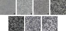
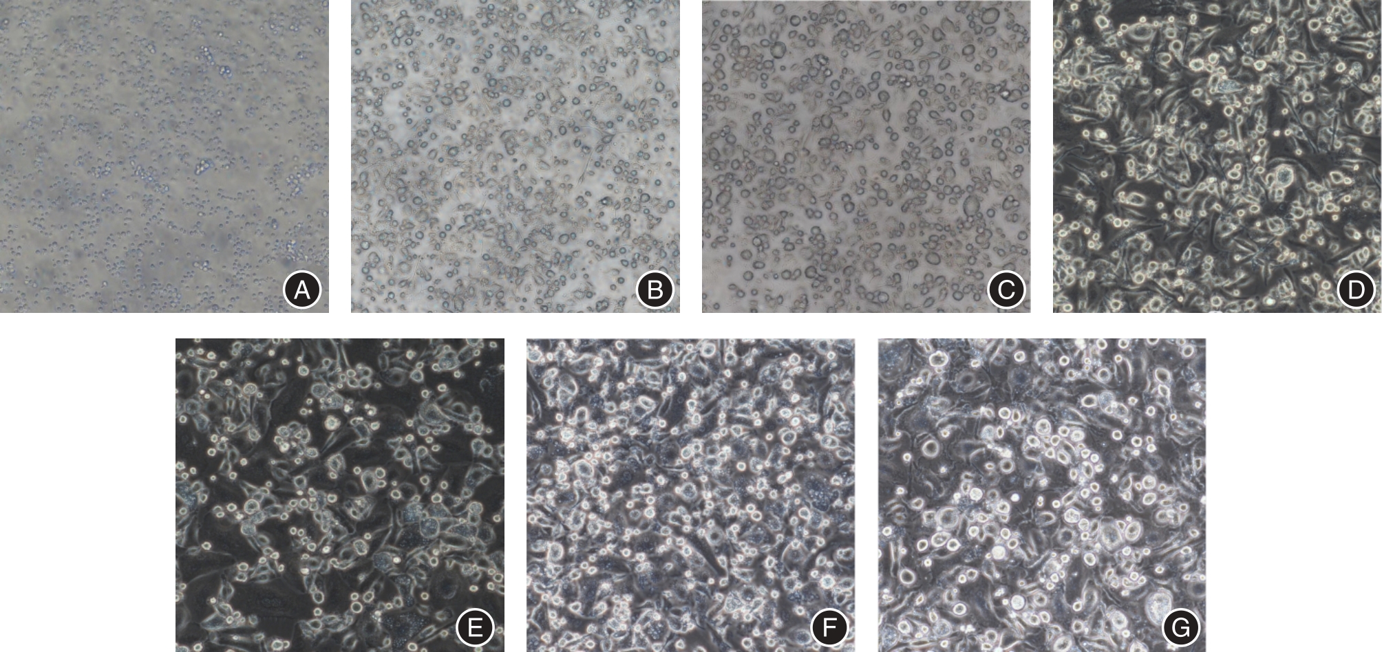
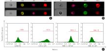
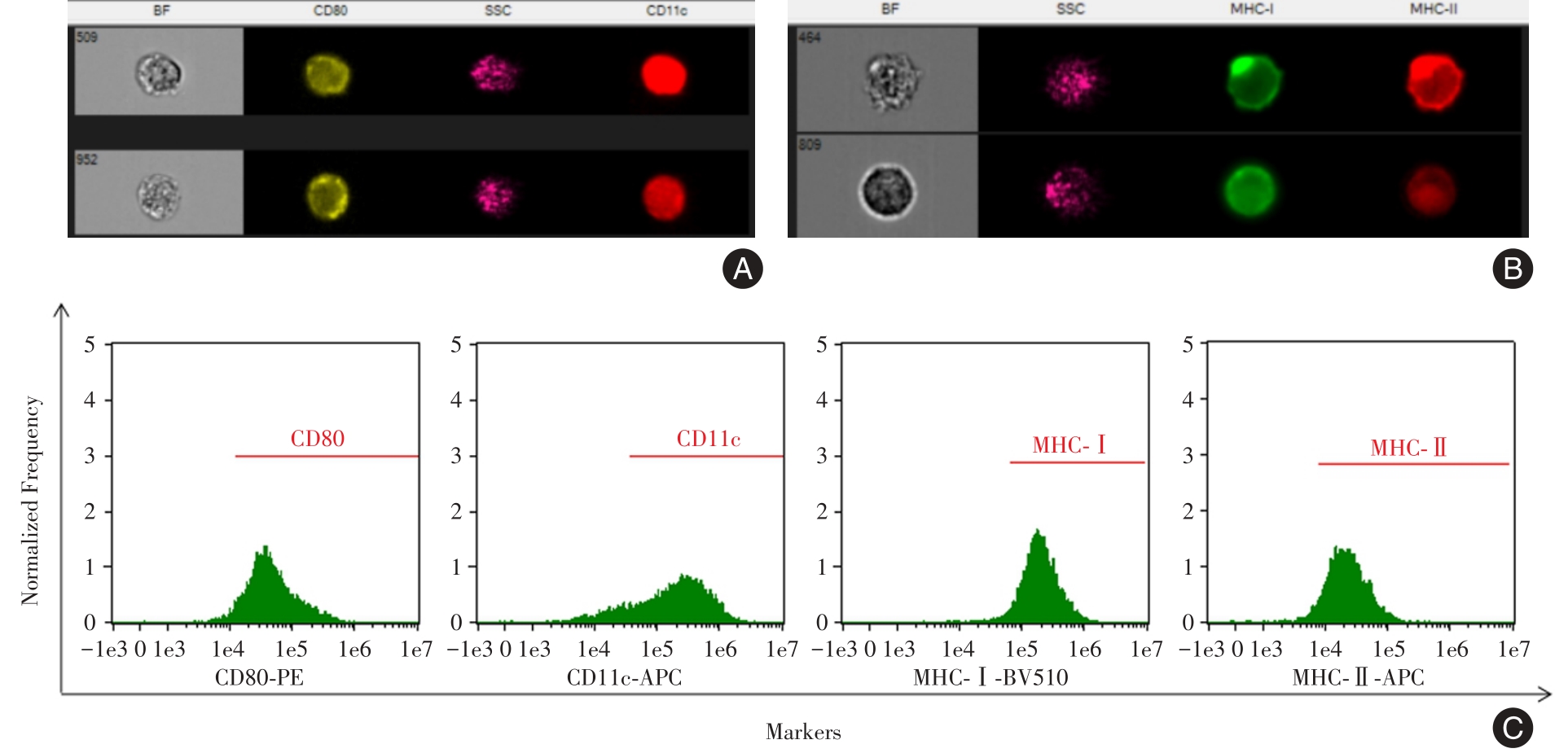
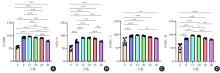
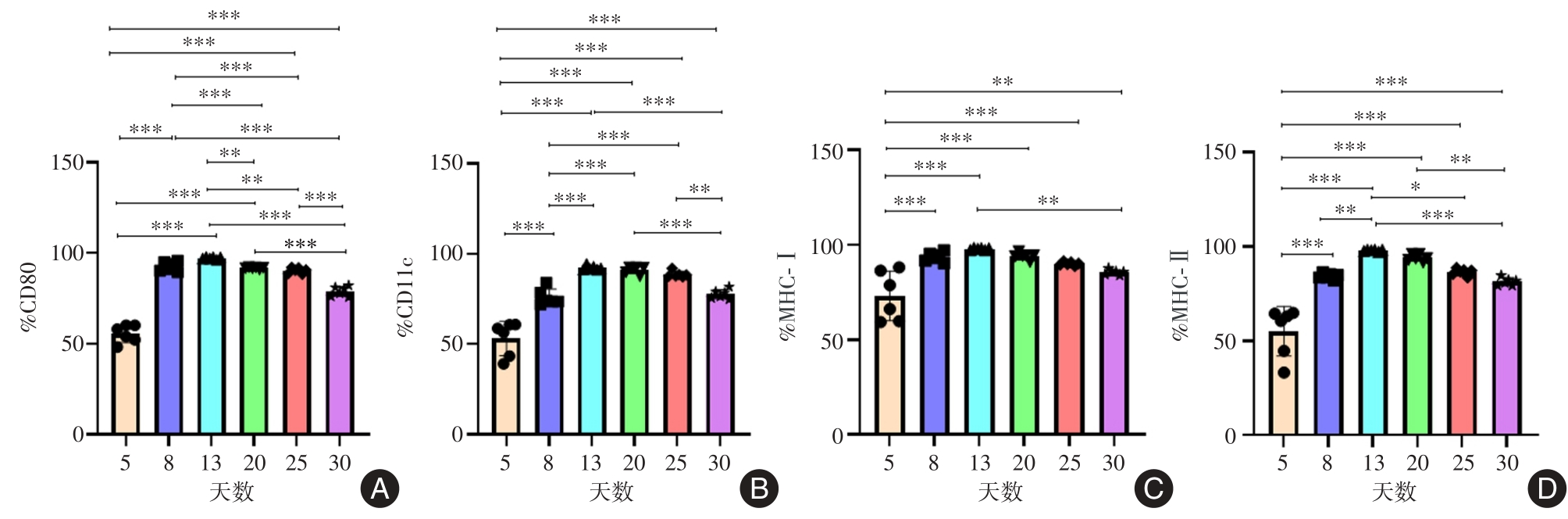
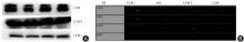
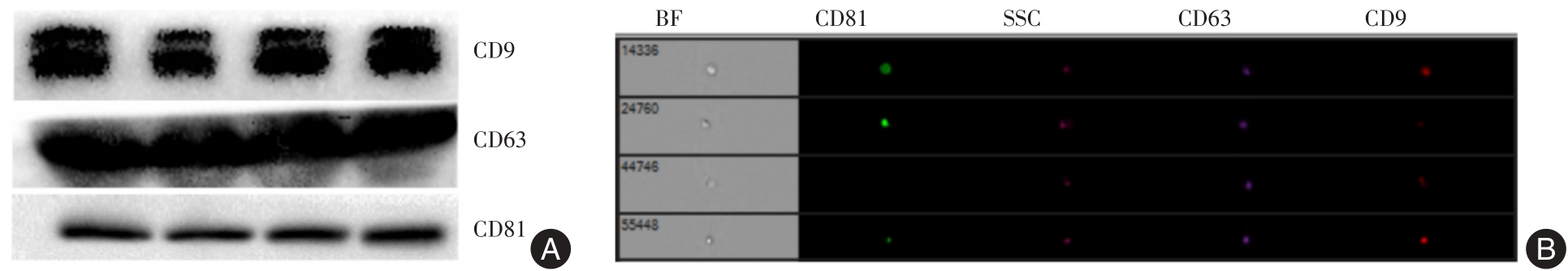
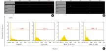
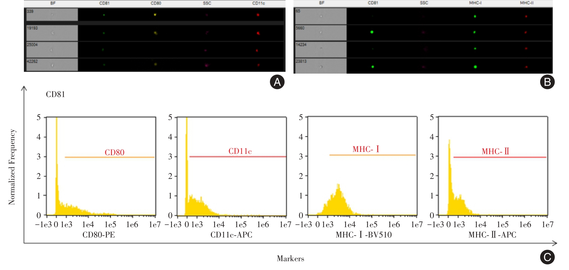
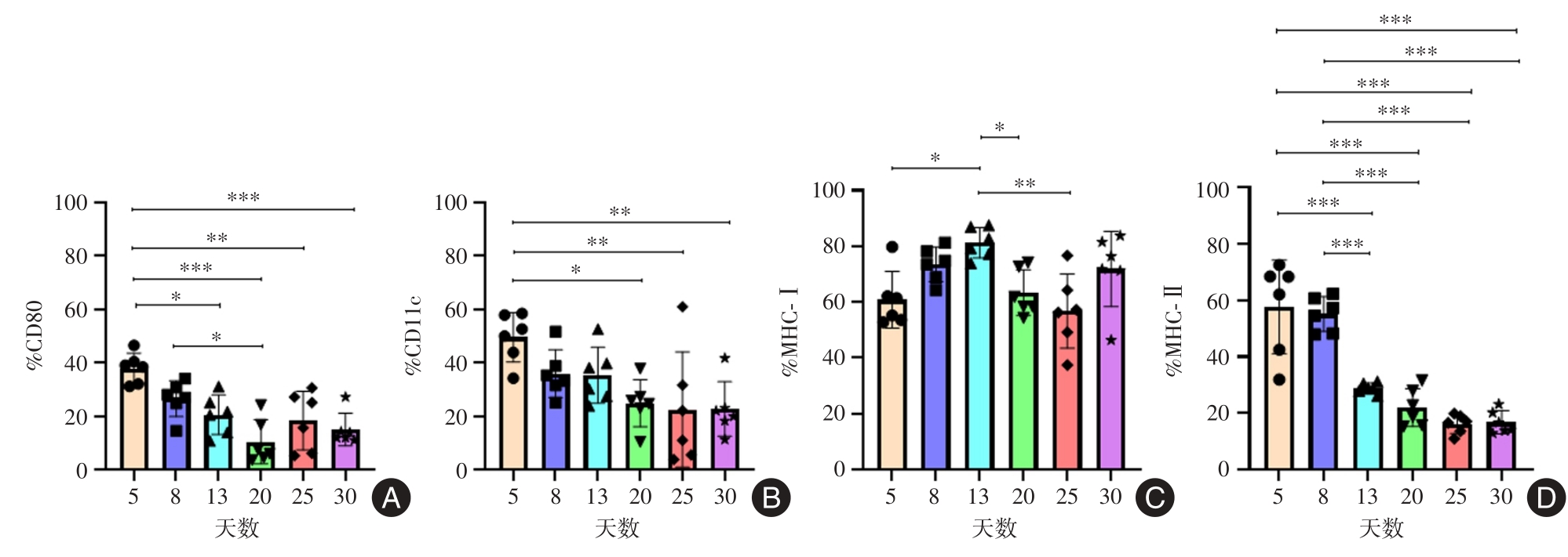
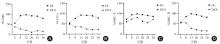
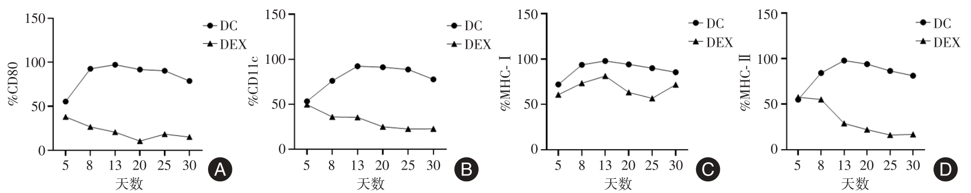
目的 明确培养时间对树突状细胞及其外泌体免疫相关膜蛋白(CD80、MHC-Ⅰ、MHC-Ⅱ)的影响,为今后相关研究提供实验依据。 方法 小鼠骨髓细胞经重组粒细胞-巨噬细胞集落刺激因子(GM-CSF)和白介素4(IL-4)诱导分化成树突状细胞,再加入肿瘤坏死因子α(TNF-α)诱导为成熟树突状细胞;超速离心法提取外泌体;蛋白印迹法和Amnis量化成像流式细胞仪对外泌体进行鉴定;Amnis量化成像流式细胞仪检测小鼠树突状细胞以及树突状细胞外泌体膜上免疫相关蛋白CD80、CD11c、MHC-Ⅰ、MHC-Ⅱ的表达情况。 结果 体外培养第5天后,约50%以上的树突状细胞表达CD80、CD11c、MHC-Ⅰ、MHC-Ⅱ,第13天达最高水平,其中CD80阳性率为(97.29 ± 0.63)%,CD11c阳性率为(92.31 ± 1.18)%,MHC-Ⅰ阳性率为(97.91 ± 0.49)%,MHC-Ⅱ阳性率为(97.91 ± 0.49)%,差异具有统计学意义(P < 0.001)。第13天后逐渐减少,至第30天,仍有约80%的树突状细胞表达MHC-Ⅰ和MHC-Ⅱ免疫分子。外泌体膜上CD80、CD11c、和MHC-Ⅱ表达水平以第5天最高,然后随培养时间延长,总体呈下降趋势,除外MHC-Ⅰ分子,差异具有统计学意义(P < 0.01)。 结论 体外培养的小鼠树突状细胞表达较高的免疫相关膜蛋白,在合适的培养条件下可长时间稳定维持。其分泌的外泌体表面也携带有丰富的免疫相关膜蛋白,但树突状细胞与外泌体膜表面的免疫相关蛋白未发现明显相关性。

关键词: 小鼠树突状细胞, 外泌体, 免疫膜蛋白

Abstract:

Objective This study aims to investigate the impact of cultivation time on dendritic cells (DCs) and their derived exosomes' expression of immune?related membrane proteins (CD80, MHC?Ⅰ, MHC?Ⅱ) and provides experimental evidence for future research. Methods Mouse bone marrow cells were induced to differentiate into DCs using GM?CSF and IL?4, followed by maturation stimulation withTNF?α. Exosomes were extracted using ultracentrifugation.Western blot and Amnis image flow cytometry were used to identify exosomes derived from mouse DCs. Amnis image flow cytometry was used to detect the expression of immune?related proteins CD80, CD11c, MHC?Ⅰ, and MHC?Ⅱ in mouse DCs and their exosomes. Results After 5 days of in vitro cultivation, more than 50% of dendritic cells expressed CD80, CD11c, MHC?Ⅰ, and MHC?Ⅱ, reaching the highest level on day 13. The positivity rates were as follows: CD80 (97.29 ± 0.63) %, CD11c (92.31 ± 1.18) %, MHC?Ⅰ(97.91 ± 0.49) %, and MHC?Ⅱ(97.91 ± 0.49) %. The differences were statistically significant (P < 0.001). The expression gradually decreased after day 13, but approximately 80% of DC cells still expressed MHC?Ⅰ and MHC?Ⅱ immune molecules on day 30. The expression levels of CD80, CD11c, and MHC?Ⅱ on the exosome membrane were highest on day 5 and then decreased overall with prolonged cultivation time, except for MHC?Ⅰ molecules. The differences were statistically significant (P < 0.01). Conclusions In vitro?cultured mouse dendritic cells express high levels of immune?related membrane proteins and can be stably maintained for a long time under suitable culture conditions. The secreted exosomes also carry abundant immune?related membrane proteins, but no significant correlation was found between the immune?related proteins on the dendritic cell surface and the exosome membrane surface.

Key words: mouse dendritic cells, exosomes, immune membrane proteins

中图分类号: