实用医学杂志 ›› 2024, Vol. 40 ›› Issue (6): 850-856.doi: 10.3969/j.issn.1006-5725.2024.06.020

• 医学检查与临床诊断 • 上一篇    下一篇

孤立性直肠溃疡综合征患者的X线排粪造影和MR排粪造影的影像学表现

李桂婷,胡美玉,曾志明,谢佩怡,邸小慧()   

  1. 中山大学附属第六医院放射诊断科、广东省结直肠盆底疾病研究重点实验室 (广州 510650 )
  • 收稿日期:2023-09-26 出版日期:2024-03-25 发布日期:2024-04-08
  • 通讯作者: 邸小慧 E-mail:dixh@mail.sysu.edu.cn
  • 基金资助:
    广东省科技创新战略专项资金(临床医学研究中心)项目(2020B1111170004);广东省医学影像智能分析与应用重点实验室开放课题项目(2022-MKF-05)

Imaging findings of X⁃ray defecography and MR defecography in patients with solitary rectal ulcer syndrome

Guiting LI,Meiyu HU,Zhiming ZENG,Peiyi XIE,Xiaohui DI()   

  1. Department of Radiology,Guangdong Provincial Key Laboratory of Colorectal and Pelvic Floor Diseases,Sixth Affiliated Hospital,Sun Yat?sen University,Guangzhou 510650,China
  • Received:2023-09-26 Online:2024-03-25 Published:2024-04-08
  • Contact: Xiaohui DI E-mail:dixh@mail.sysu.edu.cn

摘要:

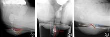
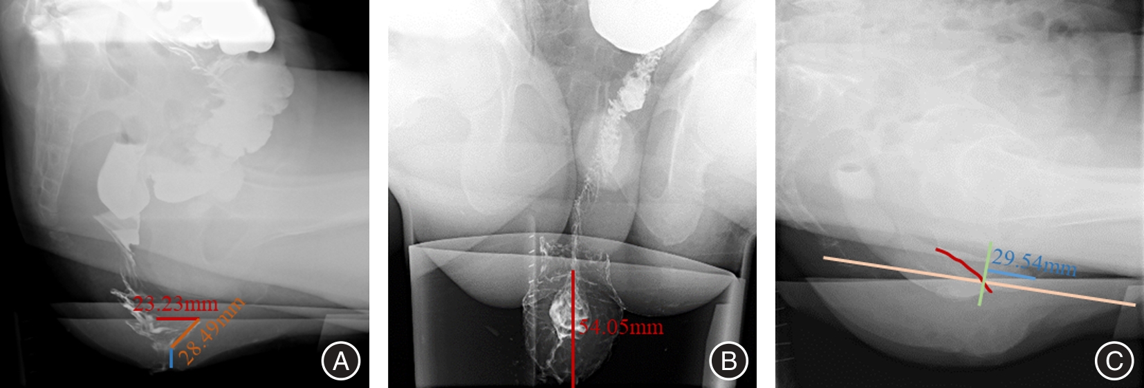
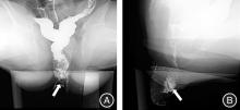
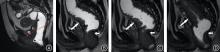
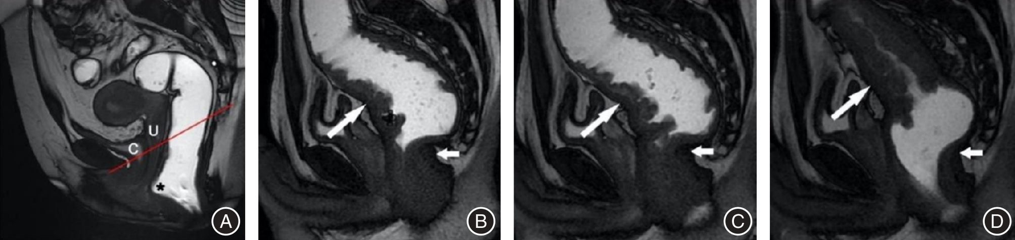
目的 回顾性总结分析孤立性直肠溃疡综合征(SRUS)患者的X线排粪造影(XRD)和MR排粪造影(MRD)的影像学表现,为临床诊疗提供重要信息。 方法 19例经临床、病理和肠镜检查证实的SRUS患者纳入本研究,15例行XRD,7例行MRD,3例患者同时进行了XRD和MRD检查。收集所有患者的数据并进行盆底功能测量。 结果 在XRD结果中,3例(20%)表现直肠内套叠,8例(53.3%)直肠外脱垂,2例(13.3%)中度直肠前突。另外耻骨直肠肌肥厚患者有2例,膀胱脱垂和子宫脱垂患者各有1例。在MRD结果中,3例(42.9%)表现直肠黏膜脱垂(部分性脱垂),4例(57.1%)直肠前突患者中,3例(均为女性)为中度直肠前突,1例为轻度直肠前突。3例患者同时观察到相关的前、中腔室器官下降,2例耻骨直肠肌肥厚,没有患者表现乙状结肠疝。 结论 排粪造影可以评估SRUS患者的直肠外脱垂、直肠前突、直肠黏膜脱垂、直肠内套叠等盆底结构及功能异常,对SRUS患者的治疗具有指导意义。

关键词: 孤立性直肠溃疡综合征, X线排粪造影, MR排粪造影, 结构异常, 功能异常

Abstract:

Objective The imaging findings of X?ray defecography (XRD) and magnetic resonance defecography (MRD) of patients with Solitary Rectal Ulcer syndrome (SRUS) were retrospectively analyzed to provide important information for clinical diagnosis and treatment. Methods 19 patients with SRUS confirmed by clinical, pathological and colonoscopy were included in this study. Among them, 15 patients underwent XRD and 7 patients underwent MRD, and 3 patients underwent both XRD and MRD. Data of all enrolled patients were collected and pelvic floor function was measured. Results In the results of XRD, 3 patients (20%) showed rectal intussusception. 8 patients (53.3%) showed external rectal prolapse and 2 patients (13.3%) showed moderate rectocele. In addition, there were 2 patients of puborectal muscle hypertrophy, and 1 patient of bladder prolapse and uterine prolapse, respectively. For MRD, 3 patients (42.9%) showed rectal mucosal prolapse (partial prolapse). At 4 patients (57.1%) with rectocele, 3 patients (all female) had moderate rectocele, 1 patient had mild rectocele. 3 patients were also observed related anterior and middle compartment organ descent. 2 patients of pubulorectal muscle hypertrophy, no sigmoidocoele. Conclusion Defecography can evaluate the structural and functional abnormalities of pelvic floor in SRUS patients, such as external rectal prolapse, rectal protrusion, rectal mucosal prolapse, and rectal intussusception, which has guiding significance for the treatment of SRUS patients。

Key words: solitary rectal ulcer syndrome, X?ray defecography, magnetic resonance defecography, structural abnormalities, functional abnormalities

中图分类号: