实用医学杂志 ›› 2023, Vol. 39 ›› Issue (21): 2743-2749.doi: 10.3969/j.issn.1006-5725.2023.21.009

• 基础研究 • 上一篇    下一篇

外周血Tfh细胞比例及其胞内IL-21含量与蓝光视网膜损伤的相关性

章梦一1,余洋洋1,2,李征亚1,俞永珍1,3,张春丽1,程天豪1,3,雷亦笑1,周文杰1,邹秀兰1,3(),邹玉平1,2,3   

  1. 1.中国人民解放军南部战区总医院 (广州 510010 )
    2.南方医科大学第一临床学院 (广州 510515 )
    3.广州中医药大学 (广州 510405 )
  • 收稿日期:2023-09-12 出版日期:2023-11-10 发布日期:2023-12-19
  • 通讯作者: 邹秀兰 E-mail:149642427@qq.com
  • 基金资助:
    广东省自然科学基金项目(2019A1515011732);广州市科技计划项目(202002030413)

Correlation of Tfh cell content in peripheral blood proportion and intracellular IL⁃21 with retinal damage caused by blue light

Mengyi ZHANG1,Yangyang YU1,2,Zhengya LI1,Yongzhen YU1,3,Chunli ZHANG1,Tianhao CHENG1,3,Yixiao LEI1,Wenjie ZHOU1,Xiulan ZOU1,3(),Yuping ZOU1,2,3   

  1. *.General Hospital of Southern Theatre Command,Guangzhou 510010,China
  • Received:2023-09-12 Online:2023-11-10 Published:2023-12-19
  • Contact: Xiulan ZOU E-mail:149642427@qq.com

摘要:

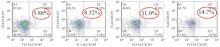
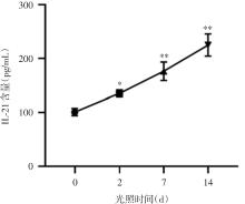
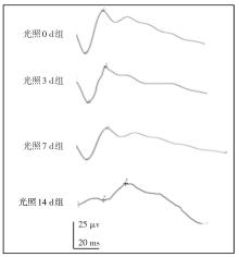
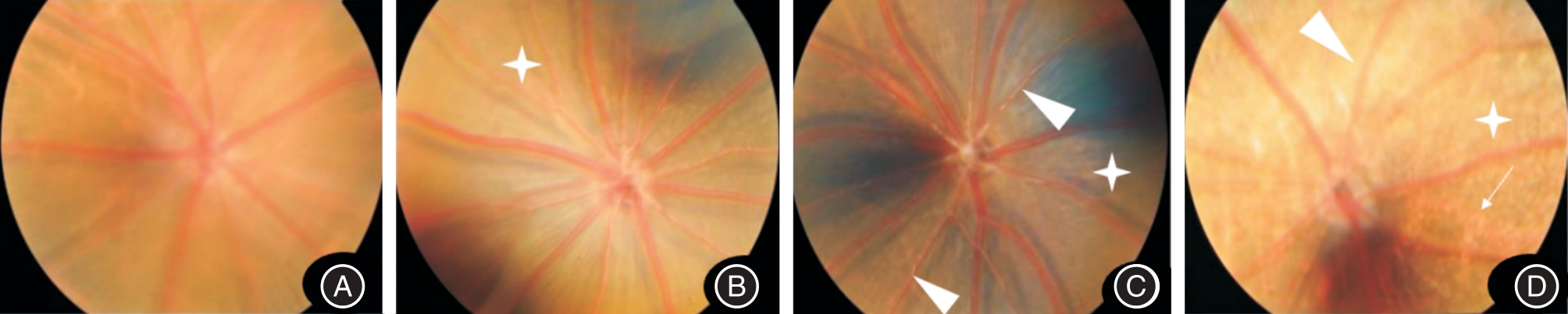
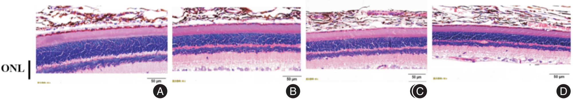
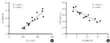
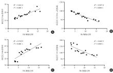
目的 探讨外周血滤泡辅助性T细胞(Tfh细胞)比例及其胞内白介素-21(IL-21)含量与蓝光视网膜损伤的相关性。 方法 随机将 Brown Norway(BN)大鼠均分为4组,采用蓝光全身每天照射3小时建立大鼠视网膜光损伤模型。按照射时间分为光照0天组即对照组(0 d组)、3天组(3 d组)、7天组(7 d组)和14天组(14 d组)。光照后采用流式细胞术和ELISA法分别检测大鼠外周血中Tfh细胞的比例及其胞内白介素21(IL-21)含量;视网膜电流图(ERG)检查评价视网膜功能;眼底照相和HE染色分别检查眼底改变和视网膜外核层厚度变化。 结果 与对照组相比,视网膜蓝光损伤后外周血Tfh细胞比例和胞内IL-21含量均随光照时间延长显著增加(均P < 0.05);ERG显示随光照时间延长视网膜功能损害加重,a、b波潜伏期和振幅分别随光照时间延长而延长和降低(均P < 0.01);大鼠眼底在光照3 d出现色素脱失样改变,随光照时间延长出现视网膜血管变细及渗出样改变;HE染色显示随光照时间延长ONL厚度显著变薄(均P < 0.05)。相关性分析提示外周血Tfh细胞比例及其胞内IL-21含量可共同反映损伤程度(P < 0.000 1),外周血Tfh细胞比例与ERG的a波和b波振幅及外核层(ONL)厚度呈负相关、与ERG的a波和b波峰时呈正相关(均P < 0.000 1)。 结论 视网膜蓝光损伤后大鼠外周血Tfh细胞比例及其胞内IL-21含量升高,并随光照时间延长显著升高,且二者存在一定的相关性。

关键词: 视网膜光损伤, 滤泡辅助T细胞, 细胞内白介素21, 光感受器细胞

Abstract:

Objective To investigate the correlation between the proportion of peripheral blood follicular T helper cells (Tfh cells) and intracellular interleukin-21(IL-21) content with blue light retinal injury. Methods Brown Norway(BN) rats were randomly divided into 4 groups and were exposed to blue light for 3 hours a day to establish retinal light damage model. According to the duration of illumination, the rats was divided into 0 days (control group), 3 days (3 d group), 7 days (7 d group) and 14 days (14 d group). The proportion of Tfh cells and content of IL-21 in Tfh cells in peripheral blood of each group was detected by flow cytometry and ELISA separately after illumination. Electroretinogram (ERG) was used to evaluate retinal function. The changes of fundus in rats were observed by fundus photography. The thickness of outer nuclear layer of retina was analyzed by HE staining. Results After retinal blue light injury, with the extension of illumination time,the proportion of Tfh cells in peripheral blood and intracellular IL-21 content both increased (P < 0.05). ERG showed that retinal function decreased after light damage and aggravated with the extension of illumination time, the latency and amplitudes of A-wave and B-wave increased and decreased respectively (P < 0.05). The retinal fundus of rats showed depigmentation in 3 d, and the retinal vessels became thinner and exudate with the extension of illumination time. HE staining showed that the outer nuclear layer of retina (ONL) became thinner (P < 0.05). Correlation analysis indicated that the proportion of Tfh cells in peripheral blood and the intracellular IL-21 content could jointly reflect the degree of injury (P < 0.000 1), and the proportion of Tfh cells in peripheral blood was negatively correlated with ONL thickness and the amplitude of a and b waves, positively correlated with the peak time of a and b waves, (P < 0.0001). Conclusion The proportion of Tfh cells in peripheral blood and the intracellular IL-21 content were increased after blue light damage to retina, and were significantly increased with the extension of light time with a certain correlation.

Key words: retinal photoinjury, follicle-assisted T cells, intracellular IL-21, photoreceptor cells

中图分类号: