| [1] |
MIN K, BEOM J, KIM B R, et al. Clinical Practice Guideline for Postoperative Rehabilitation in Older Patients With Hip Fractures[J]. Ann Rehabil Med, 2021,45(3): 225-259. doi:10.5535/arm.21110
doi: 10.5535/arm.21110
|
| [2] |
CHANG H, LUAN C, LI C. Effect of Comprehensive Rehabilitation Training Based on Balance Function on Postoperative Recovery and Function of Hip Fracture in the Elderly: A Systematic Review and Meta-Analysis[J]. Geriatr Orthop Surg Rehabil, 2024,15: 21514593241261506. doi:10.1177/21514593241261506
doi: 10.1177/21514593241261506
|
| [3] |
GOMEZ ALCARAZ J, PARDO GARCIA J M, SEVILLA FERNANDEZ J, et al. Primary total hip arthroplasty in elderly patients over 85 years old: Risks, complications and medium-long term results[J]. Rev Esp Cir Ortop Traumatol (Engl Ed), 2021,65(1): 13-23. doi:10.1016/j.recote.2020.12.006
doi: 10.1016/j.recote.2020.12.006
|
| [4] |
TURNBULL G, BLACKLOCK C, AKHTAR A, et al. Experience of an anatomic femoral stem in a UK orthopaedic centre beyond 20 years of follow-up[J]. Eur J Orthop Surg Traumatol, 2024,34(4): 2155-2162. doi:10.1007/s00590-024-03901-1
doi: 10.1007/s00590-024-03901-1
|
| [5] |
PINTO D, ALSHAHRANI M, CHAPURLAT R, et al. The Global Approach to Rehabilitation Following an Osteoporotic Fragility Fracture: A Review of the Rehabilitation Working Group of the International Osteoporosis Foundation (IOF) Committee of Scientific Advisors[J]. Osteoporos Int, 2022, 33: 527-540. doi:10.1007/s00198-021-06240-7
doi: 10.1007/s00198-021-06240-7
|
| [6] |
REGIS D, CASON M, MAGNAN B. Dislocation of Primary Total Hip Arthroplasty: Analysis of Risk Factors and Preventive Options[J]. World J Orthop, 2024,15(6): 501-511. doi:10.5312/wjo.v15.i6.501
doi: 10.5312/wjo.v15.i6.501
|
| [7] |
陈检文, 董立明, 蒋科, 等. 髋臼假体安装位置与无菌性松动的相关分析[J]. 中国矫形外科杂志, 2022, 30(1): 28-32.
|
| [8] |
MEERMANS G, GRAMMATOPOULOS G, INNMANN M, et al. Cup Placement in Primary Total Hip Arthroplasty: How to Get It Right Without Navigation or Robotics[J]. EFORT Open Rev, 2022,7(6): 365-374. doi:10.1530/eor-22-0025
doi: 10.1530/eor-22-0025
|
| [9] |
BOSKER B H, VERHEYEN C C P M, HORSTMANN W G, et al. Poor accuracy of freehand cup positioning during total hip arthroplasty[J]. Arch Orthop Trauma Surg, 2007,127(5): 375-379. doi:10.1007/s00402-007-0294-y
doi: 10.1007/s00402-007-0294-y
|
| [10] |
LEARMONTH I D, YOUNG C, RORABECK C. The operation of the century: Total hip replacement[J]. Lancet, 2007, 370(9597): 1508-1519. doi:10.1016/s0140-6736(07)60457-7
doi: 10.1016/s0140-6736(07)60457-7
|
| [11] |
BEVERLAND D E, O'NEILL C K J, RUTHERFORD M, et al. Placement of the acetabular component[J]. Bone Joint J, 2016, 98-B(1 ): 37-43. doi:10.1302/0301-620x.98b1.36343
doi: 10.1302/0301-620x.98b1.36343
|
| [12] |
ABDEL M P, von ROTH P, JENNINGS M T, et al. What Safe Zone? The Vast Majority of Dislocated THAs Are Within the Lewinnek Safe Zone for Acetabular Component Position[J]. Clin Orthop Relat Res, 2016, 474(2): 386-391. doi:10.1007/s11999-015-4432-5
doi: 10.1007/s11999-015-4432-5
|
| [13] |
崔晓荣, 兰丽华, 李向阳, 等. 基于MSCT三维扫描加重建对成人髋关节发育不良全髋关节置换术中髋臼假体的选择及截骨的准确性预测价值[J]. 实用医学杂志, 2024, 40(9): 1309-1313.
|
| [14] |
GRAMMATOPOULOS G, INNMANN M, PHAN P, et al. Spinopelvic challenges in primary total hip arthroplasty[J]. EFORT Open Rev, 2023,8(5): 298-312. doi:10.1530/eor-23-0049
doi: 10.1530/eor-23-0049
|
| [15] |
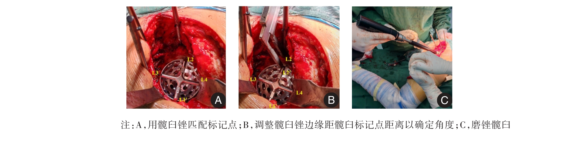
VAN DUREN B H, ROYECA J M, CUNNINGHAM C M, et al. Can the use of an inclinometer improve acetabular cup inclination in total hip arthroplasty? A review of the literature[J]. Hip Int, 2021,31(5): 609-617. doi:10.1177/1120700020946716
doi: 10.1177/1120700020946716
|
| [16] |
DARRITH B, BELL J A, CULVERN C, et al. Can the use of an inclinometer improve the positioning of the acetabular component in total hip arthroplasty?[J]. Bone Joint J, 2018, 100-B(7): 862-866. doi:10.1302/0301-620x.100b7.bjj-2017-1607.r1
doi: 10.1302/0301-620x.100b7.bjj-2017-1607.r1
|
| [17] |
BRUCE-BRAND R, MAGILL P, O'NEILL C, et al. Mechanical and Anatomical Alignment Guide Techniques Are Superior to Freehand in Achieving Target Orientation of an Acetabular Component[J]. Arthroplast Today, 2021, 11: 222-228. doi:10.1016/j.artd.2021.08.016
doi: 10.1016/j.artd.2021.08.016
|
| [18] |
GOLDSTEIN K, TYNDALL W, NICKOL M E, et al. Inclinometer use in primary total hip arthroplasty does not improve acetabular component positioning: A non-randomized control trial[J]. Arthroplasty, 2024,6(1): 41. doi:10.1186/s42836-024-00258-y
doi: 10.1186/s42836-024-00258-y
|
| [19] |
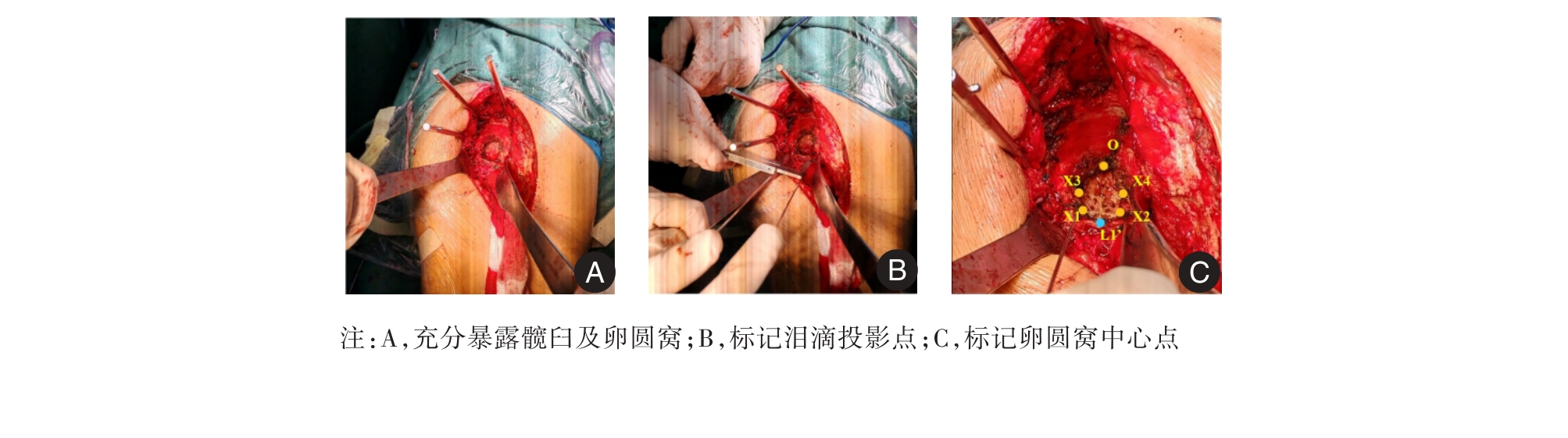
周建生, 凌遵龙, 王志岩, 等. Harris窝及髋臼切迹与髋臼中心点的应用解剖和相关性研究[J]. 解剖与临床, 2010, 15(4): 231-234.
|
| [20] |
ZHANG H, ZHOU J, LING X, et al. Determining the Orientation of Acetabular Prosthesis in Total Hip Arthroplasty by Refering to the Anatomical Landmarker of Acetabular Notches[J]. Sci Rep, 2023, 13(1):6185. doi:10.1038/s41598-023-33501-8
doi: 10.1038/s41598-023-33501-8
|
| [21] |
ARCHBOLD H A P, SLOMCZYKOWSKI M, CRONE M, et al. The Relationship of the Orientation of the Transverse Acetabular Ligament and Acetabular Labrum to the Suggested Safe Zones of Cup Positioning in Total Hip Arthroplasty[J]. Hip Int, 2008,18(1): 1-6. doi:10.5301/hip.2008.1755
doi: 10.5301/hip.2008.1755
|
| [22] |
PORTINARO N M, MATTHEWS S J, BENSON M K. The acetabular notch in hip dysplasia[J]. J Bone Joint Surg Br, 1994,76(2): 271-273. doi:10.1302/0301-620x.76b2.8113290
doi: 10.1302/0301-620x.76b2.8113290
|


 ),唐昊旭,梁英杰,丁鹏霖,钱闵龙