实用医学杂志 ›› 2023, Vol. 39 ›› Issue (17): 2176-2182.doi: 10.3969/j.issn.1006-5725.2023.17.005

• 基础研究 • 上一篇    下一篇

TMEM39A在原发性胆汁性胆管炎中的作用机制

孙晓彤1,韩崇旭1,2(),王婵1,任传利2,张明明3   

  1. 1.扬州大学医学院 (江苏 扬州 225009 )
    2.扬州大学临床医学院,江苏省苏北人民医院医学检验科 (江苏 扬州 225009 )
    3.东南大学生命科学与技术学院发育基因与人类疾病重点实验室 (南京 210096 )
  • 收稿日期:2023-03-27 出版日期:2023-09-10 发布日期:2023-09-27
  • 通讯作者: 韩崇旭 E-mail:hanchongxu@126.com
  • 基金资助:
    国家重点基础项目研究发展计划(973计划)子课题基金(2015CB755402)

Preliminary study on the mechanism of TMEM39A in primary biliary cholangitis

Xiaotong SUN1,Chongxu HAN1,2(),Chan WANG1,Chuanli REN2,Mingming. ZHANG3   

  1. Medical College of Yangzhou University,Yangzhou 225009,China
  • Received:2023-03-27 Online:2023-09-10 Published:2023-09-27
  • Contact: Chongxu HAN E-mail:hanchongxu@126.com

摘要:

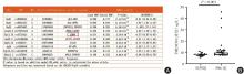
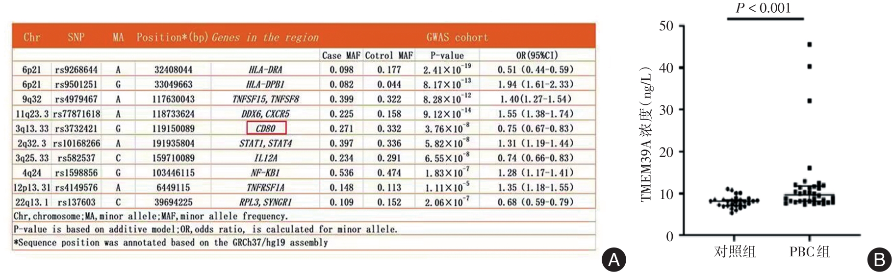
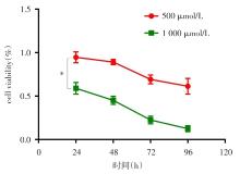
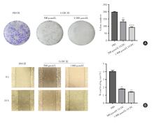
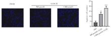
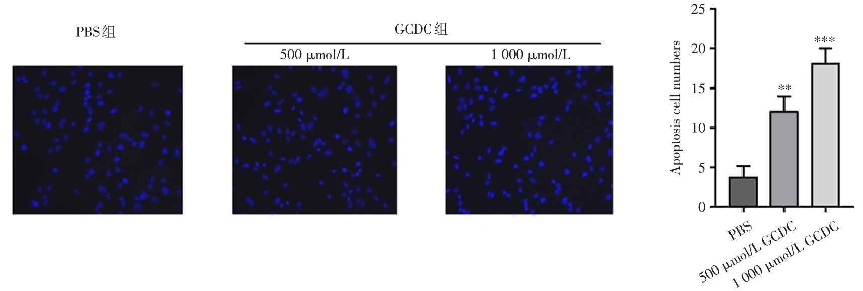
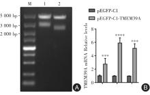
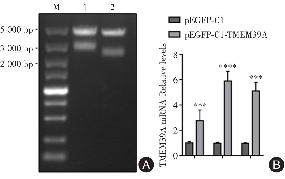
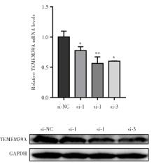
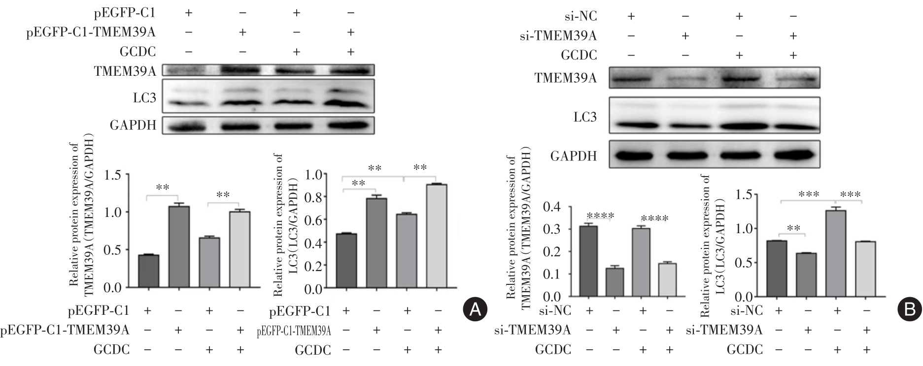
目的 探讨TMEM39A在原发性胆汁性胆管炎中的作用机制。 方法 为研究TMEM39A在原发性胆汁性胆管炎中如何调节自噬的表达,选用人肝胆管癌细胞RBE来进行研究。并用疏水性胆汁酸甘氨鹅脱氧胆酸(GCDC)处理人肝胆管癌细胞RBE分为3组:control组、500 μmol/L组和1 000 μmol/L组。细胞计数试剂盒(CCK-8)、细胞克隆、细胞划痕和Hoechst染色实验检测细胞活力、增殖、伤口愈合率和凋亡。qPCR检测细胞中TMEM39A mRNA表达量;Western blot检测TMEM39A和LC3的蛋白表达量。 结果 与control组相比较,1 000 μmol/L组的细胞活力、增殖和伤口愈合率降低,细胞凋亡现象最明显。在构建的原发性胆汁性胆管炎的模型中,过表达TMEM39A会促进LC3的表达。 结论 TMEM39A的表达增高引起胆管细胞自噬增强使胆管受损,可能是引起原发性胆汁性胆管炎的机制之一。

关键词: TMEM39A, 原发性胆汁性胆管炎, 人肝胆管癌细胞, 微管相关蛋白3β, 甘氨鹅脱氧胆酸

Abstract:

Objective To explore the mechanism of TMEM39A in primary biliary cholangitis. Method In order to study how TMEM39A regulates the expression of autophagy in primary biliary cholangitis, human hepatobiliary cancer cell RBE was selected for study. Human hepatobiliary cancer cells were treated with the hydrophobic bile acid glycine deoxycholic acid (GCDC) into three groups: control group, 500 μmol/L group and 1 000 μmol/L group. Cell counting kits (CCK-8), cell cloning, cell scratching, and Hoechst staining assays were used to detect cell viability, proliferation, wound healing rates, and apoptosis. qPCR detected TMEM39A mRNA expression in cells; Western Blot detects protein expression of TMEM39A and LC3. Results Compared with control group, cell viability, proliferation and wound healing rate were decreased in 1 000 μmol/L group, while apoptosis was the most obvious. In the constructed model of primary biliary cholangitis, overexpression of TMEM39A promoted LC3 expression. Conclusion The increased expression of TMEM39A leads to enhanced autophagy of bile duct cells and bile duct damage, which may be one of the mechanisms causing primary biliary cholangitis.

Key words: TMEM39A, primary biliary cholangitis, human hepatobiliary duct cancer cells, LC3, glycochenodeoxycholic acid

中图分类号: