实用医学杂志 ›› 2025, Vol. 41 ›› Issue (21): 3449-3454.doi: 10.3969/j.issn.1006-5725.2025.21.022

• 医学检查与临床诊断 • 上一篇    

光谱CT虚拟平扫联合碘图定量鉴别诊断血管内治疗术后早期脑出血与对比剂渗出

谭云1,孔钟仪2,曹希明1,王振邦1,郑君惠1,罗维1   

  1. 1.南方医科大学附属广东省人民医院(广东省医学科学院)放射科 (广东 广州 518000 )
    2.梅州市中医医院医学影像科 (广东 梅州 514500 )
  • 收稿日期:2025-07-09 出版日期:2025-11-10 发布日期:2025-11-13

Diagnostic efficacy of spectral CT virtual non⁃contrast imaging combined with iodine mapping for differentiating early postoperative intracerebral hemorrhage from contrast extravasation after endovascular therapy

Yun TAN1,Zhongyi KONG2,Ximing CAO1,Zhenbang WANG1,Junhui ZHENG1,Wei. LUO1   

  1. *.Department of Radiology,Guangdong Provincial People's Hospital Affiliated to Southern Medical University (Guangdong Academy of Medical Sciences),Guangzhou 518000,Guangdong,China
  • Received:2025-07-09 Online:2025-11-10 Published:2025-11-13

摘要:

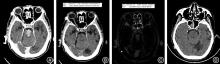
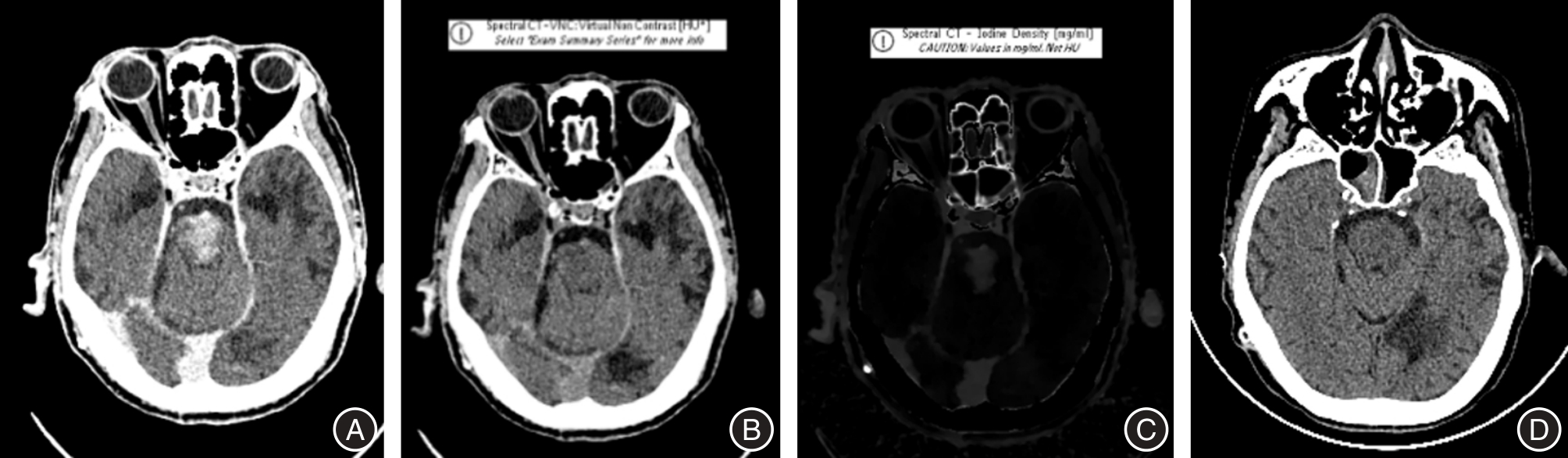
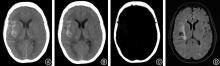
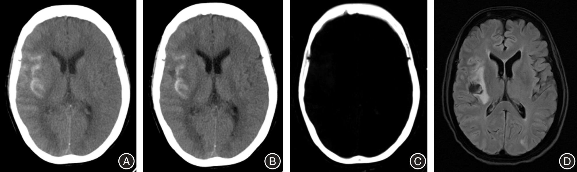
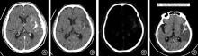
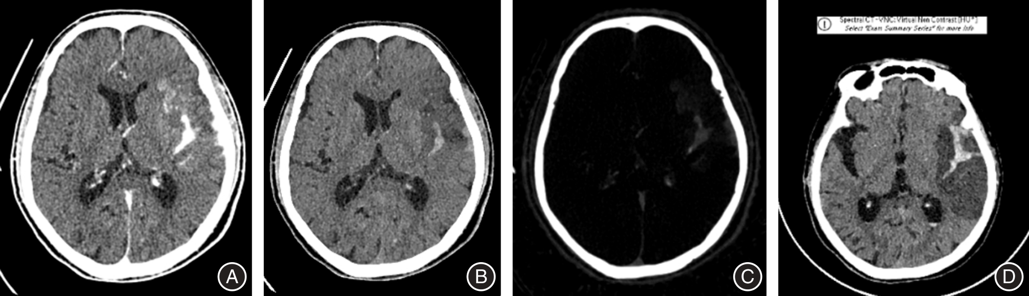
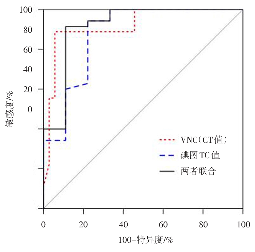
目的 探讨双层探测器光谱CT(DLCT)虚拟平扫(VNC)联合碘图对血管内治疗(EVT)术后早期脑出血与对比剂渗出的鉴别诊断价值。 方法 回顾性分析EVT术后即刻完成DLCT检查的患者资料。以术后24 h CT/MRI为金标准分为非出血组和出血组,比较两组临床资料的差异。测量VNC CT值和碘浓度(IC)值,对VNC CT值和碘图IC值进行多因素二项logistic回归,分析鉴别非出血和出血的独立指标。利用Spearman秩相关检验分析VNC CT值和碘图IC值之间的相关性。采用R统计软件绘制ROC曲线分析,评估VNC、碘图及两者联合的判别效能。 结果 共97例患者接受DLCT检查,51例(52.6%)各序列均未发现脑内高密度灶,46例(47.4%)发现异常密度灶纳入最终分析。以术后24 h CT/MRI为金标准,最终纳入分析的46例患者中,非出血38例(82.6%),出血8例(17.4%)。组间年龄、性别及治疗方式差异均无统计学意义(P > 0.05)。经Spearman秩相关检验结果显示,VNC CT值与碘图IC值呈负相关(r = -0.537, P < 0.01)。ROC曲线分析显示,利用VNC CT值诊断对比剂渗出的AUC为0.917(95%CI:0.786 ~ 0.999),碘图IC值的AUC为0.878(95%CI:0.719 ~ 0.999),两者联合的AUC为0.919(95%CI:0.812 ~ 0.999),其中两者联合的AUC明显大于VNC和碘图的AUC(P < 0.05)。VNC CT值诊断的截断值为53.6 HU,碘图IC值诊断的截断值为0.605 mg/mL。基于最终纳入分析的46例患者,VNC、碘图及两者联合诊断早期脑出血与对比剂渗出的敏感度分别为88.9%、94.3%、91.4%,特异度分别为94.3%、77.8%、88.9%,准确率分别为90.9%、90.9%、93.2%。 结论 DLCT的VNC联合碘图可显著提高EVT术后脑出血与对比剂渗出的鉴别诊断准确性,推荐作为常规影像学评估方案。

关键词: 双层探测器光谱CT, 血管内治疗, 虚拟平扫, 碘密度图

Abstract:

Objective To evaluate the diagnostic value of dual?layer spectral CT (DLCT) virtual non?contrast (VNC) imaging combined with iodine maps in differentiating early post?endovascular therapy (EVT) intracranial hemorrhage from contrast extravasation. Methods Retrospective analysis of 97 patients who underwent DLCT immediately after EVT was conducted. Taking 24?hour follow?up CT/MRI as the gold standard, patients were divided into hemorrhage and non?hemorrhage groups, and their clinical data were compared. VNC CT values and iodine concentration (IC) were measured. Spearman's rank correlation was used to analyze the relationship between VNC CT and IC values, and ROC curve analysis using R software to evaluate the diagnostic performance of VNC, iodine maps, and their combination. Results Among 97 patients, 51 (52.6%) showed no intracranial hyperdense lesions, while 46 (47.4%) with abnormal densities were analyzed. Using 24?hour postoperative CT/MRI as reference standard, among the 46 patients ultimately included in the analysis, 38 cases (82.6%) were non?hemorrhagic and 8 cases (17.4%) hemorrhagic. No significant differences existed in age, sex, or treatment methods (all P > 0.05). VNC CT values and IC showed significantly negative correlation (r = -0.537, P < 0.01). ROC analysis revealed AUCs of 0.917 (95%CI: 0.786 ~ 0.999) for VNC, 0.878 (95%CI: 0.719 ~ 0.999) for IC, and 0.919 (95%CI: 0.812 ~ 0.999) for the combination of the two (P < 0.05 for combined vs. individual methods). Optimal thresholds were 53.6 HU for VNC and 0.605 mg/ml for IC. Based on the final analysis of 46 enrolled patients, the sensitivity of VNC, iodine map, and their combination in differentiating early cerebral hemorrhage from contrast extravasation was 88.9%, 94.3%, and 91.4%, respectively; the specificity 94.3%, 77.8%, and 88.9%, respectively; and the accuracy 90.9%, 90.9%, and 93.2%, respectively. Conclusion The DLCT VNC?iodine map combination significantly improves differentiation between post?EVT hemorrhage and contrast extravasation, and it is recommended for routine clinical application.

Key words: dual-layer spectral CT, endovascular therapy, virtual non-contrast, iodine density map

中图分类号: