

The Journal of Practical Medicine ›› 2025, Vol. 41 ›› Issue (22): 3598-3608.doi: 10.3969/j.issn.1006-5725.2025.22.019
• Medical Examination and Clinical Diagnosis • Previous Articles
Dan ZHU1,Wanjun LU1( ),Ying ZHU1,Jinlu CAO1,Yingzi CHEN2
Received:2025-08-01
Online:2025-11-25
Published:2025-11-26
Contact:
Wanjun LU
E-mail:xue1203@sina.com
CLC Number:
Dan ZHU,Wanjun LU,Ying ZHU,Jinlu CAO,Yingzi CHEN. Developing an unsupervised deep learning model for diabetic nephropathy prediction using panoramic fundus retinal images[J]. The Journal of Practical Medicine, 2025, 41(22): 3598-3608.
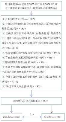
Fig.1
Participant screening and enrollment flowchart"
Tab.1
Baseline clinical characteristics of the training and validation sets"
| 项目 | 训练集 | t/Z/χ2值 | P值 | 验证集 | t/Z/χ2值 | P值 | ||
|---|---|---|---|---|---|---|---|---|
| NDKD组 | DKD组 | NDKD组 | DKD组 | |||||
| 例数 | 167 | 80 | 72 | 34 | ||||
| 年龄/岁 | 57.00(53.00,64.00) | 60.05(56.00,69.00) | -2.994 | 0.003 | 60.08 ± 10.79* | 61.82 ± 11.59* | -0.757 | 0.451 |
| 性别/[例(%)] | 0.095 | 0.758 | 0.027 | 0.868 | ||||
| 男 | 80(47.9) | 40(50.0) | 33(45.8) | 15(44.1) | ||||
| 女 | 87(52.1) | 40(50.0) | 39(54.2) | 19(55.9) | ||||
| 糖尿病病程/年 | 10(6.00,17.00) | 12(8.25,20.00) | -2.282 | 0.022 | 10(6.25,15.00) | 12(6.75,20.00) | -1.305 | 0.192 |
| BMI/(kg/m2) | 23.78(21.63,25.40) | 23.59(22.20,25.67) | -0.249 | 0.803 | 23.79(21.39,25.15) | 25.25(22.97,28.15) | -2.670 | 0.008 |
| BF/% | 29.19(24.03,36.13) | 30.34(24.45,37.41) | -0.939 | 0.348 | 28.46 ± 10.81* | 33.33 ± 8.65* | -2.295 | 0.024 |
| 吸烟史(有)/[例(%)] | 26(15.6) | 38(47.5) | 28.728 | 0.000 | 14(19.4) | 18(52.9) | 12.295 | < 0.001 |
| 高血压病史(有)/[例(%)] | 80(47.9) | 56(70.0) | 10.672 | 0.001 | 33(45.8) | 21(61.8) | 2.345 | 0.126 |
| 冠心病病史(有)/[例(%)] | 34(20.4) | 26(32.5) | 4.335 | 0.037 | 16(22.2) | 13(38.2) | 2.980 | 0.084 |
| 主动脉弓钙化(有)/[例(%)] | 78(46.7) | 46(57.5) | 2.521 | 0.112 | 33(45.8) | 19(55.9) | 0.933 | 0.334 |
| 冠状动脉钙化(有)/[例(%)] | 68(40.7) | 40(50.0) | 1.894 | 0.169 | 26(36.1) | 16(47.1) | 1.157 | 0.282 |
糖尿病周围神经病变(有) /[例(%)] | 66(39.5) | 33(41.3) | 0.067 | 0.795 | 44(61.1) | 13(38.2) | 4.862 | 0.027 |
| 糖化血红蛋白 /% | 9.70(8.40,11.00) | 9.70(8.00,11.00) | -0.469 | 0.639 | 9.83 ± 1.78* | 9.63 ± 2.72* | 0.451 | 0.653 |
| 尿酸/(mg/dL) | 281.90(229.60,329.90) | 340.85(278.53,401.33) | -4.702 | 0.000 | 261.5(207.93,335.25) | 305.75(259.78,382.40) | -2.156 | 0.031 |
| 总胆红素/(mg/dL) | 9.90(7.30,12.80) | 8.75(5.20,11.75) | -2.180 | 0.029 | 9.20(6.90,11.98) | 8.75(6.45,15.80) | -0.108 | 0.914 |
| 直接胆红素/(mg/dL) | 4.00(3.10,4.80) | 3.90(2.43,4.60) | -2.060 | 0.039 | 3.80(2.93,4.48) | 4.00(3.05,5.30) | -0.928 | 0.353 |
| 总胆汁酸/(μmol/L) | 3.25(2.69,4.07) | 3.25(2.63,3.92) | -0.354 | 0.723 | 3.45(2.85.4.23) | 3.74(2.74,4.52) | -0.798 | 0.430 |
| 白细胞计数/(109·L -1) | 6.15(5.21,7.38) | 6.68(5.44,7.70) | -1.336 | 0.182 | 6.45(5.57,7.54) | 6.11(5.19,7.92) | -0.690 | 0.490 |
| 血红蛋白/(g/dL) | 137.00(126.00,147.00) | 131.00(115.25,139.50) | -3.165 | 0.002 | 135.50(122.76,146.50) | 132.35(119.50,140.00) | -1.107 | 0.268 |
| 中性粒细胞计数/(109·L -1) | 3.89(3.08,4.92) | 4.29(3.23,5.37) | -1.400 | 0.162 | 4.23(3.27,5.14) | 3.82(2.90,5.74) | -0.042 | 0.675 |
| 淋巴细胞计数(x ± s)/(109·L -1) | 1.64 ± 0.61 | 1.63 ± 0.69 | 0.061 | 0.951 | 1.67(1.27,2.10) | 1.50(1.12,1.84) | -1.570 | 0.116 |
| 中性粒细胞/淋巴细胞比值 | 2.43(1.75,3.55) | 2.65(1.97,4.15) | -1.358 | 0.174 | 2.31(1.70,3.49) | 2.51(1.80,4.44) | -0.745 | 0.457 |
| 总胆固醇/(mg/dL) | 4.15(3.62,4,79) | 4.23(3.74,5.10) | -1.071 | 0.284 | 4.43(3.59,5.13) | 4.64(4.05,5.19) | -0.755 | 0.450 |
| 甘油三酯/(mg/dL) | 1.43(1.04,2.04) | 1.74(1.08,2.22) | -1.285 | 0.199 | 1.36(0.86,2.14) | 1.99(1.08,3.25) | -2.342 | 0.019 |
| 低密度脂蛋白/(mg/dL) | 2.27(1.79,2.81) | 2.40(1.85,3.04) | -0.914 | 0.361 | 2.42(1.91,3.08) | 2.46(1.81,3.19) | -0.203 | 0.839 |
| 高密度脂蛋白/(mg/dL) | 1.18(1.04,1.33) | 1.19(1.01,1.37) | -0.088 | 0.930 | 1.24(1.08,1.47) | 1.26(1.03,1.46) | -0.328 | 0.743 |
| LDL/HDL比值 | 1.92(1.42,2.46) | 1.94(1.46,2.50) | -0.482 | 0.629 | 1.92(1.51,2.70) | 1.89(1.30,2.32) | -0.647 | 0.518 |
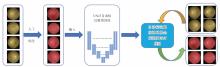
Fig.2
Flowchart for automatic segmentation of panoramic retinal fundus images"
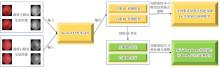
Fig.3
Flowchart of the monocular DL model development, binocular DL result integration, and binocular DL feature fusion model construction"

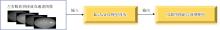
Fig. 4
Process flowchart for constructing the binocular fundus image fusion model"

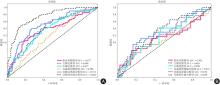
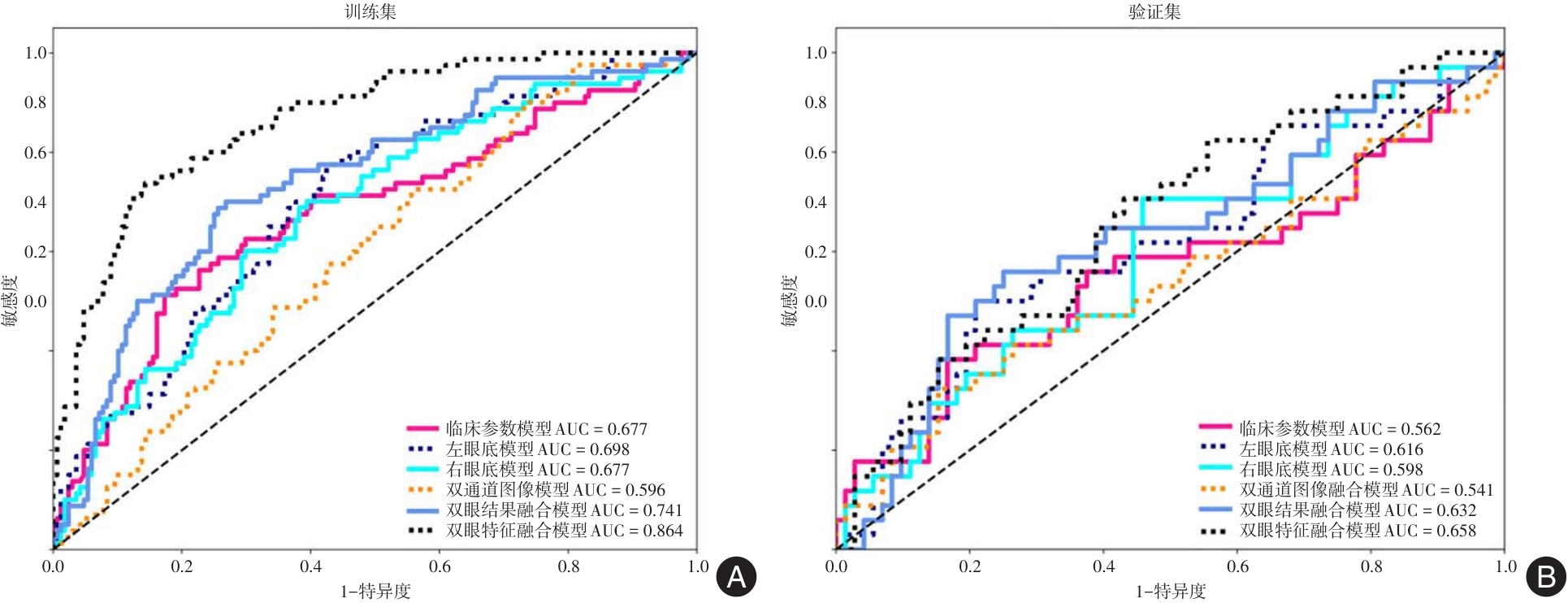
Fig.5
Comparison of the area under the curve for predicting diabetic kidney disease models"
Tab.2
Comparison of the accuracy, sensitivity and sspecificity of the models for predicting diabetic kidney disease"
| 队列 | 模型算法 | 模型名称 | 准确度 | AUC(95%CI) | 敏感度 | 特异度 |
|---|---|---|---|---|---|---|
| 训练集 | SVM | 临床参数模型 | 0.725 | 0.677(0.602 ~ 0.752) | 0.512 | 0.626 |
| ResNet152 | 左眼底模型 | 0.640 | 0.698(0.630 ~ 0.767) | 0.705 | 0.575 | |
| 右眼底模型 | 0.636 | 0.677(0.606 ~ 0.749) | 0.700 | 0.605 | ||
| 双通道图像融合模型 | 0.534 | 0.596(0.523 ~ 0.669) | 0.725 | 0.443 | ||
| ResNet152 + Ensemble | 双眼结果融合模型 | 0.721 | 0.741(0.674 ~ 0.808) | 0.700 | 0.731 | |
| ResNet152 + Transformer | 双眼特征融合模型 | 0.818 | 0.864(0.817 ~ 0.911) | 0.737 | 0.856 | |
| 验证集 | SVM | 临床参数模型 | 0.689 | 0.562(0.434 ~ 0.689) | 0.382 | 0.633 |
| ResNet152 | 左眼底模型 | 0.698 | 0.616(0.497 ~ 0.736) | 0.500 | 0.692 | |
| 右眼底模型 | 0.594 | 0.598(0.481 ~ 0.716) | 0.706 | 0.542 | ||
| 双通道图像融合模型 | 0.679 | 0.541(0.415 ~ 0.667) | 0.324 | 0.647 | ||
| ResNet152 + Ensemble | 双眼结果融合模型 | 0.689 | 0.632(0.514 ~ 0.750) | 0.559 | 0.750 | |
| ResNet152 + Transformer | 双眼特征融合模型 | 0.713 | 0.658(0.547 ~ 0.768) | 0.706 | 0.769 |

Fig.6
Comparison of predictive models for DKD using DeLong's test for ROC curves"
Fig.7
Comparison of the net reclassification index results for the diabetic kidney disease prediction models"
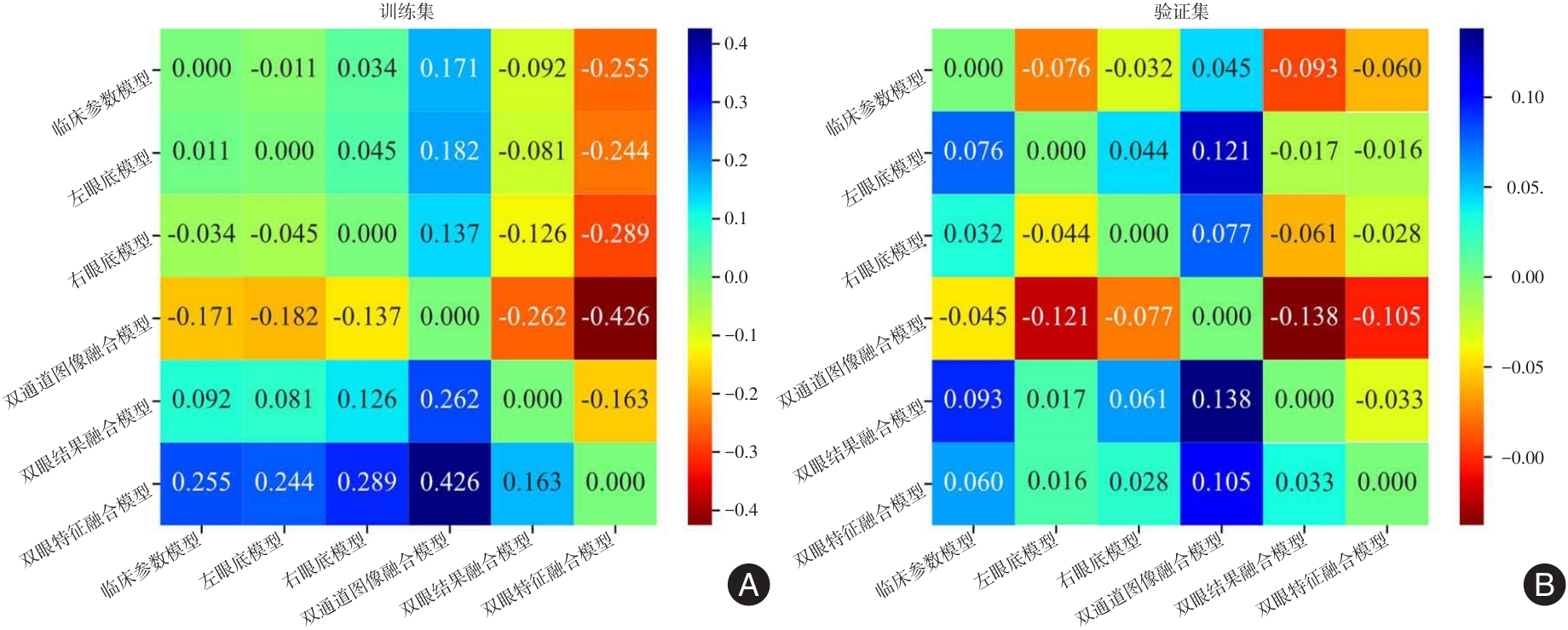
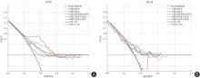
Fig.8
Comparison of the decision curve analysis results for the diabetic kidney disease prediction models"
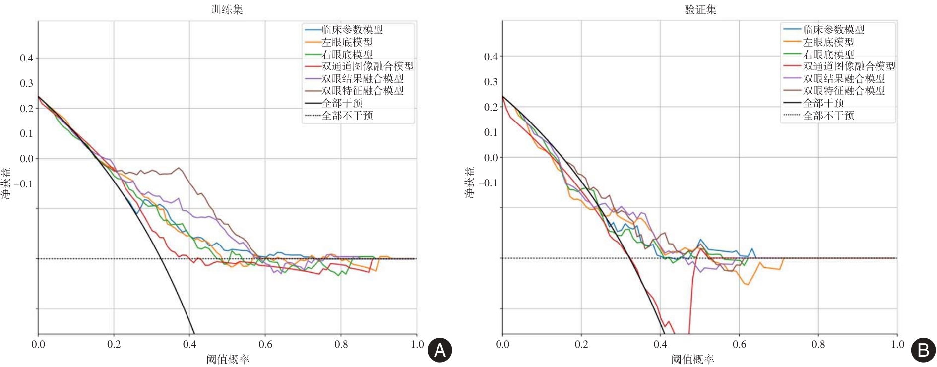

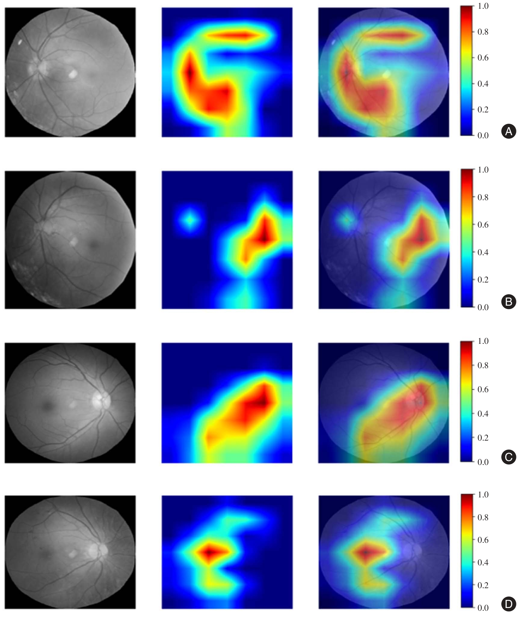
Fig. 9
Visualization results of the monocular DL model (Grad-CAM)"
| [1] | Disease Kidney : Improving Global Outcomes (KDIGO) Diabetes Work Group. KDIGO 2022 clinical practice guideline for diabetes management in chronic kidney disease[J]. Kidney Int, 2022, 102(5S): S1-S127. |
| [2] |
SUN H, SAEEDI P, KARURANGA S, et al. IDF Diabetes Atlas: Global, regional and country-level diabetes prevalence estimates for 2021 and projections for 2045[J]. Diabetes Res Clin Pract, 2022, 183:109119. doi:10.1016/j.diabres.2023.110945
doi: 10.1016/j.diabres.2023.110945 |
| [3] | 莫泽纬,陈道雄,高勇义,等. 艾塞那肽治疗早期糖尿病肾病患者的疗效研究[J]. 中国热带医学,2017,17(7):714-717. |
| [4] |
ROSSING P, GROOP P H, SINGH R, et al. Prevalence of chronic kidney disease in type 1 diabetes among adults in the U.S[J]. Diabetes Care, 2024, 47(8): 1395-1399. doi:10.2337/dc24-0335
doi: 10.2337/dc24-0335 |
| [5] |
SABANAYAGAM C, XU D, TING D S W, et al. A deep learning algorithm to detect chronic kidney disease from retinal photographs in community-based populations[J]. Lancet Digit Health, 2020, 2(6): e295-e302. doi:10.1016/s2589-7500(20)30063-7
doi: 10.1016/s2589-7500(20)30063-7 |
| [6] |
CHEN J, ZHANG Q, LIU D, et al. Exosomes: Advances, development and potential therapeutic strategies in diabetic nephropathy[J]. Metabolism, 2021, 122:154834. doi:10.1016/j.metabol.2021.154834
doi: 10.1016/j.metabol.2021.154834 |
| [7] |
SUN Z, WANG K, MILLER J D, et al. External validation of the risk prediction model for early diabetic kidney disease in Taiwan population: A retrospective cohort study[J]. BMJ Open, 2022,12(12):e059139. doi:10.1136/bmjopen-2021-059139
doi: 10.1136/bmjopen-2021-059139 |
| [8] |
LIU X Z, DUAN M, HUANG H D, et al. Predicting diabetic kidney disease for type 2 diabetes mellitus by machine learning in the real world: A multicenter retrospective study[J]. Front Endocrinol (Lausanne), 2023, 14: 1184190. doi:10.3389/fendo.2023.1184190
doi: 10.3389/fendo.2023.1184190 |
| [9] |
LI Y, JIN N, ZHAN Q, et al. Machine learning-based risk predictive models for diabetic kidney disease in type 2 diabetes mellitus patients: A systematic review and meta-analysis[J]. Front Endocrinol (Lausanne), 2025, 16: 1495306. doi:10.3389/fendo.2025.1495306
doi: 10.3389/fendo.2025.1495306 |
| [10] |
BERMEJO S, GONZÁLEZ E, LÓPEZ-REVUELTA K, et al. The coexistence of diabetic retinopathy and diabetic nephropathy is associated with worse kidney outcomes[J]. Clin Kidney J, 2023, 16(10): 1656-1663. doi:10.1093/ckj/sfad142
doi: 10.1093/ckj/sfad142 |
| [11] |
LEE G W, LEE C H, KIM S G. Association of advanced chronic kidney disease with diabetic retinopathy severity in older patients with diabetes: A retrospective cross-sectional study[J]. J Yeungnam Med Sci, 2023, 40(2): 146-155. doi:10.12701/jyms.2022.00206
doi: 10.12701/jyms.2022.00206 |
| [12] |
ZHAO X, LIU Y, ZHANG W, et al. Relationships between retinal vascular characteristics and renal function in patients with type 2 diabetes mellitus[J]. Transl Vis Sci Technol, 2021,10(2):1-11. doi:10.1167/tvst.10.2.20
doi: 10.1167/tvst.10.2.20 |
| [13] |
YAN Y, YU L, SUN C, et al. Retinal microvascular changes in diabetic patients with diabetic nephropathy[J]. BMC Endocr Disord, 2023, 23(1):101. doi:10.1186/s12902-022-01250-w
doi: 10.1186/s12902-022-01250-w |
| [14] |
ALJEFRI S, ADEL F AL. The validity of diabetic retinopathy screening using nonmydriatic fundus camera and optical coherence tomography in comparison to clinical examination[J]. Saudi J Ophthalmol, 2021, 34(4):266-272. doi:10.4103/1319-4534.322617
doi: 10.4103/1319-4534.322617 |
| [15] |
IRODI A, ZHU Z, GRZYBOWSKI A, et al. The evolution of diabetic retinopathy screening[J]. Eye (Lond), 2025, 39(6):1040-1046. doi:10.1038/s41433-025-03633-4
doi: 10.1038/s41433-025-03633-4 |
| [16] |
MESSICA S, PRESIL D, HOCH Y, et al. Enhancing stroke risk and prognostic timeframe assessment with deep learning and a broad range of retinal biomarkers[J]. Artif Intell Med, 2024, 154:102927. doi:10.1016/j.artmed.2024.102927
doi: 10.1016/j.artmed.2024.102927 |
| [17] | WHITE T, SELVARAJAH V, WOLFHAGEN-SAND F, et al. Prediction of cardiovascular risk factors from retinal fundus photographs: Validation of a deep learning algorithm in a prospective non-interventional study in Kenya[J]. Diabetes Obes Metab, 2024, 26(7):2722-2731. |
| [18] | 何雪,马欢,李聪,等. 冠心病伴抑郁患者视网膜神经血管特征及其与抑郁严重程度的相关性[J]. 实用医学杂志,2025,41(4):515-521. |
| [19] |
ZHANG K, LIU X, XU J, et al. Deep-learning models for the detection and incidence prediction of chronic kidney disease and type 2 diabetes from retinal fundus images[J]. Nat Biomed Eng, 2021, 5(6):533-545. doi:10.1038/s41551-021-00745-6
doi: 10.1038/s41551-021-00745-6 |
| [20] |
BETZLER B K, CHEE E Y L, HE F, et al. Deep learning algorithms to detect diabetic kidney disease from retinal photographs in multiethnic populations with diabetes[J]. J Am Med Inform Assoc, 2023, 30(12):1904-1914. doi:10.1093/jamia/ocad179
doi: 10.1093/jamia/ocad179 |
| [21] |
MENG Z, GUAN Z, YU S, et al. Non-invasive biopsy diagnosis of diabetic kidney disease via deep learning applied to retinal images: A population-based study[J]. Lancet Digit Health, 2025, 7(5):100868. doi:10.1016/j.landig.2025.02.008
doi: 10.1016/j.landig.2025.02.008 |
| [22] |
DONG Z, WANG X, PAN S, et al. A multimodal transformer system for noninvasive diabetic nephropathy diagnosis via retinal imaging[J]. NPJ Digit Med, 2025, 8(1):50. doi:10.1038/s41746-024-01393-1
doi: 10.1038/s41746-024-01393-1 |
| [23] |
AMIRHAMZAH N A, WANZAKI W M D, WANABDULHALIM W H, et al. Evaluating the potential of retinal photography in chronic kidney disease detection: A review[J]. PeerJ, 2024, 12:e17786. doi:10.7717/peerj.17786
doi: 10.7717/peerj.17786 |
| [24] |
SONG J, ZHENG J, LI P, et al. An Effective Multimodal Image Fusion Method Using MRI and PET for Alzheimer's Disease Diagnosis[J]. Front Digit Health, 2021, 3:637386. doi:10.3389/fdgth.2021.637386
doi: 10.3389/fdgth.2021.637386 |
| [25] |
DE BOER I H, KHUNTI K, SADUSKY T, et.al. Diabetes management in chronic kidney disease: A consensus report by the American Diabetes Association (ADA) and Kidney Disease: Improving Global Outcomes (KDIGO)[J]. Kidney Int, 2022, 102(5):974-989. doi:10.1016/j.kint.2022.08.012
doi: 10.1016/j.kint.2022.08.012 |
| [26] |
COLCOMBE J, SOLLI E, KAISER A, et al. The Use of Retinal Imaging Including Fundoscopy, OCT, and OCTA for Cardiovascular Risk Stratification and the Detection of Subclinical Atherosclerosis[J]. Curr Atheroscler Rep, 2025, 27(1):23. doi:10.1007/s11883-024-01268-6
doi: 10.1007/s11883-024-01268-6 |
| [27] |
LEE S J V, GOH Y Q, ROJAS-CARABALI W, et al. Association between retinal vessels caliber and systemic health: A comprehensive review[J]. Surv Ophthalmol, 2025, 70(2):184-199. doi:10.1016/j.survophthal.2024.11.009
doi: 10.1016/j.survophthal.2024.11.009 |
| [28] | 张锐,周颖,倪文吉,等. 人工智能视网膜微血管分析在糖尿病并发症中的应用价值[J]. 实用医学杂志,2024,40(8):1142-1147. |
| [29] |
BAKER J, SAFARZADEH M A, INCOGNITO A V,et al. Functional optical coherence tomography at altitude: Retinal microvascular perfusion and retinal thickness at 3,800 meters[J]. J Appl Physiol (1985), 2022, 133(3): 534-545. doi:10.1152/japplphysiol.00132.2022
doi: 10.1152/japplphysiol.00132.2022 |
| [30] |
EL-HAG N A, SEDIK A, EL-SHAFAI W, et al. Classification of retinal images based on convolutional neural network[J]. Microsc Res Tech, 2021, 84(3): 394-414. doi:10.1002/jemt.23596
doi: 10.1002/jemt.23596 |
| [31] |
MUKHERJEE N, SENGUPTA S. Application of deep learning approaches for classification of diabetic retinopathy stages from fundus retinal images: A survey[J]. Multimed Tools Appl, 2024, 83(14): 43115-43175. doi:10.1007/s11042-023-17254-0
doi: 10.1007/s11042-023-17254-0 |
| [32] |
ERGUN O N, ILHAN H O. Advancing Diabetic Retinopathy Severity Classification Through Stacked Generalization in Ensemble Deep Learning Models[J]. Traitement du Signal, 2023, 40(6): 2495. doi:10.18280/ts.400614
doi: 10.18280/ts.400614 |
| [33] |
BALA R, SHARMA A, GOEL N. CTNet: Convolutional transformer network for diabetic retinopathy classification[J]. Neural Comput Appl, 2024, 36(9): 4787-4809. doi:10.1007/s00521-023-09304-3
doi: 10.1007/s00521-023-09304-3 |
| [34] |
LIU C, WANG W, LIAN J, et al. Lesion classification and diabetic retinopathy grading by integrating softmax and pooling operators into vision transformer[J]. Front Public Health, 2025, 12: 1442114. doi:10.3389/fpubh.2024.1442114
doi: 10.3389/fpubh.2024.1442114 |
| [35] |
REZAEE K, FARNAMI F. Innovative Approach for Diabetic Retinopathy Severity Classification: An AI-Powered Tool using CNN-Transformer Fusion[J]. J Biomed Phys Eng, 2025, 15(2): 137-158. doi:10.31661/jbpe.v0i0.2408-1811
doi: 10.31661/jbpe.v0i0.2408-1811 |
| [36] |
MAHMOOD M A, JAMEL L, ALTURKI N, et al. Leveraging artificial intelligence for diagnosis of children autism through facial expressions[J]. Sci Rep, 2025, 15(1): 11945. doi:10.1038/s41598-025-96014-6
doi: 10.1038/s41598-025-96014-6 |
| [37] |
MORANO J, ARESTA G, GRECHENIG C, et al. Deep Multimodal Fusion of Data With Heterogeneous Dimensionality via Projective Networks[J]. IEEE J Biomed Health Inform, 2024, 28(4): 2235-2246. doi:10.1109/jbhi.2024.3352970
doi: 10.1109/jbhi.2024.3352970 |
| [38] |
LIU Y, MU F, SHI Y, et al. Brain tumor segmentation in multimodal MRI via pixel-level and feature-level image fusion[J]. Front Neurosci, 2022, 16: 1000587. doi:10.3389/fnins.2022.1000587
doi: 10.3389/fnins.2022.1000587 |
| [1] | Mingmei MA,Xiaochun MA,Shenghua MA,Yijin LI,Guifang JI. Effect of semaglutide injection on glycolipid metabolism and adipokine in the treatment of type 2 diabetes mellitus with different body mass index [J]. The Journal of Practical Medicine, 2025, 41(9): 1394-1400. |
| [2] | Yingmei HAN,Yijie LI,Heng ZHANG,Weiqing LI,Ze FENG,Feng WANG. The application value of deep learning in imaging studies for predicting the conversion of Alzheimer′s disease [J]. The Journal of Practical Medicine, 2025, 41(9): 1413-1424. |
| [3] | Gaokai HU,Ya'nan NIU,Yukang GONG,Yang HU,Ruixuan XU,Wenshan. GAO. Research progress on the application of deep learning in lumbar spine disease [J]. The Journal of Practical Medicine, 2025, 41(6): 921-928. |
| [4] | Jing WANG,Hui WANG,Yukuan MIAO,Yingqun. NI. Comparison of plantar dynamic and static pressure parameters in patients with type 2 diabetes mellitus combined with diabetic peripheral neuropathy and analysis of related factors [J]. The Journal of Practical Medicine, 2025, 41(4): 522-528. |
| [5] | Wei LIU,Jun HOU,Longquan TANG,Peng ZHOU,Yan ZHONG,Qinyan LUO,Xiaoyu KUANG,Hua LIU,Ziqing XIONG,Wei XIONG,Chenggao WU,Aiping. LE. Analysis of influencing factors of blood transfusion in children with traumatic brain injury and construction of prediction model: A multi⁃center retrospective study [J]. The Journal of Practical Medicine, 2025, 41(4): 553-560. |
| [6] | Le WU,Runlu GENG,Jingjiang ZHOU,Li LI,Lei XU,Jie KANG,Bin LU,Ying YE,Hongning YANG,Weichao. DING. Efficacy of HP in series with CRRT integrated combined blood purification under extracorporeal citrate anticoagulation in the treatment of severe HTG⁃AP [J]. The Journal of Practical Medicine, 2025, 41(4): 561-568. |
| [7] | Xiuli FENG,Zhichen ZHENG,Tongyu ZHANG,Li ZHOU,Ning XU,Renhao ZHAO,Teng YANG,Na WANG,Guofeng. WANG. Study on the correlation between blood glucose fluctuations and type 2 diabetic foot based on flash glucose monitoring technology [J]. The Journal of Practical Medicine, 2025, 41(4): 569-574. |
| [8] | Yufen LU,Xiaoming ZHENG,Shaojuan WEI,Liqin CHEN,Tongtong XU,Xiangwei. LÜ. Effects of miR⁃483⁃3p on hypoxia/reoxygenation⁃induced apoptosis and pyroptosis in cardiomyocytes [J]. The Journal of Practical Medicine, 2025, 41(3): 339-346. |
| [9] | Yan LI,Ziran JIN,Haotian SUN,Xuan LIU,Jing. GAO. Assessment of left ventricular systolic function in type 2 diabetes patients with renal insufficiency by aCMQ technique [J]. The Journal of Practical Medicine, 2025, 41(3): 414-421. |
| [10] | Congshun MA,Yuanyuan CUI,Wanshan ZHU,Xuejun ZHAN,Ying TAN. Clinical outcomes of three treatment protocols for frozen⁃thawed embryo transfer in patients with thin endometrium [J]. The Journal of Practical Medicine, 2025, 41(22): 3474-3479. |
| [11] | Xiaomeng YU,Ning. WANG. Association between novel insulin resistance indices and lower extremity atherosclerotic disease in patients with type 2 diabetes mellitus [J]. The Journal of Practical Medicine, 2025, 41(21): 3371-3377. |
| [12] | Yujian CUI,Yuke LI,Sainan ZHU,Shuangling LI,Nan. LI. The association between early peripheral perfusion and acute kidney injury in patients admitted to the intensive care unit following major noncardiac surgery [J]. The Journal of Practical Medicine, 2025, 41(2): 195-201. |
| [13] | Wei LU,Pan ZHANG,Yushu. QIN. Clinical significance of CT perfusion imaging combined with artificial intelligence in evaluating reperfusion injury after cerebral infarction [J]. The Journal of Practical Medicine, 2025, 41(2): 264-270. |
| [14] | Xiaokun GAO,Ziming XIE,Guangyu TAO,Yanbing SUN,Hua REN,Jiahui YU,Lin ZHU,Hong YU,Qiming. NI. The application value of multi⁃parameter quantitative analysis of spectral and perfusion CT in differentiating pathological types of lung cancer [J]. The Journal of Practical Medicine, 2025, 41(19): 3096-3105. |
| [15] | Mengying ZHANG,Yaokai MA,Yifan DU,Wei ZHANG,Bo ZHOU,Xiyi YANG. A Randomized controlled study on the efficacy of HAIC sequential DEB⁃TACE in the treatment of colorectal cancer liver metastasis [J]. The Journal of Practical Medicine, 2025, 41(17): 2721-2728. |
| Viewed | ||||||
|
Full text |
|
|||||
|
Abstract |
|
|||||