

The Journal of Practical Medicine ›› 2025, Vol. 41 ›› Issue (22): 3529-3536.doi: 10.3969/j.issn.1006-5725.2025.22.009
• Basic Research • Previous Articles
Shaochu CHEN1,2,Ming GONG2,Wang ZHANG2,Jiawen WU2,Guangxin HUANG3,Yadong ZHANG1( )
)
Received:2025-08-01
Online:2025-11-25
Published:2025-11-26
Contact:
Yadong ZHANG
E-mail:zhangyadong6@126.com
CLC Number:
Shaochu CHEN,Ming GONG,Wang ZHANG,Jiawen WU,Guangxin HUANG,Yadong ZHANG. Effects of exosomes secreted from mesenchymal stem cells on chondrocyte injury under hypoxia[J]. The Journal of Practical Medicine, 2025, 41(22): 3529-3536.
Tab.1
qPCR primer sequence"
| 基因名称 | 引物序列 |
|---|---|
| GPX4 | F: 5′-GAGGCAAGACCGAAGTAAACTAC-3′ |
| R: 5′-CCGAACTGGTTACACGGGAA-3′ | |
| SLC7A11 | F: 5′-TGCTGGGCTGATTTTATCTTCG-3′ |
| R: 5′-GAAAGGGCAACCATGAAGAGG-3′ | |
| ACSL4 | F: 5′-TCTGCTTCTGCTGCCCAATT-3′ |
| R: 5′-CGCCTTCTTGCCAGTCTTTT-3′ | |
| COL2A1 | F: 5′-TGCATGAGGGCGCGGTA-3′ |
| R: 5′-GGTCCTGGTTGCCGGACAT-3′ | |
| MMP13 | F: 5′-ATGACTATGCGTGGCTGGAA-3′ |
| R: 5′-TGTCCCATTTGTGGTGTGGG-3′ | |
| ADAMTS5 | F: 5′-TGGCTCACGAAATCGGACAT-3′ |
| R: 5′-TTGGACCAGGGCTTAGATGC-3′ | |
| GAPDH | F: 5'-CGCATCTTCTTTTGCGTCG-3' |
| R: 5'-TTGAGGTCAATGAAGGGGTCA-3' |

Fig. 1
Exosomes secreted by hypoxia-treated mesenchymal stem cells promote chondrocyte viability"
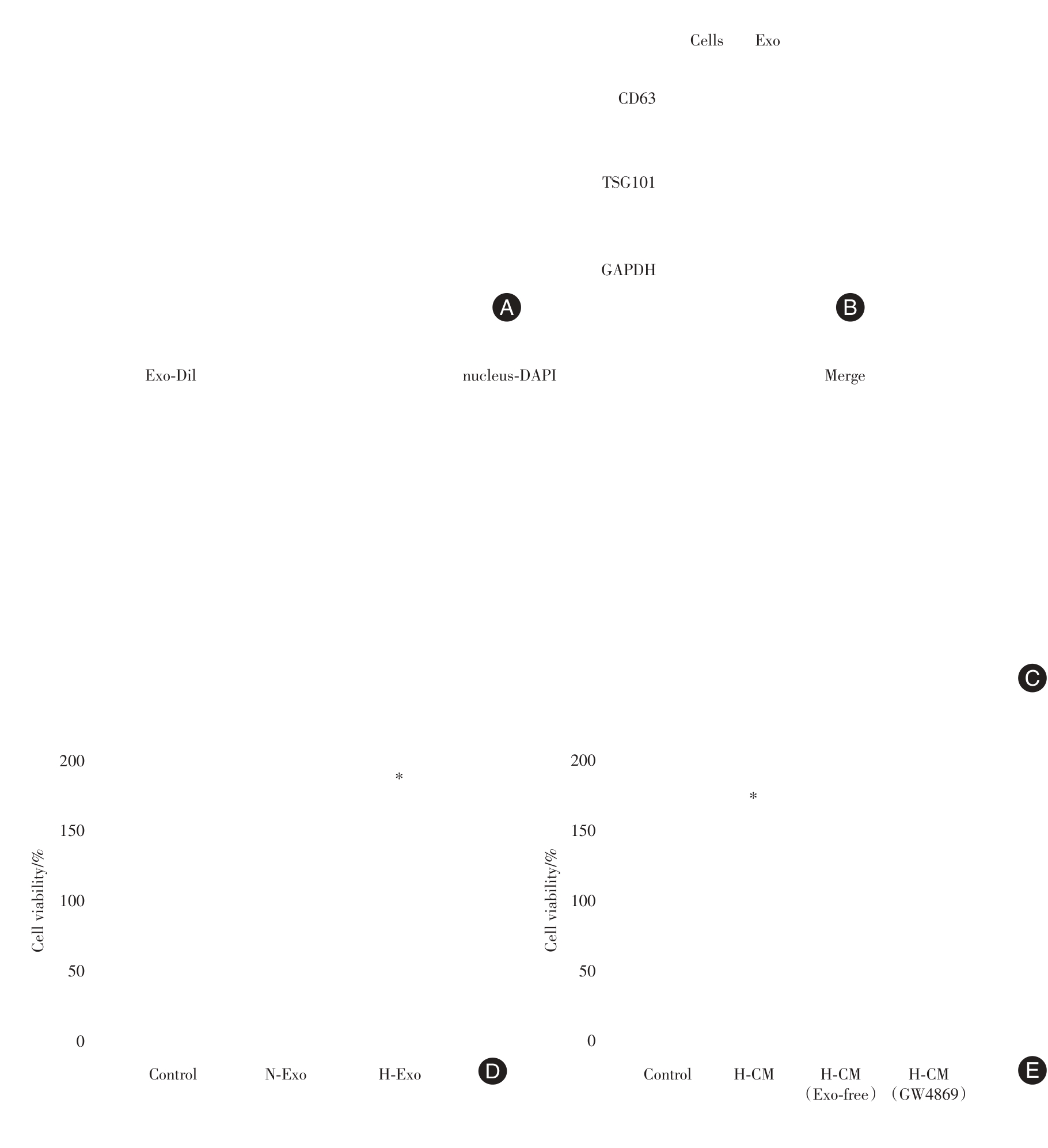

Fig. 2
The effect of exosomes secreted by hypoxia-treated mesenchymal stem cells on apoptosis of IL-1β-treated chondrocytes"
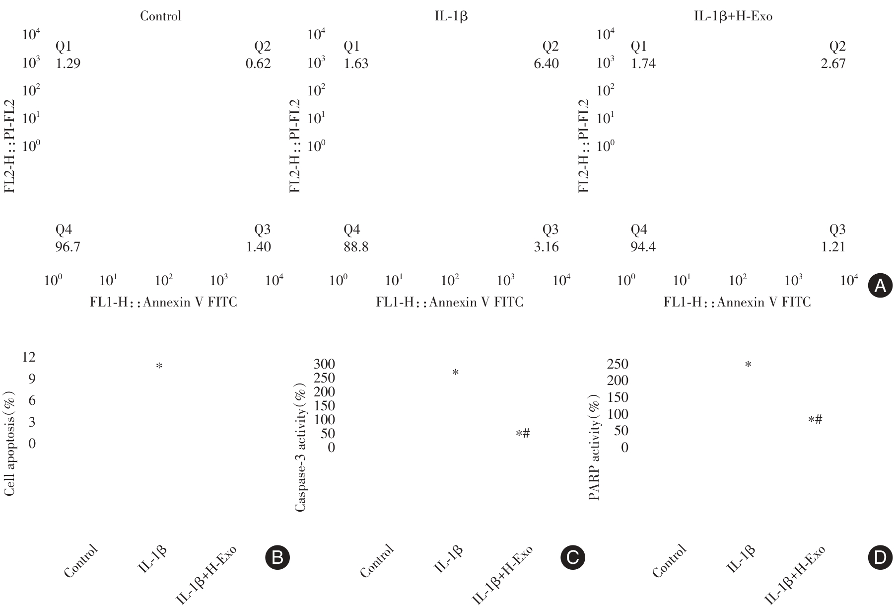
Fig. 3
The effect of exosomes secreted by hypoxia-treated mesenchymal stem cells on ferroptosis of IL-1β-treated chondrocytes"
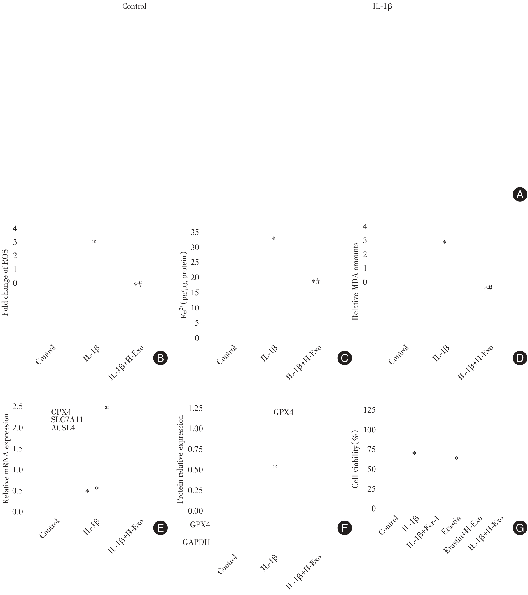
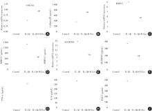
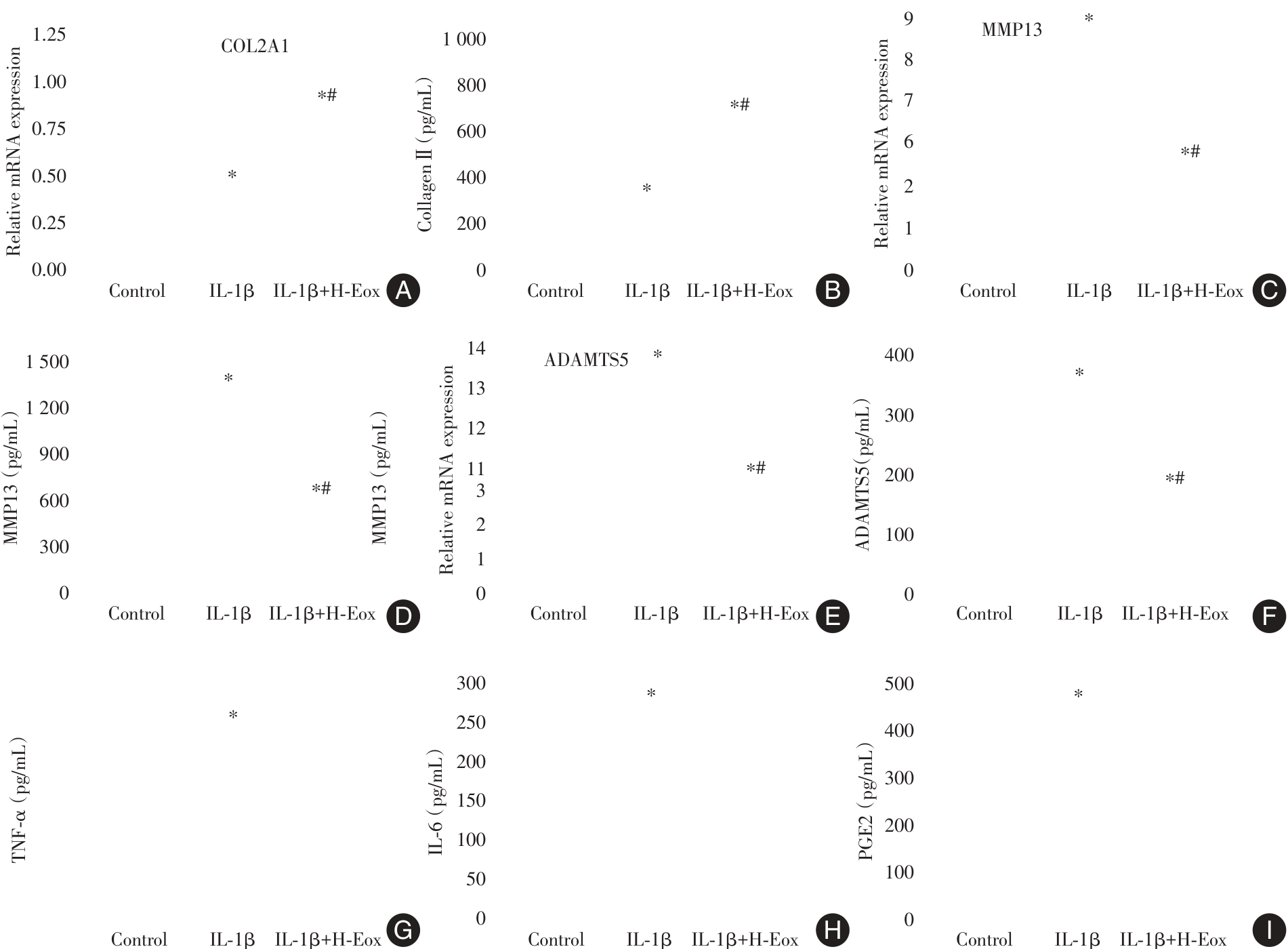
Fig. 4
The effects of exosomes secreted by hypoxia-treated mesenchymal stem cells on the degradation and inflammation of IL-1β-treated chondrocytes"
| [1] | 洪坤豪,吴淮,刘文刚,等. 血清五聚素3和C1q/TNF相关蛋白3对膝关节骨性关节炎患者合并骨质疏松的诊断价值[J]. 实用医学杂志,2022,38(16):2046-2050. |
| [2] | 贾祥,徐田杰,樊佳欣,等. 二甲双胍通过激活SIRT1/p53信号通路对骨关节炎大鼠关节软骨发挥保护作用[J]. 实用医学杂志,2024,40(23):3306-3316. |
| [3] |
ZHANG W, LUO M, XING Y, et al. M2 Macrophage-Derived Extracellular Vehicles-Loaded Hyaluronic Acid-Alginate Hydrogel for Treatment of Osteoarthritis[J]. Orthop Surg, 2025,17(6):1867-1881. doi:10.1111/os.70059
doi: 10.1111/os.70059 |
| [4] |
MAMACHAN M, SHARUN K, BANU S A, et al. Mesenchymal stem cells for cartilage regeneration: Insights into molecular mechanism and therapeutic strategies[J]. Tissue Cell, 2024, 88:102380. doi:10.1016/j.tice.2024.102380
doi: 10.1016/j.tice.2024.102380 |
| [5] |
TAN F, LI X, WANG Z, et al. Clinical applications of stem cell-derived exosomes[J]. Signal Transduct Target Ther, 2024, 9(1):17. doi:10.1038/s41392-023-01704-0
doi: 10.1038/s41392-023-01704-0 |
| [6] |
YU H, HUANG Y, YANG L. Research progress in the use of mesenchymal stem cells and their derived exosomes in the treatment of osteoarthritis[J]. Ageing Res Rev, 2022, 80:101684. doi:10.1016/j.arr.2022.101684
doi: 10.1016/j.arr.2022.101684 |
| [7] |
HAO W, ZHU R, ZHANG H, et al. NS8593 inhibits chondrocyte ferroptosis and alleviates cartilage injury in rat adjuvant arthritis through TRPM7/HO-1 pathway[J]. Int J Biochem Cell Biol, 2024, 174:106618. doi:10.1016/j.biocel.2024.106618
doi: 10.1016/j.biocel.2024.106618 |
| [8] |
XU W, ZHANG B, XI C, et al. Ferroptosis Plays a Role in Human Chondrocyte of Osteoarthritis Induced by IL-1β In Vitro[J]. Cartilage, 2023, 14(4):455-466. doi:10.1177/19476035221142011
doi: 10.1177/19476035221142011 |
| [9] |
VINCENT T L, ALLISTON T, KAPOOR M, et al. Osteoarthritis Pathophysiology: Therapeutic Target Discovery may Require a Multifaceted Approach[J]. Clin Geriatr Med, 2022, 38(2):193-219. doi:10.1016/j.cger.2021.11.015
doi: 10.1016/j.cger.2021.11.015 |
| [10] |
TUNCAY DURUÖZ M, ÖZ N, GÜRSOY D E, et al. Clinical aspects and outcomes in osteoarthritis[J]. Best Pract Res Clin Rheumatol, 2023, 37(2):101855. doi:10.1016/j.berh.2023.101855
doi: 10.1016/j.berh.2023.101855 |
| [11] |
XIAO S Q, CHENG M, WANG L, et al. The role of apoptosis in the pathogenesis of osteoarthritis[J]. Int Orthop, 2023, 47(8):1895-1919. doi:10.1007/s00264-023-05847-1
doi: 10.1007/s00264-023-05847-1 |
| [12] |
AL-HETTY H R A K, ABDULAMEER S J, ALGHAZALI M W, et al. The Role of Ferroptosis in the Pathogenesis of Osteoarthritis[J]. J Membr Biol, 2023, 256(3):223-228. doi:10.1007/s00232-023-00282-0
doi: 10.1007/s00232-023-00282-0 |
| [13] |
LIU Y, ZHANG Z, FANG Y, et al. Ferroptosis in Osteoarthritis: Current Understanding[J]. J Inflamm Res, 2024, 17:8471-8486. doi:10.2147/jir.s493001
doi: 10.2147/jir.s493001 |
| [14] |
PAN X, KONG X, FENG Z, et al. 4-Octyl itaconate protects chondrocytes against IL-1β-induced oxidative stress and ferroptosis by inhibiting GPX4 methylation in osteoarthritis[J]. Int Immunopharmacol, 2024, 137:112531. doi:10.1016/j.intimp.2024.112531
doi: 10.1016/j.intimp.2024.112531 |
| [15] |
LIU J, ZHOU H, CHEN J, et al. Baicalin inhibits IL-1β-induced ferroptosis in human osteoarthritis chondrocytes by activating Nrf-2 signaling pathway[J]. J Orthop Surg Res, 2024, 19(1):23. doi:10.1186/s13018-023-04483-0
doi: 10.1186/s13018-023-04483-0 |
| [16] |
TIAN R, SU S, YU Y, et al. Revolutionizing osteoarthritis treatment: How mesenchymal stem cells hold the key[J]. Biomed Pharmacother, 2024, 173:116458. doi:10.1016/j.biopha.2024.116458
doi: 10.1016/j.biopha.2024.116458 |
| [17] |
HADE M D, SUIRE C N, SUO Z. Mesenchymal Stem Cell-Derived Exosomes: Applications in Regenerative Medicine[J]. Cells, 2021, 10(8):1959. doi:10.3390/cells10081959
doi: 10.3390/cells10081959 |
| [18] |
SHEN K, DUAN A, CHENG J, et al. Exosomes derived from hypoxia preconditioned mesenchymal stem cells laden in a silk hydrogel promote cartilage regeneration via the miR-205-5p/PTEN/AKT pathway[J]. Acta Biomater, 2022, 143:173-188. doi:10.1016/j.actbio.2022.09.005
doi: 10.1016/j.actbio.2022.09.005 |
| [19] |
ZHAO J, SUN Y, SHENG X, et al. Hypoxia-treated adipose mesenchymal stem cell-derived exosomes attenuate lumbar facet joint osteoarthritis[J]. Mol Med, 2023, 29(1):120. doi:10.1186/s10020-023-00709-3
doi: 10.1186/s10020-023-00709-3 |
| [20] |
XU W, ZHANG B, XI C, et al. Ferroptosis Plays a Role in Human Chondrocyte of Osteoarthritis Induced by IL-1β In Vitro[J]. Cartilage, 2023, 14(4):455-466. doi:10.1177/19476035221142011
doi: 10.1177/19476035221142011 |
| [21] |
PENG S, SUN C, LAI C, et al. Exosomes derived from mesenchymal stem cells rescue cartilage injury in osteoarthritis through Ferroptosis by GOT1/CCR2 expression[J]. Int Immunopharmacol, 2023, 122:110566. doi:10.1016/j.intimp.2023.110566
doi: 10.1016/j.intimp.2023.110566 |
| [22] |
LIU Y, WANG Y, YANG M, et al. Exosomes from hypoxic pretreated ADSCs attenuate ultraviolet light-induced skin injury via GLRX5 delivery and ferroptosis inhibition[J]. Photochem Photobiol Sci, 2024, 23(1):55-63. doi:10.1007/s43630-023-00498-y
doi: 10.1007/s43630-023-00498-y |
| [23] |
SHAO M, YE S, CHEN Y, et al. Exosomes from hypoxic ADSCs ameliorate neuronal damage post spinal cord injury through circ-Wdfy3 delivery and inhibition of ferroptosis[J]. Neurochem Int, 2024, 177:105759. doi:10.1016/j.neuint.2024.105759
doi: 10.1016/j.neuint.2024.105759 |
| [1] | Zhicong LIU,Daiyin LIU,Juntian LONG,Chixing CHENG,Jian HUANG. Exosomal lncRNA CIAT1 promotes collective invasion of bladder cancer [J]. The Journal of Practical Medicine, 2025, 41(9): 1299-1308. |
| [2] | Li TANG,Yurong GONG,Liye ZENG,Yanfang GAO,Chengzhe. DENG. Application value of 3.0T magnetic resonance imaging T2 mapping sequence combined with serum nesfatin⁃1 level detection in the diagnosis of elderly knee early osteoarthritis [J]. The Journal of Practical Medicine, 2025, 41(8): 1238-1242. |
| [3] | Xi LI,Xiaoying REN,Yongwei JIAO,Zhipeng SUN,Shilin YIN,Zekun ZHANG,Tianci GAO,Jingxi WANG,Yongwang ZHANG,Lu LIU,Shuangqing. DU. The effect of hip⁃knee⁃ankle active and passive movement therapy on joint function in early and intermediate⁃stage knee osteoarthritis patients [J]. The Journal of Practical Medicine, 2025, 41(6): 829-837. |
| [4] | Junjie ZHAI,Shaoying WEN,Xinru LI,Rui SUN,Ning QI,Qifan ZHANG,Li YANG,Hui HUANG,Lingju MA,Yinju HAO,Yideng JIANG,Guizhong LI,Shengchao. MA. Role of Toll⁃like receptor 4 in regulation of homocysteine⁃induced ferroptosis in macrophages [J]. The Journal of Practical Medicine, 2025, 41(3): 313-321. |
| [5] | Yijia LIU,Bin LIU,Kui HU,Yanfang CHEN,De. CAI. Regulatory effects and mechanisms of exosomes derived from stem cells and macrophages on colitis [J]. The Journal of Practical Medicine, 2025, 41(3): 447-453. |
| [6] | Haiyan LI,Mengzhu LI,Mengxuan CHEN,Da GAO,Kexin DUAN,Lijun ZHAO,Meiling ZHU. The comparison of ferroptosis characteristics and motor deficits in Parkinson′s disease mouse models [J]. The Journal of Practical Medicine, 2025, 41(22): 3501-3509. |
| [7] | Chengcheng LI,Anli LI,Yuhong LIU,Lu WANG,Hong PENG,Hong LI. MCT1⁃mediated lactic acid accumulation and ferroptosis in acute liver failure: A positive feedback loop [J]. The Journal of Practical Medicine, 2025, 41(22): 3520-3528. |
| [8] | Chunhui LIU,Ruipeng WU,Zhiqiang WANG,Wensheng SHAN,Shaojun. LI. Molecular mechanisms and diagnostic value of seminal plasma exosomal miR-26a-5p targeting PTEN in idiopathic teratozoospermia [J]. The Journal of Practical Medicine, 2025, 41(20): 3256-3266. |
| [9] | Chao WANG,Qianqian XU,Shujuan ZHANG,Yanping ZHU. Effect of human umbilical cord mesenchymal stem cell-derived exosomes on microglial polarization in neonatal rats with white matter injury [J]. The Journal of Practical Medicine, 2025, 41(16): 2447-2454. |
| [10] | Yuancheng LI,Li. LI. Androgen‑mediated DGAT2 upregulation promotes ferroptosis in granulosa cells in polycystic ovary syndrome [J]. The Journal of Practical Medicine, 2025, 41(16): 2498-2506. |
| [11] | Huizhen LIU,Xiaonan LU,Ge LIU,Guanqing CAI,Pingan LI,Yingjian ZENG,Guangbin SHANG. The effects and mechanism of total flavonoids of Sarcandra glabra in modulating bone marrow mesenchymal stem cells and their exosomes to promote megakaryocyte differentiation [J]. The Journal of Practical Medicine, 2025, 41(11): 1618-1626. |
| [12] | Guizhi KE,Yu HUANG,Liping FU,Binhua ZOU,Gang. LIU. Research progress on matrix⁃chondrocyte interactions in osteoarthritis [J]. The Journal of Practical Medicine, 2025, 41(10): 1590-1596. |
| [13] | Jing LIU,Chuntao LENG,Yan. WANG. Study on the angiogenic ability of exosomes derived from dental pulp stem cells modified by circRNA SIPA1L1 [J]. The Journal of Practical Medicine, 2024, 40(9): 1211-1217. |
| [14] | Shangzeng WANG,Bei ZHANG,Zhen WANG,Shang MA,Deyang RUANGZHANG,Zhiying YIN,Yunqi ZHU,Kunpeng HU,Shao CHENG. Comparison of the effects of CR and PS prostheses in the treatment of knee osteoarthritis [J]. The Journal of Practical Medicine, 2024, 40(9): 1251-1256. |
| [15] | Fu CHEN,Bin LIU,Shuaijun HE,Yong ZHAO,Weizhou. WANG. The relationship between semen quality and trace element levels in seminal plasma and miR⁃184 levels in seminal vesicles of male infertility patients [J]. The Journal of Practical Medicine, 2024, 40(7): 930-935. |
| Viewed | ||||||
|
Full text |
|
|||||
|
Abstract |
|
|||||